Antibodies directed against phosphorylated proteins often cross-react with related proteins, resulting in cross-reacting bands or a false positive signal. We compared two XP® monoclonal antibodies, Phospho-p44/42 MAPK (Erk1/2) (Thr202/Tyr204) (D13.14.4E) XP® Rabbit mAb #4370 and Phospho-Met (Tyr1234/1235) (D26) XP® Rabbit mAb #3077, to competing products in a number of relevant research applications.
| CST #4370 | Competitor 1 | Competitor 2 | |
|---|---|---|---|
| Western Blot Dilution | 1:2000 | 1:100 | 1:100 |
| Assay Concentration (µg/mL) | 0.222 | 1 | 10 |
| Recommended Applications | W, IP, IHC-P, IF-IC, F | W, IP, IF | W, IHC-P, IHC-F |
| Species Cross-reactivity | H, M, R, Hm, Mk, Mi, Dm, Z, B, Dg, Pg, Sc, (Ce) | H, M, R, Dg | H, M, R, (C, X, Z) |
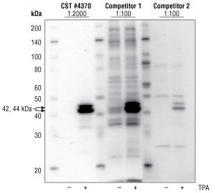
Figure 1. Jurkat cells were either left untreated or treated with the phorbol ester TPA #4174 (200 nM,10 min), following overnight serum-deprivation, to induce phosphorylation of p44/42 protein. #4370 was used at the recommended dilution and both competitor antibodies were used at the highest concentration of the manufacturer’s recommended dilution range. All antibodies showed the expected signal induction upon TPA treatment. Both competitor antibodies showed greater background staining, with competitor 1 showing a number of cross-reacting bands and competitor 2 also showing weaker overall staining.
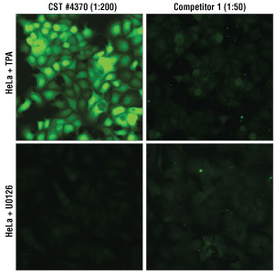
Figure 2. HeLa cells were treated with the MEK 1/2 inhibitor U0126 #9903 (10 µM, 2 hr) or treated with TPA #4174 (200 nM, 30 min), following overnight serum deprivation. #4370 was used at the optimal IF-IC recommended dilution of 1:200 and competitor 1 antibody was tested at a dilution range of 1:50-1:500 per the manufacturer’s suggestion (data not shown). At the concentration determined optimal, the competitor antibody exhibited more background staining, both nuclear and cytoplasmic, in the U0126-treated cells and only weak, diffuse cytoplasmic staining in the stimulated cells. The expected fold-induction following TPA treatment was observed with #4370, but very little induction was observed in the competitor-stained cells and the antibody appeared “dirtier” overall.
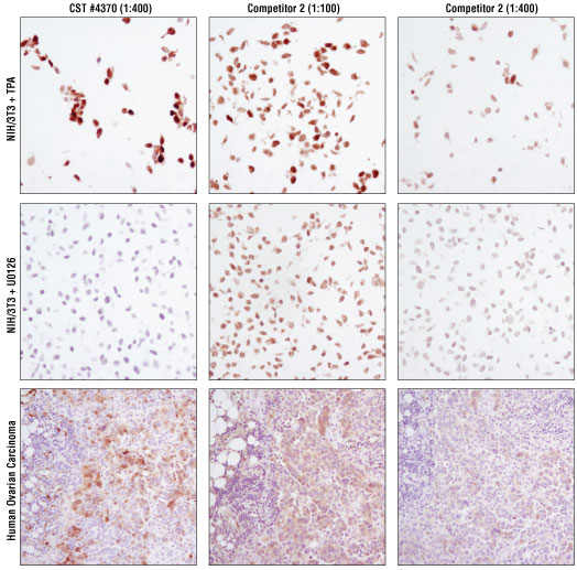
Figure 3. #4370 was compared to a competitor’s IHC-approved antibody tested at two concentrations. IHC analysis was performed on #8103 [paraffin-embedded NIH/3T3 cell pellets, treated with either TPA #4174 (upper) or U0126 #9903 (middle)] and paraffin-embedded human ovarian carcinoma (lower). At the optimal recommended dilution of 1:400, #4370 showed the appropriate staining in the cell pellet system. The competitor 2 antibody required a 1:400 dilution to eliminate staining in the negative control U0126-treated cells. However, signal in the positive control TPA-treated cells was considerably reduced at this dilution. At 1:100 (lowest suggested dilution), the competitor 2 antibody staining was weak in human ovarian carcinoma tissue and cannot be considered specific given the staining observed in U0126-treated cells. Tissue staining was barely present at a 1:400 dilution of the competitor 2 antibody. In contrast, #4370 demonstrated strong specific staining in human ovarian carcinoma.
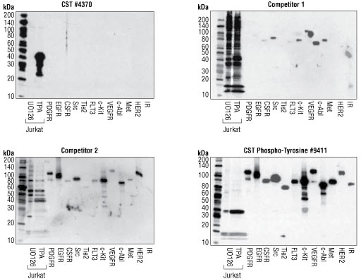
Figure 4. Western blot analysis using a panel of recombinant tyrosine-phosphorylated proteins shows no detectable cross-reactivity using #4370, and significant cross-reactivity with other tyrosine phosphorylated proteins using both competitor antibodies tested. Phospho-Tyrosine Mouse mAb (P-Tyr-100) #9411 was used to demonstrate protein loading and verify molecular weight of the tagged recombinant proteins. These results demonstrate that #4370 displays exceptional specificity.
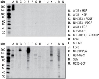
Figure 5. A single band at 145 kDa was observed by western blotting in HGF-stimulated, but not in unstimulated A431 cells using #3077 (upper). Extracts treated with growth factors that activate other RTKs or that overexpress other RTKs or cytoplasmic tyrosine kinases were negative. By comparison, the competitor phospho-Met antibody recognizes several non-specific bands (lower). Both membranes were developed on the same film with the same exposure time (10 seconds).
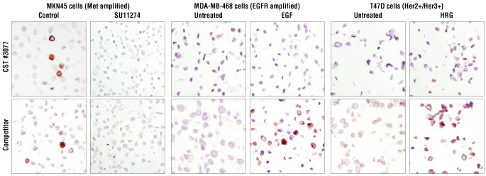
Figure 6. Immunohistochemical analysis of paraffin-embedded MKN45 cells treated with the Met inhibitor SU11274 showed no staining as compared to the control using both #3077 and the competitor’s antibody. EGF treatment and HRG treatment of MDA-MB-468 and T47D breast carcinoma cell lines, respectively, showed no increase in staining compared to control using #3077. In contrast, the competitor’s antibody detected non-specific staining with both treatments, indicating cross-reactivity with other RTKs.
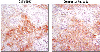
Figure 7. Immunohistochemical analysis of paraffin-embedded HCC827 xenograft comparing #3077 (left) and a competitor’s antibody (right) gives the appearance of specific staining for both products.
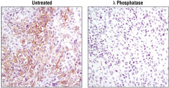
Figure 8. Phosphatase treatment of paraffin-embedded human lung carcinoma confirms phospho-specificity of #3077.