Revision 3
#94845
Store at -20C
877-616-CELL (2355)
877-678-TECH (8324)
3 Trask Lane | Danvers | Massachusetts | 01923 | USA
For Research Use Only. Not for Use in Diagnostic Procedures.
Applications:
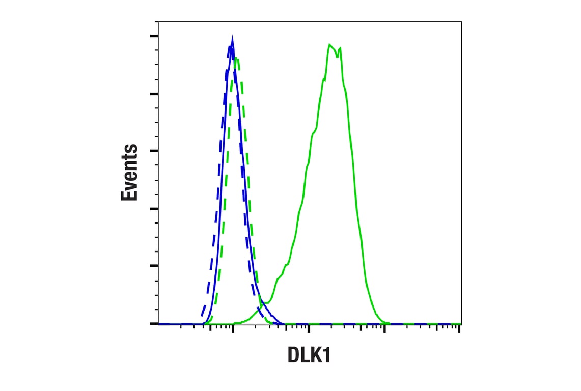
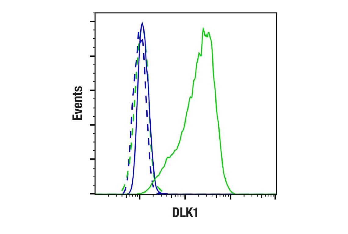
IHC-P, IF-IC, FC-FP, FC-L
Reactivity:
H
Sensitivity:
Endogenous
MW (kDa):
40-60
Source/Isotype:
Rabbit IgG
UniProt ID:
#P80370
Entrez-Gene Id:
8788
Product Usage Information
| Application | Dilution |
|---|---|
| Immunohistochemistry (Paraffin) | 1:50 - 1:200 |
| Immunofluorescence (Immunocytochemistry) | 1:400 - 1:1600 |
| Flow Cytometry (Fixed/Permeabilized) | 1:200 - 1:800 |
| Flow Cytometry (Live) | 1:1800 - 1:7200 |
Storage
For a carrier free (BSA and azide free) version of this product see product #47633.
Specificity/Sensitivity
Source / Purification
Background
Humans and rodents express multiple DLK1 isoforms, which are either membrane-bound or contain an ADAM17/TACE cleavage site for release of the soluble ectodomain (8). As high DLK1 expression is pro-oncogenic in some contexts, differential isoform expression may promote cancer cell survival, with both the ectodomain and intracellular domain having distinct functions (9). Under hypoxic conditions, HIF proteins induce ADAM17/TACE cleavage and internalization of the DLK1 intracellular domain, which localizes to the nucleus and alters Akt and p53 signaling cascades in glioma (10). Hypoxia increases DLK1 expression, and phosphorylation of the DLK1 C-terminus at Tyr339 and Ser355 increases neuronal tumor sphere growth (11). Nuclear DLK1 directly interacts with tumor suppressor NCoR1, correlating with poor prognosis in non-small cell lung cancer (NSCLC) (12). DLK1 has emerged as a target for novel antibody drug conjugates (ADCs) in neuroblastoma and adrenocortical carcinoma (13,14).
Background References
- Laborda, J. et al. (1993) J Biol Chem 268, 3817-20.
- Smas, C.M. and Sul, H.S. (1993) Cell 73, 725-34.
- Kobayashi, S. et al. (2000) Genes Cells 5, 1029-37.
- Moon, Y.S. et al. (2002) Mol Cell Biol 22, 5585-92.
- Smas, C.M. et al. (1997) Mol Cell Biol 17, 977-88.
- Mei, B. et al. (2002) Biochem J 364, 137-44.
- Wang, Y. et al. (2006) J Nutr 136, 2953-6.
- Grassi, E.S. and Pietras, A. (2022) J Histochem Cytochem 70, 17-28.
- Pittaway, J.F.H. et al. (2021) Endocr Relat Cancer 28, R271-R287.
- Grassi, E.S. et al. (2020) Oncogene 39, 4028-4044.
- Kim, Y. et al. (2009) Cancer Res 69, 9271-80.
- Tan, J. et al. (2019) Biosci Rep 39, BSR20192362. doi: 10.1042/BSR20192362.
- Hamilton, A.K. et al. (2024) Cancer Cell 42, 1970-1982.e7.
- Sun, N.Y. et al. (2024) bioRxiv , 0.09.617077. doi: 10.1101/2024.10.09.617077.
Species Reactivity
Species reactivity is determined by testing in at least one approved application (e.g., western blot).
Applications Key
IHC-P: Immunohistochemistry (Paraffin) IF-IC: Immunofluorescence (Immunocytochemistry) FC-FP: Flow Cytometry (Fixed/Permeabilized) FC-L: Flow Cytometry (Live)
Cross-Reactivity Key
H: Human
Trademarks and Patents
Cell Signaling Technology is a trademark of Cell Signaling Technology, Inc.
All other trademarks are the property of their respective owners. Visit cellsignal.com/trademarks for more information.
Limited Uses
Except as otherwise expressly agreed in a writing signed by a legally authorized representative of CST, the following terms apply to Products provided by CST, its affiliates or its distributors. Any Customer's terms and conditions that are in addition to, or different from, those contained herein, unless separately accepted in writing by a legally authorized representative of CST, are rejected and are of no force or effect.
Products are labeled with For Research Use Only or a similar labeling statement and have not been approved, cleared, or licensed by the FDA or other regulatory foreign or domestic entity, for any purpose. Customer shall not use any Product for any diagnostic or therapeutic purpose, or otherwise in any manner that conflicts with its labeling statement. Products sold or licensed by CST are provided for Customer as the end-user and solely for research and development uses. Any use of Product for diagnostic, prophylactic or therapeutic purposes, or any purchase of Product for resale (alone or as a component) or other commercial purpose, requires a separate license from CST. Customer shall (a) not sell, license, loan, donate or otherwise transfer or make available any Product to any third party, whether alone or in combination with other materials, or use the Products to manufacture any commercial products, (b) not copy, modify, reverse engineer, decompile, disassemble or otherwise attempt to discover the underlying structure or technology of the Products, or use the Products for the purpose of developing any products or services that would compete with CST products or services, (c) not alter or remove from the Products any trademarks, trade names, logos, patent or copyright notices or markings, (d) use the Products solely in accordance with CST Product Terms of Sale and any applicable documentation, and (e) comply with any license, terms of service or similar agreement with respect to any third party products or services used by Customer in connection with the Products.
Revision 3
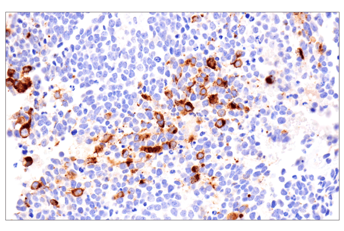
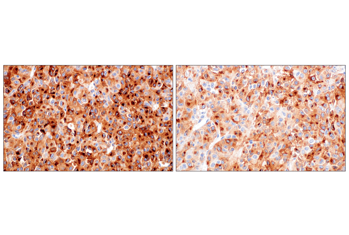
Immunohistochemical analysis of paraffin-embedded human adrenal neuroblastoma using DLK1 (F7O3Q) Rabbit mAb.
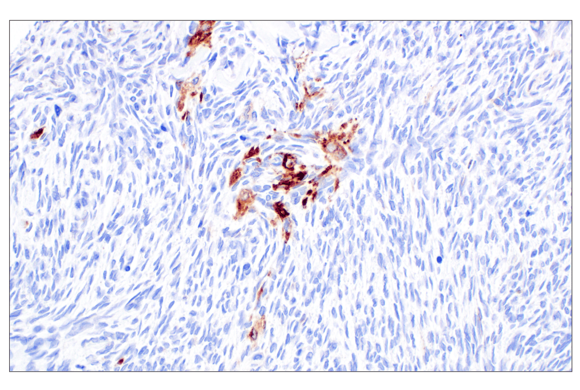
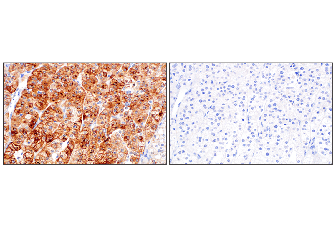
Immunohistochemical analysis of paraffin-embedded normal human testis using DLK1 (F7O3Q) Rabbit mAb.

Immunohistochemical analysis of paraffin-embedded normal human ovary using DLK1 (F7O3Q) Rabbit mAb.
Revision 3
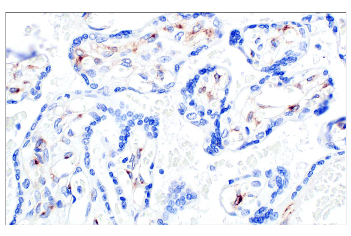
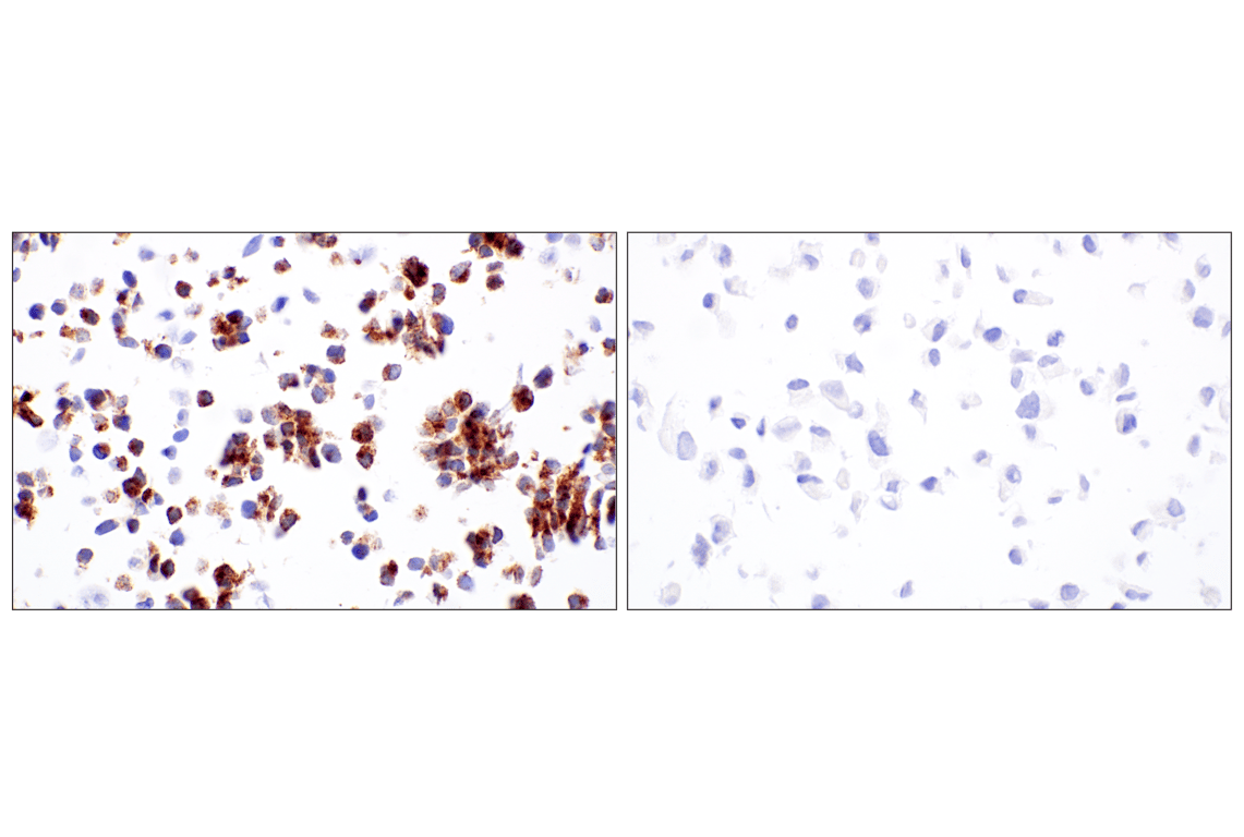
Immunohistochemical analysis of paraffin-embedded normal human placenta using DLK1 (F7O3Q) Rabbit mAb.
Revision 3

Revision 3