Revision 3
#7240
Store at +4C
PathScan® Phospho-EGF Receptor (Tyr1068) Sandwich ELISA Kit
1 Kit
(96 assays)
Species Cross Reactivity:
H
UniProt ID:
#P00533
Entrez-Gene Id:
#1956
877-616-CELL (2355)
877-678-TECH (8324)
3 Trask Lane | Danvers | Massachusetts | 01923 | USA
For Research Use Only. Not for Use in Diagnostic Procedures.
| Product Includes | Product # | Quantity | Color | Storage Temp |
|---|---|---|---|---|
| EGF Receptor Mouse mAb Coated Microwells | 91179 | 96 tests | +4C | |
| Phospho-EGF Receptor (Tyr1068) Rabbit Detection mAb | 13019 | 1 ea | Green (Lyophilized) | +4C |
| Anti-rabbit IgG, HRP-linked Antibody (ELISA Formulated) | 13272 | 1 ea | Red (Lyophilized) | +4C |
| Detection Antibody Diluent | 13339 | 11 ml | Green | +4C |
| HRP Diluent | 13515 | 11 ml | Red | +4C |
| TMB Substrate | 7004 | 11 ml | +4C | |
| STOP Solution | 7002 | 11 ml | +4C | |
| Sealing Tape | 54503 | 2 ea | +4C | |
| ELISA Wash Buffer (20X) | 9801 | 25 ml | +4C | |
| ELISA Sample Diluent | 11083 | 25 ml | Blue | +4C |
| Cell Lysis Buffer (10X) | 9803 | 15 ml | -20C |
*The microwell plate is supplied as 12 8-well modules - Each module is designed to break apart for 8 tests.
Description
CST's PathScan® Phospho-EGF Receptor (Tyr1068) Sandwich ELISA Kit is a solid phase sandwich enzyme-linked immunosorbent assay (ELISA) that detects endogenous levels of phospho-EGF Receptor (Tyr1068) protein. A EGF Receptor Mouse mAb has been coated onto the microwells. After incubation with cell lysates, both phospho- and nonphospho-EGF Receptor proteins are captured by the coated antibody. Following extensive washing, Phospho-EGF Receptor (Tyr1068) Rabbit mAb is added to detect the captured phospho-EGF Receptor protein. Anti-rabbit IgG, HRP-linked Antibody is then used to recognize the bound detection antibody. HRP substrate, TMB, is added to develop color. The magnitude of optical density for this developed color is proportional to the quantity of phospho-EGF Receptor (Tyr1068) protein.
*Antibodies in kit are custom formulations specific to kit.
Specificity/Sensitivity
CST's PathScan® Phospho-EGF Receptor (Tyr1068) Sandwich ELISA Kit detects endogenous levels of phospho-EGF Receptor (Tyr 1068) protein. As shown in Figure 1, using the Phospho-EGF Receptor (Tyr1068) ELISA Kit #7240, a significant induction of Phospho-EGF Receptor (Tyr1068) in A-431 cells treated with EGF is detected. However, levels of total EGF Receptor (phospho and non-phospho) detected by PathScan® Total EGF Receptor Sandwich ELISA Kit #7250, remain unchanged (Figure 1).
This kit detects proteins from the indicated species, as determined through in-house testing, but may also detect homologous proteins from other species.
Background
The epidermal growth factor (EGF) receptor is a transmembrane tyrosine kinase that belongs to the HER/ErbB protein family. Ligand binding results in receptor dimerization, autophosphorylation, activation of downstream signaling, internalization, and lysosomal degradation (1,2). Phosphorylation of EGF receptor (EGFR) at Tyr845 in the kinase domain is implicated in stabilizing the activation loop, maintaining the active state enzyme, and providing a binding surface for substrate proteins (3,4). c-Src is involved in phosphorylation of EGFR at Tyr845 (5). The SH2 domain of PLCγ binds at phospho-Tyr992, resulting in activation of PLCγ-mediated downstream signaling (6). Phosphorylation of EGFR at Tyr1045 creates a major docking site for the adaptor protein c-Cbl, leading to receptor ubiquitination and degradation following EGFR activation (7,8). The GRB2 adaptor protein binds activated EGFR at phospho-Tyr1068 (9). A pair of phosphorylated EGFR residues (Tyr1148 and Tyr1173) provide a docking site for the Shc scaffold protein, with both sites involved in MAP kinase signaling activation (2). Phosphorylation of EGFR at specific serine and threonine residues attenuates EGFR kinase activity. EGFR carboxy-terminal residues Ser1046 and Ser1047 are phosphorylated by CaM kinase II; mutation of either of these serines results in upregulated EGFR tyrosine autophosphorylation (10).
Background References
- Hackel, P.O. et al. (1999) Curr Opin Cell Biol 11, 184-9.
- Zwick, E. et al. (1999) Trends Pharmacol Sci 20, 408-12.
- Cooper, J.A. and Howell, B. (1993) Cell 73, 1051-4.
- Hubbard, S.R. et al. (1994) Nature 372, 746-54.
- Biscardi, J.S. et al. (1999) J Biol Chem 274, 8335-43.
- Emlet, D.R. et al. (1997) J Biol Chem 272, 4079-86.
- Levkowitz, G. et al. (1999) Mol Cell 4, 1029-40.
- Ettenberg, S.A. et al. (1999) Oncogene 18, 1855-66.
- Rojas, M. et al. (1996) J Biol Chem 271, 27456-61.
- Feinmesser, R.L. et al. (1999) J Biol Chem 274, 16168-73.
Cross-Reactivity Key
H: Human M: Mouse R: Rat Hm: Hamster Mk: Monkey Vir: Virus Mi: Mink C: Chicken Dm: D. melanogaster X: Xenopus Z: Zebrafish B: Bovine Dg: Dog Pg: Pig Sc: S. cerevisiae Ce: C. elegans Hr: Horse GP: Guinea Pig Rab: Rabbit G: Goat All: All Species Expected
Trademarks and Patents
Cell Signaling Technology is a trademark of Cell Signaling Technology, Inc.
PathScan is a registered trademark of Cell Signaling Technology, Inc.
All other trademarks are the property of their respective owners. Visit cellsignal.com/trademarks for more information.
Limited Uses
Except as otherwise expressly agreed in a writing signed by a legally authorized representative of CST, the following terms apply to Products provided by CST, its affiliates or its distributors. Any Customer's terms and conditions that are in addition to, or different from, those contained herein, unless separately accepted in writing by a legally authorized representative of CST, are rejected and are of no force or effect.
Products are labeled with For Research Use Only or a similar labeling statement and have not been approved, cleared, or licensed by the FDA or other regulatory foreign or domestic entity, for any purpose. Customer shall not use any Product for any diagnostic or therapeutic purpose, or otherwise in any manner that conflicts with its labeling statement. Products sold or licensed by CST are provided for Customer as the end-user and solely for research and development uses. Any use of Product for diagnostic, prophylactic or therapeutic purposes, or any purchase of Product for resale (alone or as a component) or other commercial purpose, requires a separate license from CST. Customer shall (a) not sell, license, loan, donate or otherwise transfer or make available any Product to any third party, whether alone or in combination with other materials, or use the Products to manufacture any commercial products, (b) not copy, modify, reverse engineer, decompile, disassemble or otherwise attempt to discover the underlying structure or technology of the Products, or use the Products for the purpose of developing any products or services that would compete with CST products or services, (c) not alter or remove from the Products any trademarks, trade names, logos, patent or copyright notices or markings, (d) use the Products solely in accordance with CST Product Terms of Sale and any applicable documentation, and (e) comply with any license, terms of service or similar agreement with respect to any third party products or services used by Customer in connection with the Products.
Revision 3
Figure 1: Treatment of A431 cells with EGF stimulates phosphorylation of EGF Receptor at Tyr1068, detected by PathScan® Phospho-EGF Receptor (Tyr1068) Sandwich ELISA kit #7240, but does not affect the level of total EGF Receptor detected by PathScan® Total EGF Receptor Sandwich ELISA kit #7250. OD 450 nm readings are shown in the top figure, while the corresponding Western blot using Phospho-EGF Receptor (Tyr1068) Antibody #2234 (right panel) or EGF Receptor Antibody #2232 (left panel), is shown in the bottom figure.
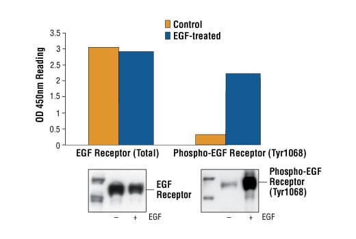
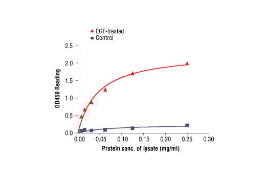
Figure 2: The relationship between protein concentration of lysates from untreated and EGF-treated A431 cells and kit assay optical density readings. After starvation, A431 cells (85% confluence) were treated with EGF (100 ng/ml) for 5 min at 37°C, and then lysed.