Revision 2
#17450
Store at +4C
PathScan® Phospho-MLKL (Ser345) Chemiluminescent Sandwich ELISA Kit
1 Kit
(96 assays)
Species Cross Reactivity:
M
UniProt ID:
#Q9D2Y4
Entrez-Gene Id:
#74568
877-616-CELL (2355)
877-678-TECH (8324)
3 Trask Lane | Danvers | Massachusetts | 01923 | USA
For Research Use Only. Not for Use in Diagnostic Procedures.
| Product Includes | Product # | Quantity | Color | Storage Temp |
|---|---|---|---|---|
| Phospho-MLKL (Ser345) Rabbit mAb Coated Microwells | 33298 | 96 tests | +4C | |
| MLKL Mouse Detection mAb | 52942 | 1 ea | Green (Lyophilized) | +4C |
| Anti-mouse IgG, HRP-linked Antibody (ELISA Formulated) | 13304 | 1 ea | Red (Lyophilized) | +4C |
| Detection Antibody Diluent | 13339 | 5.5 ml | Green | +4C |
| HRP Diluent | 13515 | 5.5 ml | Red | +4C |
| Luminol/Enhancer Solution | 84850 | 3 ml | RT | |
| Stable Peroxide Buffer | 42552 | 3 ml | RT | |
| Sealing Tape | 54503 | 2 ea | +4C | |
| ELISA Wash Buffer (20X) | 9801 | 25 ml | +4C | |
| ELISA Sample Diluent | 11083 | 25 ml | Blue | +4C |
| Cell Lysis Buffer (10X) | 9803 | 15 ml | -20C |
*The microwell plate is supplied as 12 8-well modules - Each module is designed to break apart for 8 tests.
Description
The PathScan® Phospho-MLKL (Ser345) Chemiluminescent Sandwich ELISA Kit is a solid phase sandwich enzyme-linked immunosorbent assay (ELISA) that detects endogenous levels of MLKL protein phosphorylated at Ser345. A phospho-MLKL (Ser345) rabbit antibody has been coated onto the microwells. After incubation with cell lysates, MLKL protein phosphorylated at Ser345 is captured by the coated antibody. Following extensive washing, an MLKL mouse detection antibody is added to detect the captured phospho-MLKL (Ser345) protein. Anti-mouse IgG, HRP-linked Antibody is then used to recognize the bound detection antibody. Chemiluminescent reagent is added for signal development. The magnitude of light emission, measured in relative light units (RLU), is proportional to the quantity of phospho-MLKL (Ser345) protein.
*Antibodies in this kit are custom formulations specific to kit.
Specificity/Sensitivity
The PathScan® Phospho-MLKL (Ser345) Chemiluminescent Sandwich ELISA Kit detects endogenous levels of MLKL protein phosphorylated at Ser345. The kit sensitivity is shown in Figure 1. This kit detects proteins from the indicated species, as determined through in-house testing, but may also detect homologous proteins from other species.
Background
Necroptosis, a regulated pathway for necrotic cell death, is triggered by a number of inflammatory signals including cytokines in the tumor necrosis factor (TNF) family, pathogen sensors such as toll-like receptors (TLRs), and ischemic injury (1,2). The process is negatively regulated by caspases and is initiated through a complex containing the RIP1 and RIP3 kinases, typically referred to as the necrosome. Mixed lineage kinase domain-like protein (MLKL) is a pseudokinase that was identified as a downstream target of RIP3 in the necroptosis pathway (3,4). During necroptosis RIP3 is phosphorylated at Ser227, which recruits MLKL and leads to its phosphorylation at Thr357 and Ser358 (3). Knockdown of MLKL through multiple mechanisms results in inhibition of necroptosis (3-5). While the precise mechanism for MLKL-induced necroptosis is unclear, some studies have shown that necroptosis leads to oligomerization of MLKL and translocation to the plasma membrane, where it affects membrane integrity (6-9).
Background References
- Christofferson, D.E. and Yuan, J. (2010) Curr Opin Cell Biol 22, 263-8.
- Kaczmarek, A. et al. (2013) Immunity 38, 209-23.
- Sun, L. et al. (2012) Cell 148, 213-27.
- Wang, Z. et al. (2012) Cell 148, 228-43.
- Wu, J. et al. (2013) Cell Res 23, 994-1006.
- Cai, Z. et al. (2014) Nat Cell Biol 16, 55-65.
- Chen, X. et al. (2014) Cell Res 24, 105-21.
- Wang, H. et al. (2014) Mol Cell 54, 133-46.
- Dondelinger, Y. et al. (2014) Cell Rep 7, 971-81.
Cross-Reactivity Key
H: Human M: Mouse R: Rat Hm: Hamster Mk: Monkey Vir: Virus Mi: Mink C: Chicken Dm: D. melanogaster X: Xenopus Z: Zebrafish B: Bovine Dg: Dog Pg: Pig Sc: S. cerevisiae Ce: C. elegans Hr: Horse GP: Guinea Pig Rab: Rabbit G: Goat All: All Species Expected
Trademarks and Patents
Cell Signaling Technology is a trademark of Cell Signaling Technology, Inc.
PathScan is a registered trademark of Cell Signaling Technology, Inc.
All other trademarks are the property of their respective owners. Visit cellsignal.com/trademarks for more information.
Limited Uses
Except as otherwise expressly agreed in a writing signed by a legally authorized representative of CST, the following terms apply to Products provided by CST, its affiliates or its distributors. Any Customer's terms and conditions that are in addition to, or different from, those contained herein, unless separately accepted in writing by a legally authorized representative of CST, are rejected and are of no force or effect.
Products are labeled with For Research Use Only or a similar labeling statement and have not been approved, cleared, or licensed by the FDA or other regulatory foreign or domestic entity, for any purpose. Customer shall not use any Product for any diagnostic or therapeutic purpose, or otherwise in any manner that conflicts with its labeling statement. Products sold or licensed by CST are provided for Customer as the end-user and solely for research and development uses. Any use of Product for diagnostic, prophylactic or therapeutic purposes, or any purchase of Product for resale (alone or as a component) or other commercial purpose, requires a separate license from CST. Customer shall (a) not sell, license, loan, donate or otherwise transfer or make available any Product to any third party, whether alone or in combination with other materials, or use the Products to manufacture any commercial products, (b) not copy, modify, reverse engineer, decompile, disassemble or otherwise attempt to discover the underlying structure or technology of the Products, or use the Products for the purpose of developing any products or services that would compete with CST products or services, (c) not alter or remove from the Products any trademarks, trade names, logos, patent or copyright notices or markings, (d) use the Products solely in accordance with CST Product Terms of Sale and any applicable documentation, and (e) comply with any license, terms of service or similar agreement with respect to any third party products or services used by Customer in connection with the Products.
Revision 2
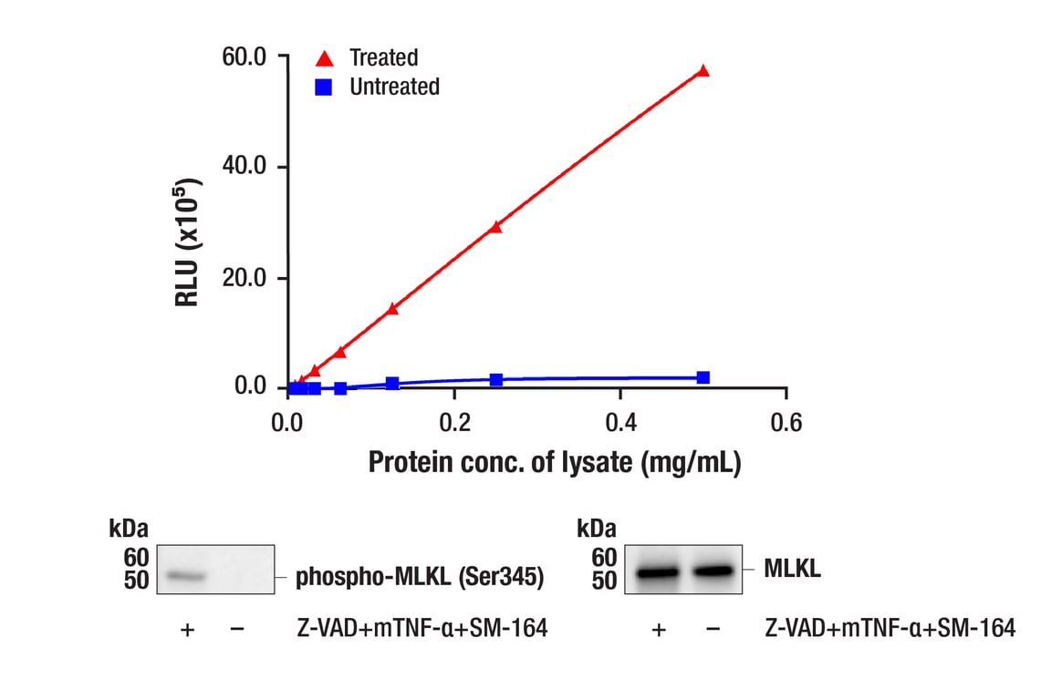
Figure 1. Treatment of L-929 cells with Z-VAD followed by addition of mouse TNF-α and SM-164 stimulates phosphorylation of MLKL protein at Ser345 but does not affect the level of total MLKL protein. The relationship between lysate protein concentration from untreated and treated L-929 cells and immediate light generation with chemiluminescent substrate using the PathScan® Phospho-MLKL (Ser345) Chemiluminescent Sandwich ELISA Kit #17450 is shown in the upper figure. The corresponding western blots using phospho-MLKL (Ser345) antibody (left panel) and MLKL antibody (right panel) are shown in the lower figure. L-929 cells were either left untreated or pre-treated with Z-VAD (20 μM, 30 min), then treated with mouse TNF-α (20 ng/mL) and SM-164 (100 nM) for 4 hr and then lysed.