Revision 1
#14511
Store at -20C
877-616-CELL (2355)
877-678-TECH (8324)
3 Trask Lane | Danvers | Massachusetts | 01923 | USA
For Research Use Only. Not for Use in Diagnostic Procedures.
| Product Includes | Product # | Quantity | Mol. Wt | Isotype/Source |
|---|---|---|---|---|
| Phospho-Tau (Thr181) (D9F4G) Rabbit mAb | 12885 | 100 µl | 50-80 kDa | Rabbit IgG |
| Tau (D1M9X) XP® Rabbit mAb | 46687 | 100 µl | 50-80 kDa | Rabbit IgG |
Please visit cellsignal.com for individual component applications, species cross-reactivity, dilutions, protocols, and additional product information.
UniProt ID:
#P10636-8
Entrez-Gene Id:
4137
Description
PhosphoPlus® Duets from Cell Signaling Technology (CST) provide a means to assess protein activation status. Each Duet contains an activation-state and total protein antibody to your target of interest. These antibodies have been selected from CST's product offering based upon superior performance in specified applications.
Storage
Background
Tau is a heterogeneous microtubule-associated protein that promotes and stabilizes microtubule assembly, especially in axons. Six isoforms with different amino-terminal inserts and different numbers of tandem repeats near the carboxy terminus have been identified, and tau is hyperphosphorylated at approximately 25 sites by Erk, glycogen synthase kinase-3 (GSK-3), and CDK5 (1,2). Phosphorylation decreases the ability of tau to bind to microtubules. Neurofibrillary tangles are a major hallmark of Alzheimer's disease (AD); these tangles are bundles of paired helical filaments (PHFs) composed of hyperphosphorylated tau. In particular, phosphorylation at Ser396 by GSK-3 or CDK5 destabilizes microtubules. Furthermore, research studies have shown that inclusions of tau are found in a number of other neurodegenerative diseases, collectively known as tauopathies (1,3).
The cerebrospinal fluid concentration of Tau phosphorylated at Thr181 has been proposed to be a biomarker for the study of neurodegenerative disorders (4).
Trademarks and Patents
Cell Signaling Technology is a trademark of Cell Signaling Technology, Inc.
PhosphoPlus is a registered trademark of Cell Signaling Technology, Inc.
XP is a registered trademark of Cell Signaling Technology, Inc.
All other trademarks are the property of their respective owners. Visit cellsignal.com/trademarks for more information.
Limited Uses
Except as otherwise expressly agreed in a writing signed by a legally authorized representative of CST, the following terms apply to Products provided by CST, its affiliates or its distributors. Any Customer's terms and conditions that are in addition to, or different from, those contained herein, unless separately accepted in writing by a legally authorized representative of CST, are rejected and are of no force or effect.
Products are labeled with For Research Use Only or a similar labeling statement and have not been approved, cleared, or licensed by the FDA or other regulatory foreign or domestic entity, for any purpose. Customer shall not use any Product for any diagnostic or therapeutic purpose, or otherwise in any manner that conflicts with its labeling statement. Products sold or licensed by CST are provided for Customer as the end-user and solely for research and development uses. Any use of Product for diagnostic, prophylactic or therapeutic purposes, or any purchase of Product for resale (alone or as a component) or other commercial purpose, requires a separate license from CST. Customer shall (a) not sell, license, loan, donate or otherwise transfer or make available any Product to any third party, whether alone or in combination with other materials, or use the Products to manufacture any commercial products, (b) not copy, modify, reverse engineer, decompile, disassemble or otherwise attempt to discover the underlying structure or technology of the Products, or use the Products for the purpose of developing any products or services that would compete with CST products or services, (c) not alter or remove from the Products any trademarks, trade names, logos, patent or copyright notices or markings, (d) use the Products solely in accordance with CST Product Terms of Sale and any applicable documentation, and (e) comply with any license, terms of service or similar agreement with respect to any third party products or services used by Customer in connection with the Products.
Revision 1
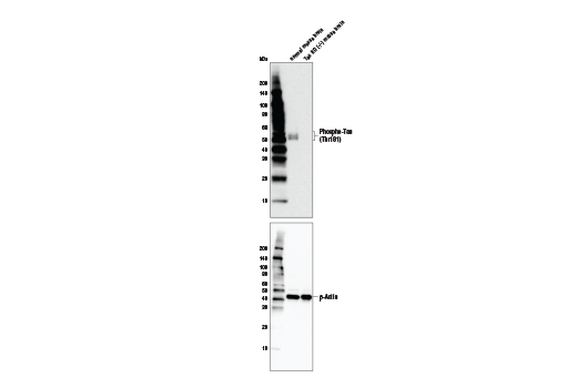
Western blot analysis of normal mouse brain and Tau KO (-/-) mouse brain with Phospho-Tau (Thr181) (D9F4G) Rabbit mAb (upper) and β-Actin (D6A8) Rabbit mAb #8457 (lower). Tau-KO mouse brain tissue was kindly provided by Dr. Dominic Walsh at Brigham and Women's Hospital and Harvard Medical School.
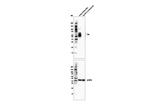
Western blot analysis of normal mouse brain and Tau KO (-/-) mouse brain with Tau (D1M9X) XP® Rabbit mAb (upper) and β-Actin (D6A8) Rabbit mAb #8457 (lower). Tau-KO mouse brain tissue was kindly provided by Dr. Dominic Walsh at Brigham and Women's Hospital and Harvard Medical School.
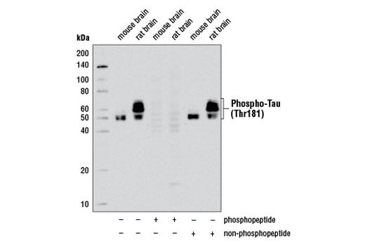
Western blot analysis of extracts from mouse and rat brain using Phospho-Tau (Thr181) (D9F4G) Rabbit mAb. The phospho-specificity of Phospho-Tau (Thr181) (D9F4G) Rabbit mAb was verified by peptide blocking using a phosphopeptide or non-phosphopeptide targeting residue Thr181.
Revision 1
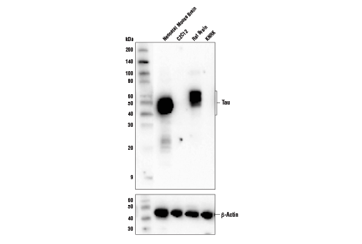
Western blot analysis of extracts from various cell lines and tissues using Tau (D1M9X) XP® Rabbit mAb (upper) and β-Actin (D6A8) Rabbit mAb #8457 (lower).
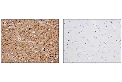
Immunohistochemical analysis of paraffin-embedded human Alzheimer's brain using Tau (D1M9X) XP® Rabbit mAb in the presence of control peptide (left) or antigen-specific peptide (right).
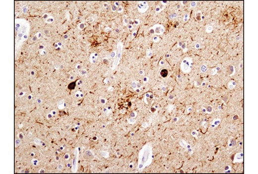
Immunohistochemical analysis of paraffin-embedded human Alzheimer's brain using Phospho-Tau (Thr181) (D9F4G) Rabbit mAb.
Revision 1
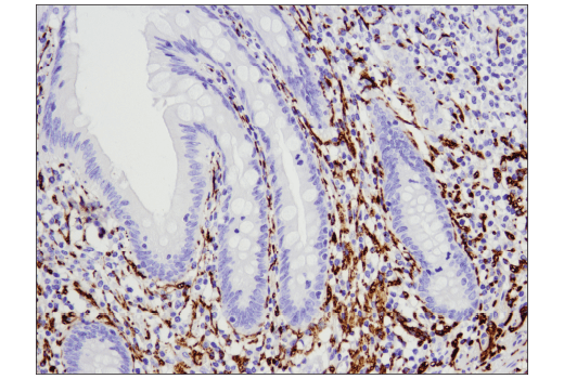
Immunohistochemical analysis of paraffin-embedded human normal appendix using Tau (D1M9X) XP® Rabbit mAb.
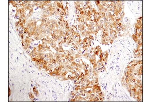
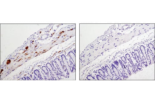
Immunohistochemical analysis of paraffin-embedded human breast carcinoma using Phospho-Tau (Thr181) (D9F4G) Rabbit mAb.
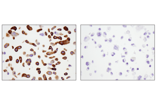
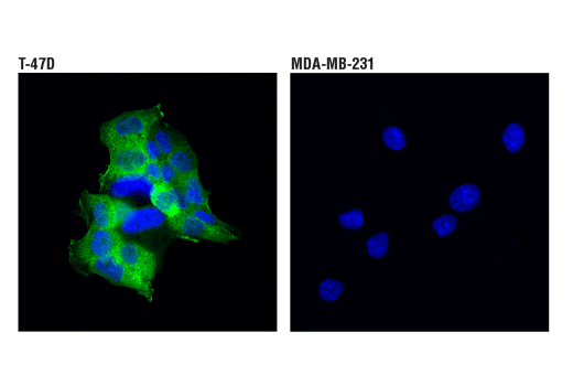
Immunohistochemical analysis of paraffin-embedded T-47D cell pellet (left, positive) or MDA-MB-231 cell pellet (right, negative) using Tau (D1M9X) XP® Rabbit mAb.
Revision 1
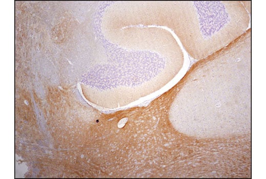
Immunohistochemical analysis of paraffin-embedded mouse brain using Phospho-Tau (Thr181) (D9F4G) Rabbit mAb.
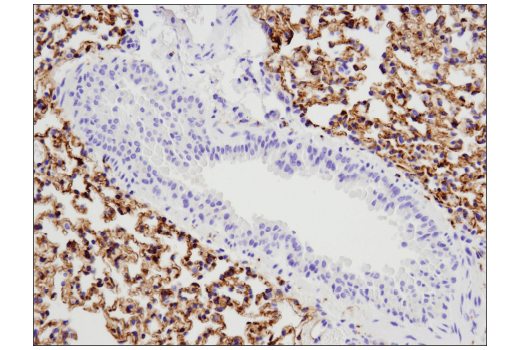
Immunohistochemical analysis of paraffin-embedded mouse lung using Tau (D1M9X) XP® Rabbit mAb.
Immunohistochemical analysis of paraffin-embedded mouse colon, control (left) or λ phosphatase-treated (right), using Phospho-Tau (Thr181) (D9F4G) Rabbit mAb.
Revision 1
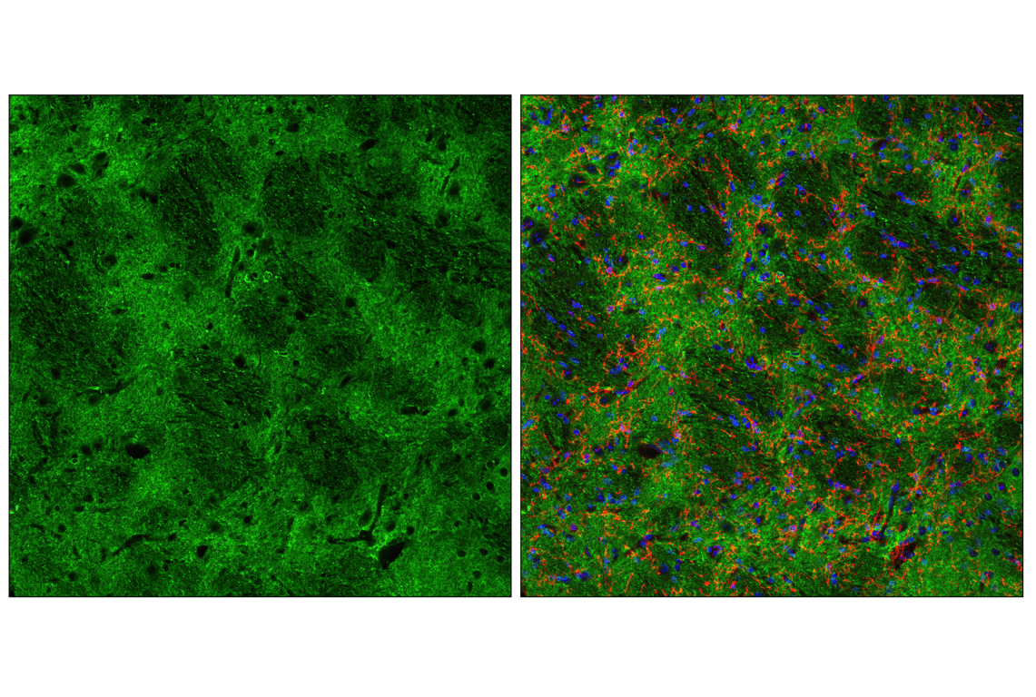
Confocal immunofluorescent analysis of fixed frozen mouse striatum using Tau (D1M9X) XP® Rabbit mAb (green), TMEM119 (E4B9S) Mouse mAb #98778 (red) and ProLong® Gold Antifade Reagent with DAPI #8961 (blue).
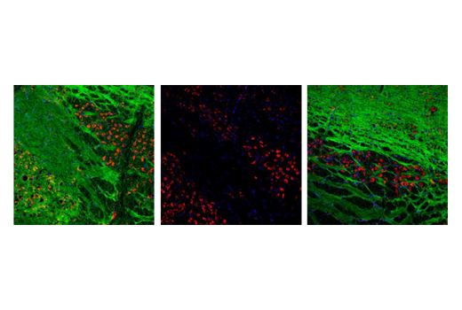
Confocal immunofluorescent analysis of mouse brainstem using Phospho-Tau (Thr181) (D9F4G) Rabbit mAb #12885 (green) and S6 Ribosomal Protein (54D2) Mouse mAb #2317 (red). Antibody was pre-incubated with non-phospho-Tau (Thr181) peptide (left), a phospho-Tau (Thr181) peptide (middle), or without peptide (right) to confirm phospho-specificity. Samples were mounted in ProLong® Gold Antifade Reagent with DAPI #8961 (blue).
Confocal immunofluorescent analysis of T-47D (positive, left) or MDA-MB-231 (negative, right) cells using Tau (D1M9X) XP® Rabbit mAb (green). Blue pseudocolor = DRAQ5® #4084 (fluorescent DNA dye).
Revision 1
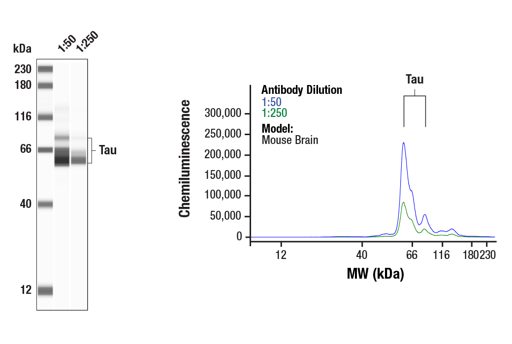
Simple Western™ analysis of lysates (0.1 mg/mL) from Mouse Brain Tissue Extracts using Tau (D1M9X) XP® Rabbit #46687. The virtual lane view (left) shows the target band (as indicated) and a band corresponding to Tau (as indicated) at 1:50 and 1:250 dilutions of primary antibody. The corresponding electropherogram view (right) plots chemiluminescence by molecular weight along the capillary at 1:50 (blue line) and 1:250 (green line) dilutions of primary antibody. This experiment was performed under reducing conditions on the Jess™ Simple Western instrument from ProteinSimple, a BioTechne brand, using the 12-230 kDa separation module.
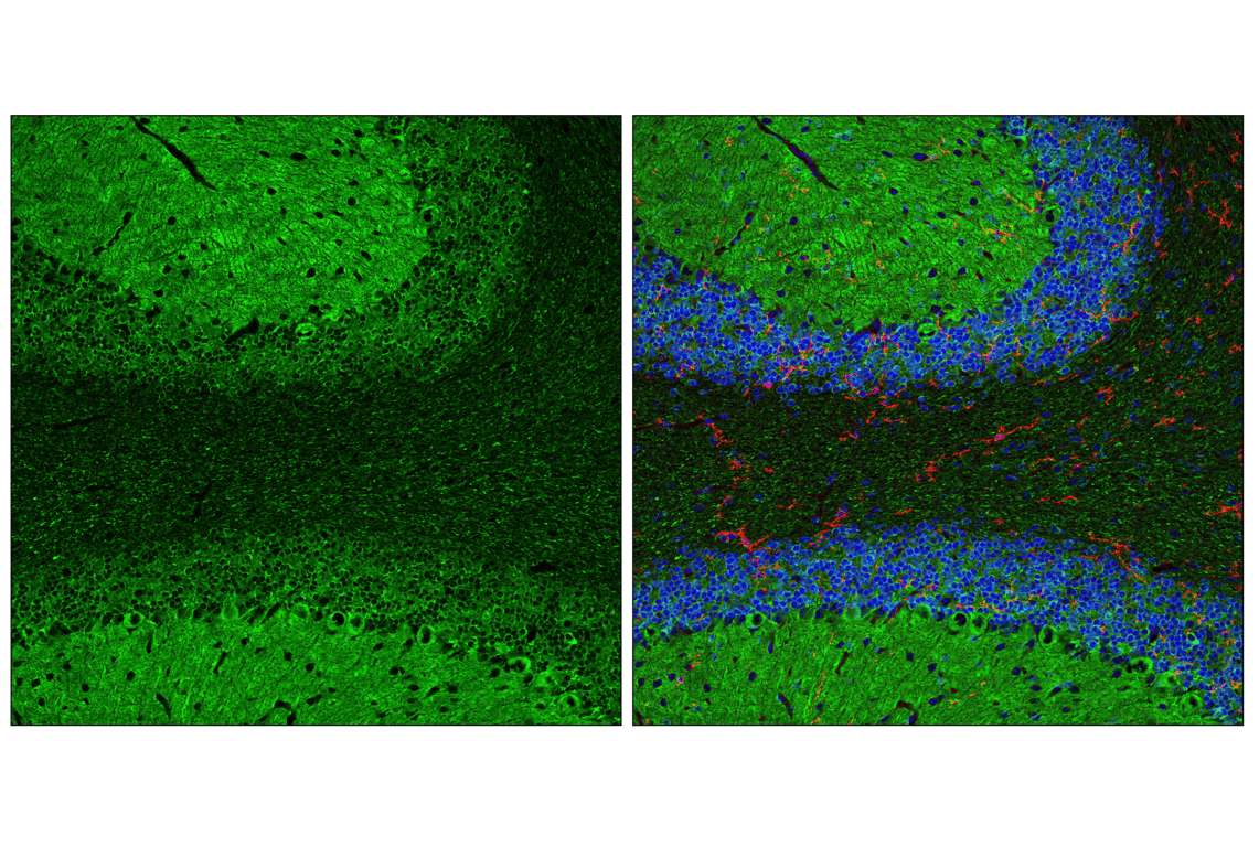
Confocal immunofluorescent analysis of fixed frozen mouse cerebellum using Tau (D1M9X) XP® Rabbit mAb (green), TMEM119 (E4B9S) Mouse mAb #98778 (red) and ProLong® Gold Antifade Reagent with DAPI #8961 (blue).
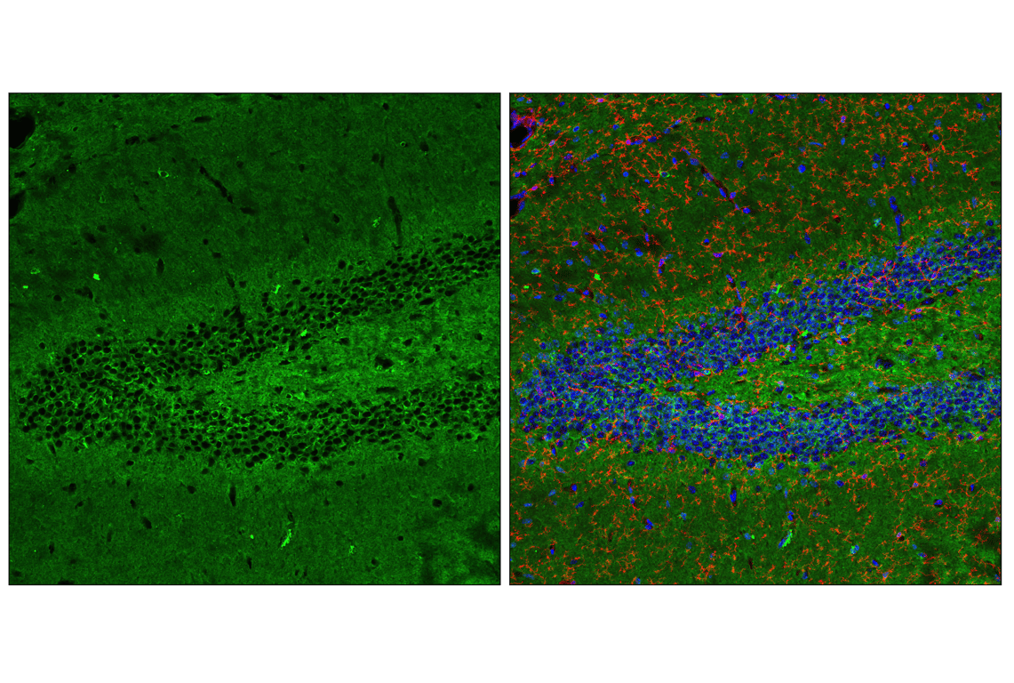
Confocal immunofluorescent analysis of fixed frozen mouse hippocampus using Tau (D1M9X) XP® Rabbit mAb (green), TMEM119 (E4B9S) Mouse mAb #98778 (red) and ProLong® Gold Antifade Reagent with DAPI #8961 (blue).