Revision 1
#38241
Store at -20C
877-616-CELL (2355)
877-678-TECH (8324)
3 Trask Lane | Danvers | Massachusetts | 01923 | USA
For Research Use Only. Not for Use in Diagnostic Procedures.
Applications:
W, IHC-Bond, IHC-P
Reactivity:
H M
Sensitivity:
Endogenous
MW (kDa):
120-135
Source/Isotype:
Mouse IgG1
UniProt ID:
#Q9BYF1
Entrez-Gene Id:
59272
Product Usage Information
| Application | Dilution |
|---|---|
| Western Blotting | 1:1000 |
| IHC Leica Bond | 1:100 - 1:400 |
| Immunohistochemistry (Paraffin) | 1:50 |
Storage
Specificity/Sensitivity
ACE2 (E9G3S) Mouse mAb recognizes endogenous levels of total ACE2 protein.
Source / Purification
Monoclonal antibody is produced by immunizing animals with a synthetic peptide corresponding to residues surrounding Asp201 of human ACE2 protein.
Background
ACE2 is a carboxypeptidase that catalyses the conversion of angiotensin I to angiotensin 1-9, or of angiotensin II to the vasodilator angiotensin 1-7 (1). ACE2 is a critical component in the renin-angiotensin system (RAS). ACE2 is predominantly expressed in vascular endothelial cells of the heart and kidney and Leydig and Sertoli cells of the testis (2,3). The unique expression pattern of ACE2 determines its essential role in the regulation of cardiovascular and kidney functions, as well as fertility. ACE2 protein is localized mainly in the extracellular space with its carboxy terminal end attached to the membrane via its transmembrane domain. Active ACE2 enzyme is secreted by cleavage at the amino terminus. Research studies have shown that ACE2 expression is elevated in human failing heart (4). ACE2 has also been identified as the receptor for SARS and SARS-CoV-2 coronaviruses (5-7).
Background References
- Schmidt, B.L. et al. (2000) J Clin Microbiol 38, 1279-82.
- Boehm, M. and Nabel, E.G. (2002) N Engl J Med 347, 1795-7.
- Douglas, G.C. et al. (2004) Endocrinology 145, 4703-11.
- Goulter, A.B. et al. (2004) BMC Med 2, 19.
- Li, W. et al. (2005) EMBO J 24, 1634-43.
- Hoffmann, M. et al. (2020) Cell 181, 271-280.e8.
- Lan, J. et al. (2020) Nature 581, 215-20.
Species Reactivity
Species reactivity is determined by testing in at least one approved application (e.g., western blot).
Western Blot Buffer
IMPORTANT: For western blots, incubate membrane with diluted primary antibody in 5% w/v BSA, 1X TBS, 0.1% Tween® 20 at 4°C with gentle shaking, overnight.
Applications Key
W: Western Blotting IHC-Bond: IHC Leica Bond
Cross-Reactivity Key
H: Human M: Mouse R: Rat Hm: Hamster Mk: Monkey Vir: Virus Mi: Mink C: Chicken Dm: D. melanogaster X: Xenopus Z: Zebrafish B: Bovine Dg: Dog Pg: Pig Sc: S. cerevisiae Ce: C. elegans Hr: Horse GP: Guinea Pig Rab: Rabbit G: Goat All: All Species Expected
Trademarks and Patents
Cell Signaling Technology is a trademark of Cell Signaling Technology, Inc.
SignalStain is a registered trademark of Cell Signaling Technology, Inc.
XP is a registered trademark of Cell Signaling Technology, Inc.
All other trademarks are the property of their respective owners. Visit cellsignal.com/trademarks for more information.
Limited Uses
Except as otherwise expressly agreed in a writing signed by a legally authorized representative of CST, the following terms apply to Products provided by CST, its affiliates or its distributors. Any Customer's terms and conditions that are in addition to, or different from, those contained herein, unless separately accepted in writing by a legally authorized representative of CST, are rejected and are of no force or effect.
Products are labeled with For Research Use Only or a similar labeling statement and have not been approved, cleared, or licensed by the FDA or other regulatory foreign or domestic entity, for any purpose. Customer shall not use any Product for any diagnostic or therapeutic purpose, or otherwise in any manner that conflicts with its labeling statement. Products sold or licensed by CST are provided for Customer as the end-user and solely for research and development uses. Any use of Product for diagnostic, prophylactic or therapeutic purposes, or any purchase of Product for resale (alone or as a component) or other commercial purpose, requires a separate license from CST. Customer shall (a) not sell, license, loan, donate or otherwise transfer or make available any Product to any third party, whether alone or in combination with other materials, or use the Products to manufacture any commercial products, (b) not copy, modify, reverse engineer, decompile, disassemble or otherwise attempt to discover the underlying structure or technology of the Products, or use the Products for the purpose of developing any products or services that would compete with CST products or services, (c) not alter or remove from the Products any trademarks, trade names, logos, patent or copyright notices or markings, (d) use the Products solely in accordance with CST Product Terms of Sale and any applicable documentation, and (e) comply with any license, terms of service or similar agreement with respect to any third party products or services used by Customer in connection with the Products.
Revision 1
#38241
ACE2 (E9G3S) Mouse mAb
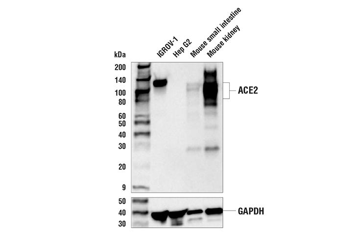
Western blot analysis of extracts from various cells and tissues using ACE2 (E9G3S) Mouse mAb (upper) or GAPDH (D16H11) XP® Rabbit mAb #5174 (lower). The absence of detectable ACE2 protein in Hep G2 cells is consistent with data from both proteomic and molecular expression profiling studies, confirming specificity of the antibody for ACE2.
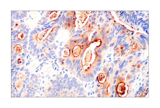
Immunohistochemical analysis of paraffin-embedded human colon adenocarcinoma using ACE2 (E9G3S) Mouse mAb performed on the Leica® BOND™ Rx.
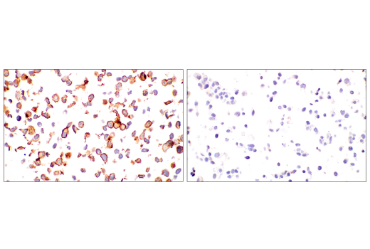
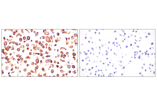
Immunohistochemical analysis of paraffin-embedded IGROV-1 cell pellet (left, positive) or Hep G2 cell pellet (right, negative) using ACE2 (E9G3S) Mouse mAb.
Revision 1
#38241
ACE2 (E9G3S) Mouse mAb
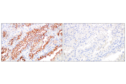
Immunohistochemical analysis of paraffin-embedded cell pellets of transgenic A549 cells stably expressing human ACE2 (left, positive) or wild type A549 cells (right, negative) using ACE2 (E9G3S) Mouse mAb. Transgenic A549 cells were generously provided by Dr. Elena Piskounova, Weill Cornell Medicine.
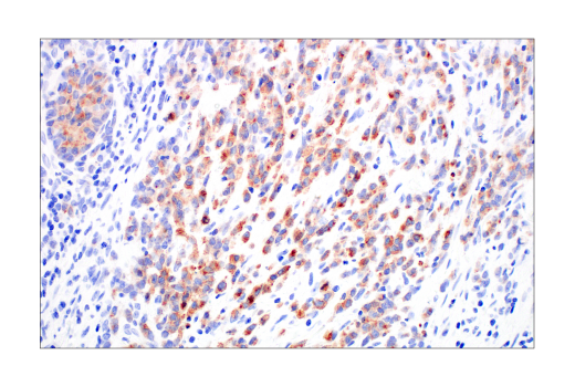
Immunohistochemical analysis of paraffin-embedded human esophageal carcinoma using ACE2 (E9G3S) Mouse mAb.
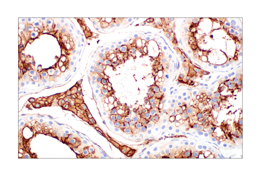
Immunohistochemical analysis of paraffin-embedded human renal cell carcinoma using ACE2 (E9G3S) Mouse mAb.
Revision 1
#38241
ACE2 (E9G3S) Mouse mAb
Immunohistochemical analysis of paraffin-embedded human renal cell carcinoma using ACE2 (E9G3S) Mouse mAb (left) compared to concentration-matched Mouse (G3A1) mAb IgG1 Isotype Control #5415 (right).
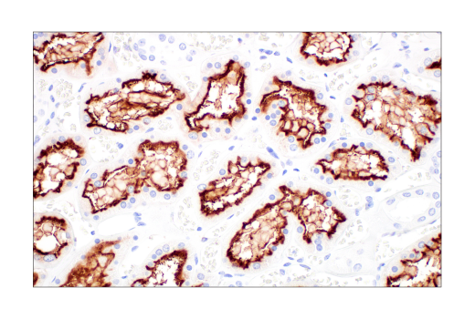
Immunohistochemical analysis of paraffin-embedded normal human kidney using ACE2 (E9G3S) Mouse mAb.
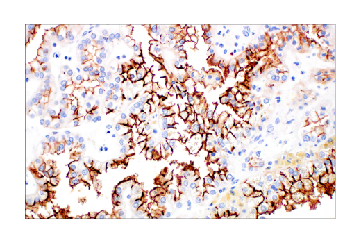
Immunohistochemical analysis of paraffin-embedded human non-small cell lung carcinoma using ACE2 (E9G3S) Mouse mAb.

Revision 1
#38241
ACE2 (E9G3S) Mouse mAb
Immunohistochemical analysis of paraffin-embedded normal human testis using ACE2 (E9G3S) Mouse mAb.