Revision 1
#29256
Store at -20C
877-616-CELL (2355)
877-678-TECH (8324)
3 Trask Lane | Danvers | Massachusetts | 01923 | USA
For Research Use Only. Not for Use in Diagnostic Procedures.
Applications:
W, IP, IHC-P
Reactivity:
H
Sensitivity:
Endogenous
MW (kDa):
42
Source/Isotype:
Rabbit IgG
UniProt ID:
#Q9UHK6
Entrez-Gene Id:
23600
Product Usage Information
| Application | Dilution |
|---|---|
| Western Blotting | 1:1000 |
| Immunoprecipitation | 1:200 |
| Immunohistochemistry (Paraffin) | 1:400 - 1:1600 |
Storage
Specificity/Sensitivity
AMACR (F7T6V) Rabbit mAb recognizes endogenous levels of total AMACR protein. Non-specific diffuse staining was observed in human skeletal muscle, breast myoepithelium, and scattered cells in thymus by immunohistochemistry.
Source / Purification
Monoclonal antibody is produced by immunizing animals with recombinant protein specific to the carboxy terminus of human AMACR protein.
Background
α-Methylacyl-CoA racemase (AMACR), an enzyme localized in peroxisomes and mitochondria, is involved in the β-oxidation of branched-chain fatty acids and fatty acid derivatives (1). AMACR has been reported to be a biomarker for prostate cancer (2-4). AMACR expression is also related to other types of cancers, such as hepatocellular carcinoma (1), noninvasive bladder cancer (5), colorectal cancer (6), and gastric adenocarcinoma (7).
Background References
- Li, W. et al. (2008) J Exp Clin Cancer Res 27, 2.
- Rubin, M.A. et al. (2002) JAMA 287, 1662-70.
- Jiang, Z. et al. (2002) Am J Surg Pathol 26, 1169-74.
- Jiang, Z. et al. (2001) Am J Surg Pathol 25, 1397-404.
- Gunia, S. et al. (2008) Virchows Arch 453, 165-70.
- Marx, A. et al. (2008) Virchows Arch 453, 243-248.
- Truong, C.D. et al. (2008) Int J Clin Exp Pathol 1, 518-523.
Species Reactivity
Species reactivity is determined by testing in at least one approved application (e.g., western blot).
Western Blot Buffer
IMPORTANT: For western blots, incubate membrane with diluted primary antibody in 5% w/v nonfat dry milk, 1X TBS, 0.1% Tween® 20 at 4°C with gentle shaking, overnight.
Applications Key
W: Western Blotting IP: Immunoprecipitation IHC-P: Immunohistochemistry (Paraffin)
Cross-Reactivity Key
H: Human M: Mouse R: Rat Hm: Hamster Mk: Monkey Vir: Virus Mi: Mink C: Chicken Dm: D. melanogaster X: Xenopus Z: Zebrafish B: Bovine Dg: Dog Pg: Pig Sc: S. cerevisiae Ce: C. elegans Hr: Horse GP: Guinea Pig Rab: Rabbit G: Goat All: All Species Expected
Trademarks and Patents
Cell Signaling Technology is a trademark of Cell Signaling Technology, Inc.
All other trademarks are the property of their respective owners. Visit cellsignal.com/trademarks for more information.
Limited Uses
Except as otherwise expressly agreed in a writing signed by a legally authorized representative of CST, the following terms apply to Products provided by CST, its affiliates or its distributors. Any Customer's terms and conditions that are in addition to, or different from, those contained herein, unless separately accepted in writing by a legally authorized representative of CST, are rejected and are of no force or effect.
Products are labeled with For Research Use Only or a similar labeling statement and have not been approved, cleared, or licensed by the FDA or other regulatory foreign or domestic entity, for any purpose. Customer shall not use any Product for any diagnostic or therapeutic purpose, or otherwise in any manner that conflicts with its labeling statement. Products sold or licensed by CST are provided for Customer as the end-user and solely for research and development uses. Any use of Product for diagnostic, prophylactic or therapeutic purposes, or any purchase of Product for resale (alone or as a component) or other commercial purpose, requires a separate license from CST. Customer shall (a) not sell, license, loan, donate or otherwise transfer or make available any Product to any third party, whether alone or in combination with other materials, or use the Products to manufacture any commercial products, (b) not copy, modify, reverse engineer, decompile, disassemble or otherwise attempt to discover the underlying structure or technology of the Products, or use the Products for the purpose of developing any products or services that would compete with CST products or services, (c) not alter or remove from the Products any trademarks, trade names, logos, patent or copyright notices or markings, (d) use the Products solely in accordance with CST Product Terms of Sale and any applicable documentation, and (e) comply with any license, terms of service or similar agreement with respect to any third party products or services used by Customer in connection with the Products.
Revision 1
#29256
AMACR (F7T6V) Rabbit mAb
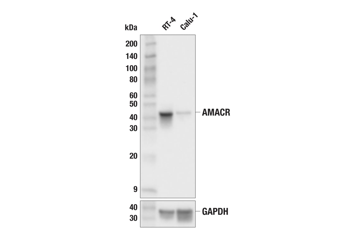
Western blot analysis of extracts from RT-4 and Calu-1 cells using AMACR (F7T6V) Rabbit mAb (upper) or GAPDH (D16H11) XP® Rabbit mAb #5174 (lower). Low expression of AMACR protein in Calu-1 cells is consistent with the predicted expression pattern.
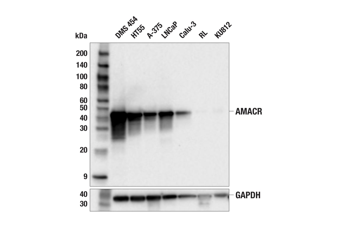
Western blot analysis of extracts from various cell lines using AMACR (F7T6V) Rabbit mAb (upper) or GAPDH (D16H11) XP® Rabbit mAb #5174 (lower). Negative expression of AMACR protein in RL and KU812 cells is consistent with the predicted expression pattern.
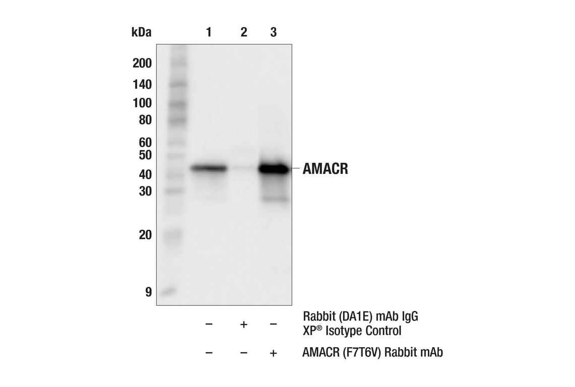
Immunoprecipitation of AMACR protein from HT55 cell extracts. Lane 1 is 10% input, lane 2 is Rabbit (DA1E) mAb IgG XP® Isotype Control #3900, and lane 3 is AMACR (F7T6V) Rabbit mAb. Western blot analysis was performed using AMACR (F7T6V) Rabbit mAb. Mouse Anti-rabbit IgG (Conformation Specific) (L27A9) mAb (HRP Conjugate) #5127 was used as a secondary antibody.
Revision 1
#29256
AMACR (F7T6V) Rabbit mAb
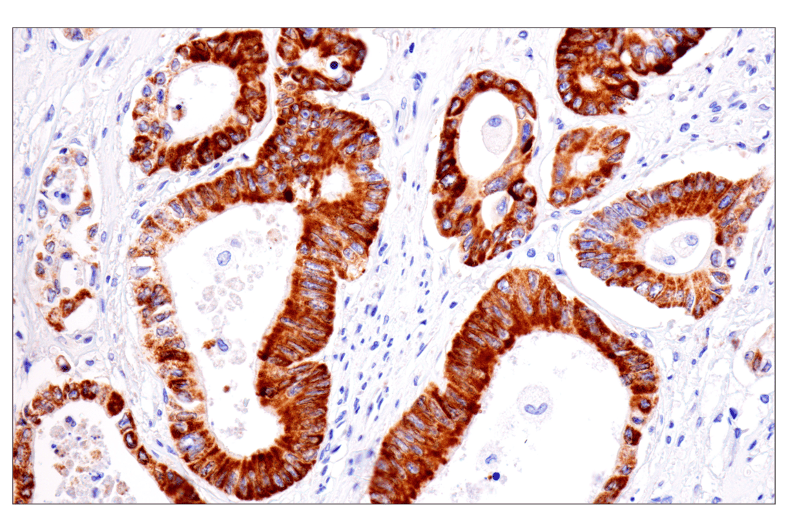
Immunohistochemical analysis of paraffin-embedded human colon adenocarcinoma using AMACR (F7T6V) Rabbit mAb.
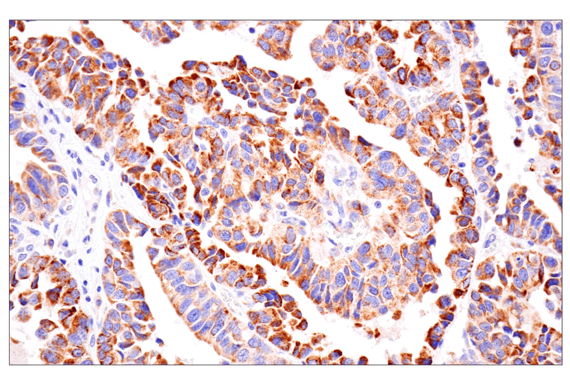
Immunohistochemical analysis of paraffin-embedded human bronchioloalveolar adenocarcinoma using AMACR (F7T6V) Rabbit mAb.
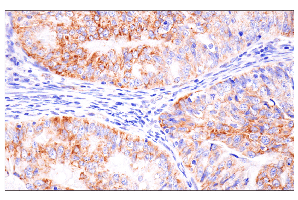
Immunohistochemical analysis of paraffin-embedded human endometrioid adenocarcinoma using AMACR (F7T6V) Rabbit mAb.
Revision 1
#29256
AMACR (F7T6V) Rabbit mAb
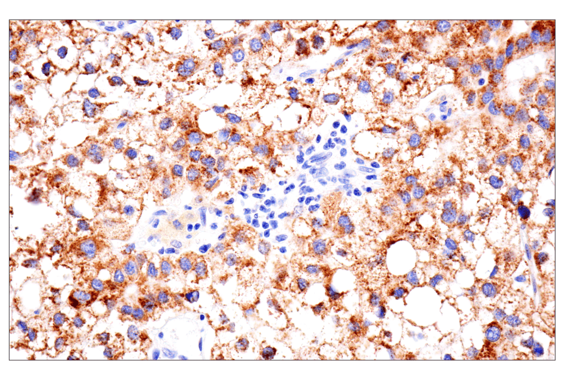
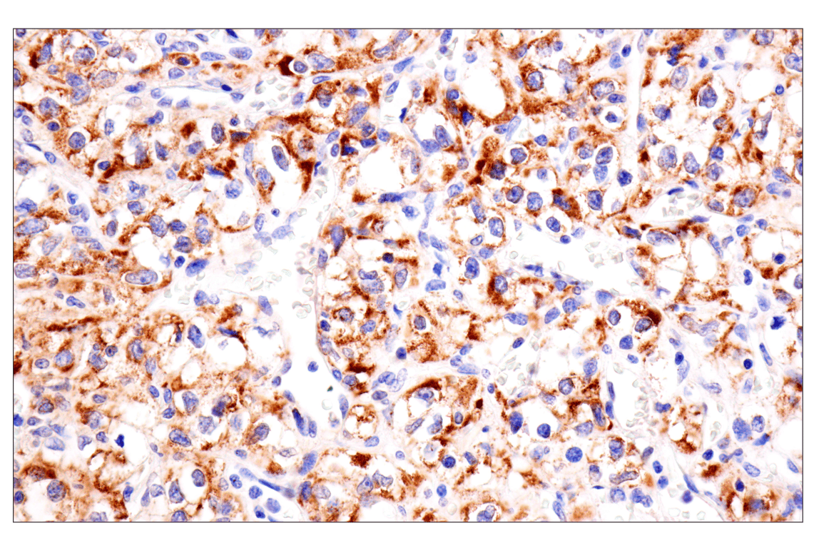
Immunohistochemical analysis of paraffin-embedded human hepatocellular carcinoma using AMACR (F7T6V) Rabbit mAb.
Immunohistochemical analysis of paraffin-embedded human renal cell carcinoma using AMACR (F7T6V) Rabbit mAb.
Immunohistochemical analysis of paraffin-embedded human urothelial carcinoma using AMACR (F7T6V) Rabbit mAb.
Revision 1
#29256
AMACR (F7T6V) Rabbit mAb
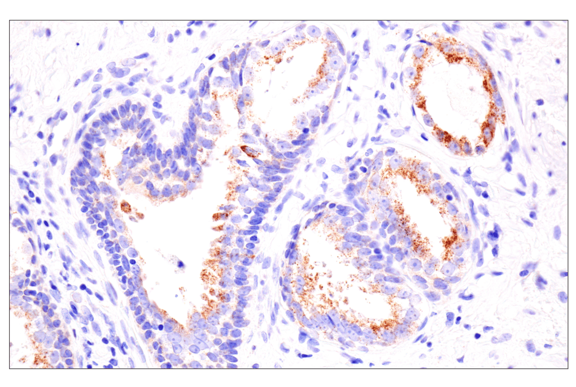
Immunohistochemical analysis of paraffin-embedded human prostate adenocarcinoma using AMACR (F7T6V) Rabbit mAb.
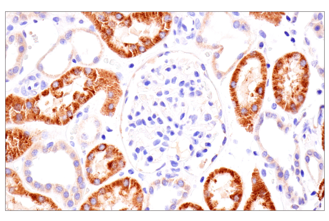
Immunohistochemical analysis of paraffin-embedded normal human kidney using AMACR (F7T6V) Rabbit mAb.
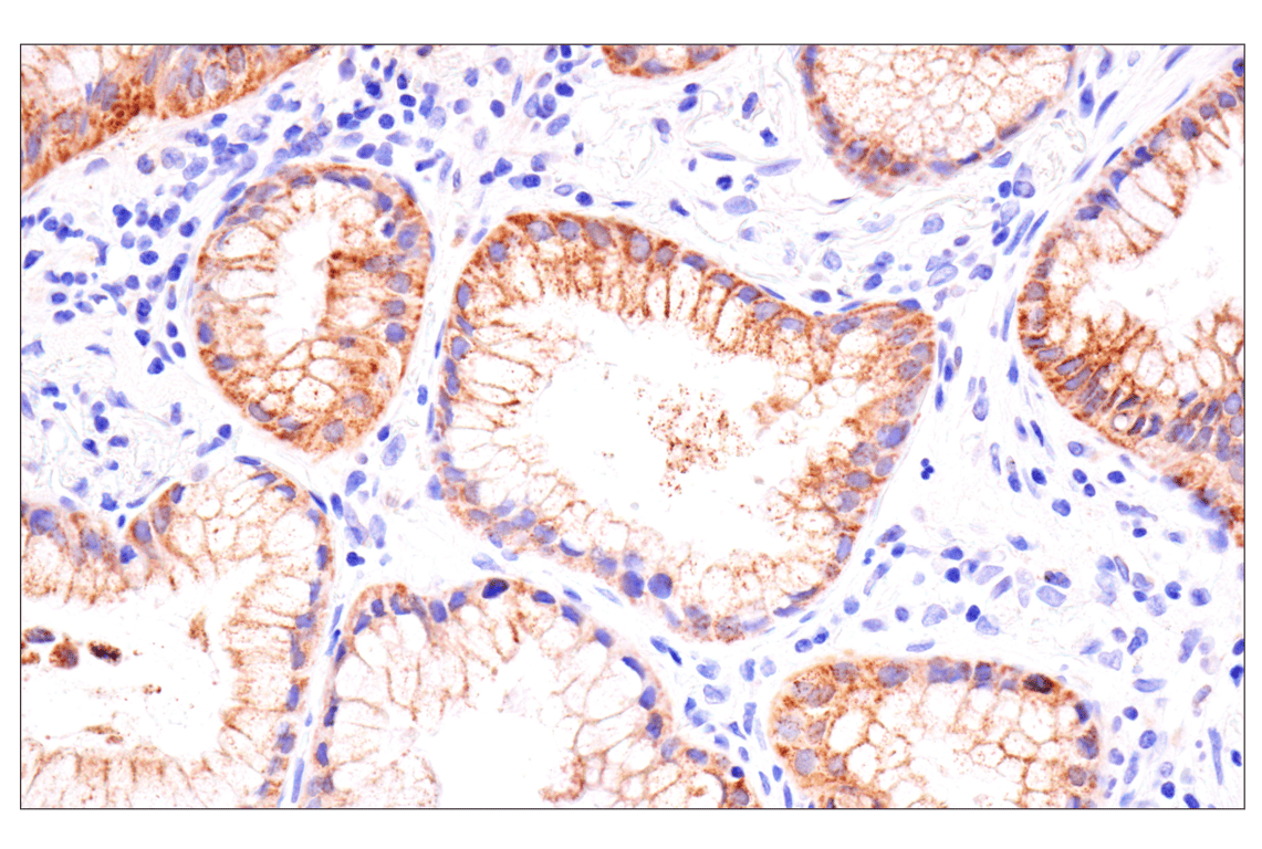
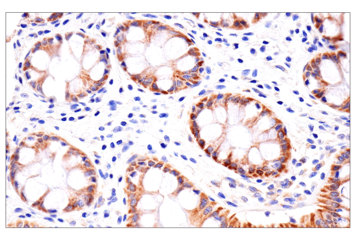
Immunohistochemical analysis of paraffin-embedded normal human colon using AMACR (F7T6V) Rabbit mAb.
Revision 1
#29256
AMACR (F7T6V) Rabbit mAb
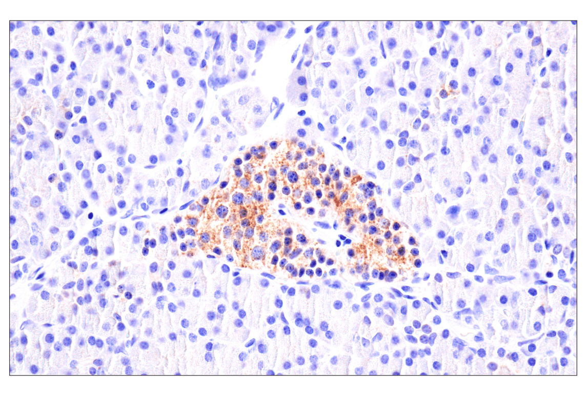
Immunohistochemical analysis of paraffin-embedded normal human pancreas using AMACR (F7T6V) Rabbit mAb.
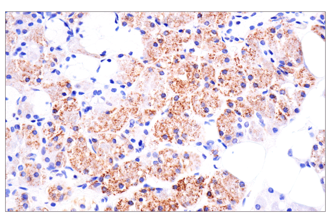
Immunohistochemical analysis of paraffin-embedded normal human salivary gland using AMACR (F7T6V) Rabbit mAb.
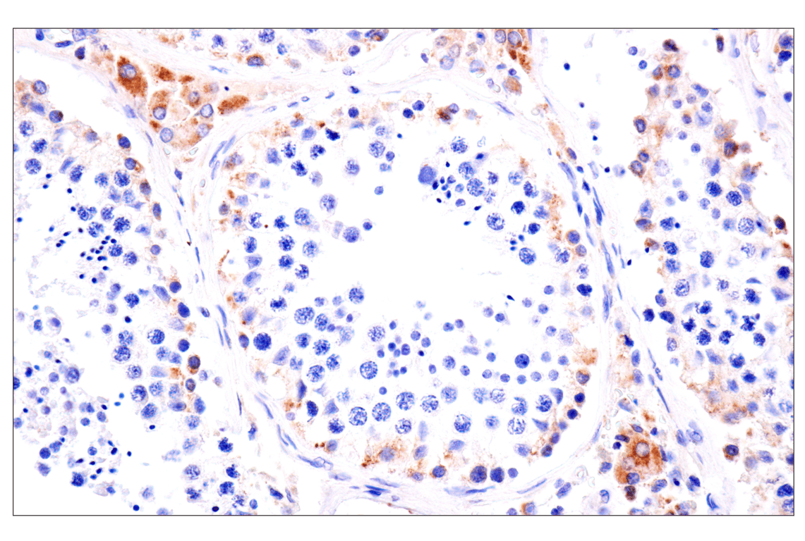
Immunohistochemical analysis of paraffin-embedded normal human testis using AMACR (F7T6V) Rabbit mAb.
Revision 1
#29256
AMACR (F7T6V) Rabbit mAb
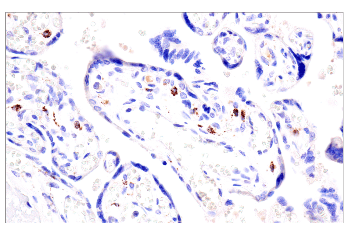
Immunohistochemical analysis of paraffin-embedded normal human placenta using AMACR (F7T6V) Rabbit mAb.
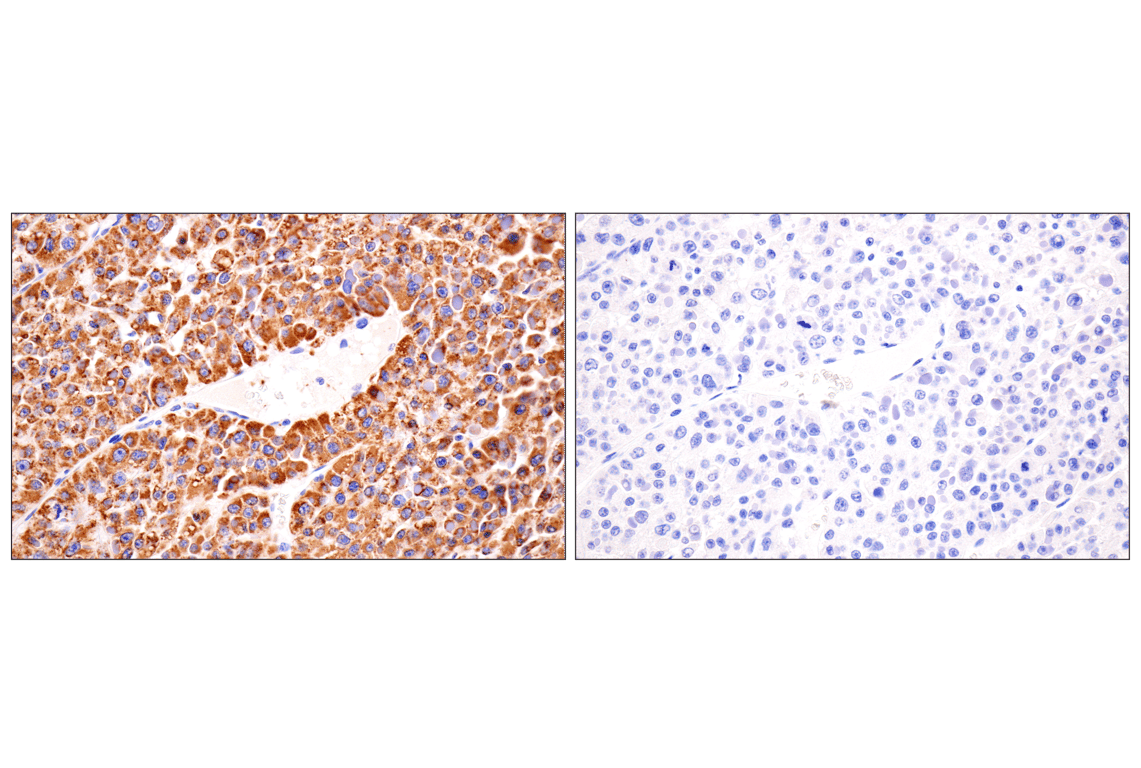
Immunohistochemical analysis of paraffin-embedded human hepatocellular carcinoma using AMACR (F7T6V) Rabbit mAb (left) compared to concentration-matched Rabbit (DA1E) mAb IgG XP® Isotype Control #3900 (right).
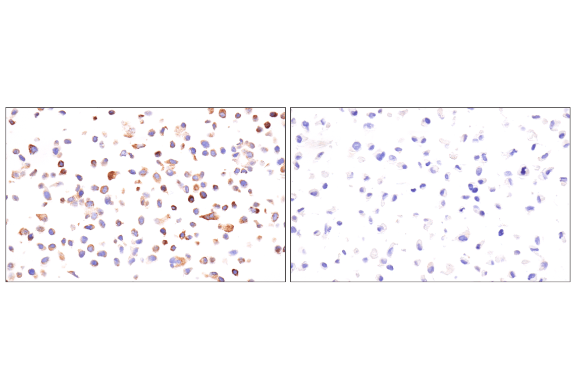
Immunohistochemical analysis of paraffin-embedded RT-4 cell pellet (left, positive) or Calu-1 cell pellet (right, negative) using AMACR (F7T6V) Rabbit mAb.