Revision 1
#30797
Store at -20C
877-616-CELL (2355)
877-678-TECH (8324)
3 Trask Lane | Danvers | Massachusetts | 01923 | USA
For Research Use Only. Not for Use in Diagnostic Procedures.
Applications:
W, IHC-P, IF-IC, FC-FP
Reactivity:
H
Sensitivity:
Endogenous
MW (kDa):
38
Source/Isotype:
Rabbit IgG
UniProt ID:
#P04083
Entrez-Gene Id:
301
Product Usage Information
This formulation is ideal for use with technologies requiring specialized or custom antibody labeling, including fluorophores, metals, lanthanides, and oligonucleotides. It is not recommended for ChIP, ChIP-seq, CUT&RUN or CUT&Tag assays. If you require a carrier free formulation for chromatin profiling, please contact us. Optimal dilutions/concentrations should be determined by the end user.
Formulation
Supplied in 1X PBS, BSA and Azide Free.
For standard formulation of this product see product #32934
Storage
Specificity/Sensitivity
Annexin A1 (D5V2T) XP® Rabbit mAb (BSA and Azide Free) recognizes endogenous levels of total annexin A1 protein.
Source / Purification
Monoclonal antibody is produced by immunizing animals with a synthetic peptide corresponding to residues near the amino terminus of human annexin A1 protein.
Background
The annexin superfamily consists of 13 calcium or calcium and phospholipid binding proteins with high biological and structural homology (1). Annexin-1 (ANXA1) is the first characterized member of the annexin family of proteins and is able to bind to cellular membranes in a calcium-dependent manner, promoting membrane fusion and endocytosis (2-4). Annexin A1 has anti-inflammatory properties and inhibits phospholipase A2 activity (5,6). Annexin A1 can accumulate on internalized vesicles after EGF-stimulated endocytosis and may be required for a late stage in inward vesiculation (7). Phosphorylation by PKC, EGFR, and Chak1 results in inhibition of annexin A1 function (8-10). Annexin A1 has also been identified as one of the 'eat-me' signals on apoptotic cells that are to be recognized and ingested by phagocytes (11). Annexin A1, as an endogenous anti-inflammatory mediator, has roles in many diverse cellular functions, such as membrane aggregation, inflammation, phagocytosis, proliferation, apoptosis, and tumorigenesis and cancer development (12-14).
Background References
- Raynal, P. and Pollard, H.B. (1994) Biochim Biophys Acta 1197, 63-93.
- Blackwell, G.J. et al. (1980) Nature 287, 147-9.
- Rothhut, B. et al. (1983) Biochem Biophys Res Commun 117, 878-84.
- Hirata, F. et al. (1981) Proc Natl Acad Sci USA 78, 3190-4.
- Kim, K.M. et al. (1994) FEBS Lett 343, 251-5.
- Kim, S.W. et al. (2001) J Biol Chem 276, 15712-9.
- White, I.J. et al. (2006) EMBO J 25, 1-12.
- Varticovski, L. et al. (1988) Biochemistry 27, 3682-90.
- Dorovkov, M.V. and Ryazanov, A.G. (2004) J Biol Chem 279, 50643-6.
- Wang, W. and Creutz, C.E. (1994) Biochemistry 33, 275-82.
- Arur, S. et al. (2003) Dev Cell 4, 587-98.
- Perretti, M. and Gavins, F.N. (2003) News Physiol Sci 18, 60-4.
- Parente, L. and Solito, E. (2004) Inflamm Res 53, 125-32.
- Lim, L.H. and Pervaiz, S. (2007) FASEB J 21, 968-75.
Species Reactivity
Species reactivity is determined by testing in at least one approved application (e.g., western blot).
Applications Key
W: Western Blotting IHC-P: Immunohistochemistry (Paraffin) IF-IC: Immunofluorescence (Immunocytochemistry) FC-FP: Flow Cytometry (Fixed/Permeabilized)
Cross-Reactivity Key
H: Human M: Mouse R: Rat Hm: Hamster Mk: Monkey Vir: Virus Mi: Mink C: Chicken Dm: D. melanogaster X: Xenopus Z: Zebrafish B: Bovine Dg: Dog Pg: Pig Sc: S. cerevisiae Ce: C. elegans Hr: Horse GP: Guinea Pig Rab: Rabbit G: Goat All: All Species Expected
Trademarks and Patents
Cell Signaling Technology is a trademark of Cell Signaling Technology, Inc.
Alexa Fluor is a registered trademark of Life Technologies Corporation.
This product is provided under an intellectual property license from Life Technologies Corporation. The transfer of this product is conditioned on the buyer using the purchased product solely in research conducted by the buyer, excluding contract research or any fee for service research, and the buyer must not (1) use this product or its components for (a) diagnostic, therapeutic or prophylactic purposes; (b) testing, analysis or screening services, or information in return for compensation on a per-test basis; or (c) manufacturing or quality assurance or quality control, and/or (2) sell or transfer this product or its components for resale, whether or not resold for use in research. For information on purchasing a license to this product for purposes other than as described above, contact Life Technologies Corporation, 5791 Van Allen Way, Carlsbad, CA 92008 USA or [email protected].
All other trademarks are the property of their respective owners. Visit cellsignal.com/trademarks for more information.
Limited Uses
Except as otherwise expressly agreed in a writing signed by a legally authorized representative of CST, the following terms apply to Products provided by CST, its affiliates or its distributors. Any Customer's terms and conditions that are in addition to, or different from, those contained herein, unless separately accepted in writing by a legally authorized representative of CST, are rejected and are of no force or effect.
Products are labeled with For Research Use Only or a similar labeling statement and have not been approved, cleared, or licensed by the FDA or other regulatory foreign or domestic entity, for any purpose. Customer shall not use any Product for any diagnostic or therapeutic purpose, or otherwise in any manner that conflicts with its labeling statement. Products sold or licensed by CST are provided for Customer as the end-user and solely for research and development uses. Any use of Product for diagnostic, prophylactic or therapeutic purposes, or any purchase of Product for resale (alone or as a component) or other commercial purpose, requires a separate license from CST. Customer shall (a) not sell, license, loan, donate or otherwise transfer or make available any Product to any third party, whether alone or in combination with other materials, or use the Products to manufacture any commercial products, (b) not copy, modify, reverse engineer, decompile, disassemble or otherwise attempt to discover the underlying structure or technology of the Products, or use the Products for the purpose of developing any products or services that would compete with CST products or services, (c) not alter or remove from the Products any trademarks, trade names, logos, patent or copyright notices or markings, (d) use the Products solely in accordance with CST Product Terms of Sale and any applicable documentation, and (e) comply with any license, terms of service or similar agreement with respect to any third party products or services used by Customer in connection with the Products.
Revision 1
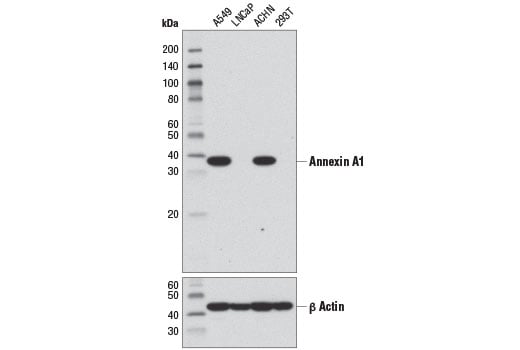
Western blot analysis of extracts from various cell lines using Annexin A1 (D5V2T) XP® Rabbit mAb (upper) and β-Actin (D6A8) Rabbit mAb #8457 (lower). Data were generated using the standard formulation of this product.
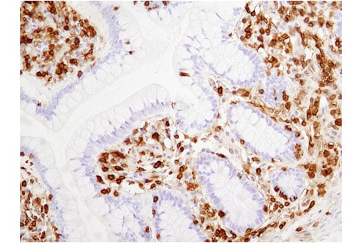
Immunohistochemical analysis of paraffin-embedded normal human colon using Annexin A1 (D5V2T) XP® Rabbit mAb. Data were generated using the standard formulation of this product.
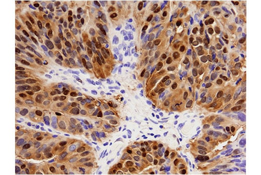
Immunohistochemical analysis of paraffin-embedded human endometrioid adenocarcinoma of the ovary using Annexin A1 (D5V2T) XP® Rabbit mAb. Data were generated using the standard formulation of this product.
Revision 1
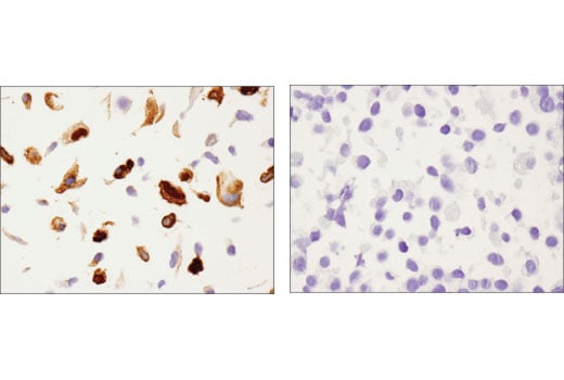
Immunohistochemical analysis of paraffin-embedded A549 (left) and LNCaP (right) cell pellets using Annexin A1 (D5V2T) XP® Rabbit mAb. Data were generated using the standard formulation of this product.
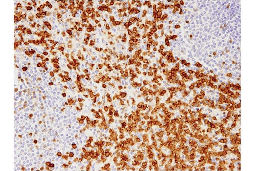
Immunohistochemical analysis of paraffin-embedded human spleen using Annexin A1 (D5V2T) XP® Rabbit mAb. Data were generated using the standard formulation of this product.
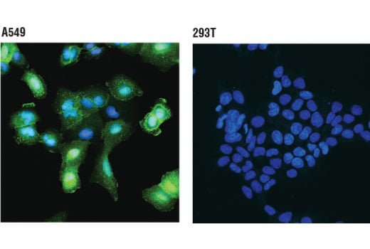
Confocal immunofluorescent analysis of A549 (positive, left) and 293T (negative, right) cells using Annexin A1 (D5V2T) XP® Rabbit mAb (green). Blue pseudocolor = DRAQ5® #4084 (fluorescent DNA dye). Data were generated using the standard formulation of this product.
Revision 1
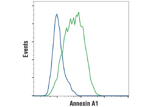
Flow cytometric analysis of RL cells (blue) and Jurkat cells (green) using Annexin A1 (D5V2T) XP® Rabbit mAb. Anti-rabbit IgG (H+L), F(ab')2 Fragment (Alexa Fluor® 488 Conjugate) #4412 was used as a secondary antibody. Data were generated using the standard formulation of this product.