Revision 1
#50563
Store at -20C
877-616-CELL (2355)
877-678-TECH (8324)
3 Trask Lane | Danvers | Massachusetts | 01923 | USA
For Research Use Only. Not for Use in Diagnostic Procedures.
Applications:
W, IP, IHC-P, IF-F, IF-IC
Reactivity:
H M R Mk
Sensitivity:
Endogenous
MW (kDa):
180
Source/Isotype:
Rabbit IgG
UniProt ID:
#Q6PL18
Entrez-Gene Id:
29028
Product Usage Information
| Application | Dilution |
|---|---|
| Western Blotting | 1:1000 |
| Immunoprecipitation | 1:50 |
| Immunohistochemistry (Paraffin) | 1:50 - 1:200 |
| Immunofluorescence (Frozen) | 1:400 - 1:1600 |
| Immunofluorescence (Immunocytochemistry) | 1:400 - 1:1600 |
Storage
Specificity/Sensitivity
ATAD2 (E8Y7F) Rabbit mAb recognizes endogenous levels of total ATAD2 protein. Presumed non-specific punctate staining has been observed by immunofluorescence in a population of cells in fixed-frozen mouse testis tissue.
Source / Purification
Monoclonal antibody is produced by immunizing animals with recombinant protein specific to the carboxy terminus of human ATAD2 protein.
Background
ATPase family AAA domain containing protein 2 (ATAD2) is an oncogenic protein that was originally identified as a coactivator for estrogen receptor (ESR1), and later identified as a coactivator for other transcription factors including c-Myc and E2F1, E2F2, and E2F3 proteins (1-4). ATAD2 is highly expressed and associated with poor prognosis in many types of cancer, including breast, uterine, colon, ovarian, stomach, non-small cell lung carcinoma, osteosarcoma, and cervical cancer (1,5-14). In cancer cells, overexpressed ATAD2 interacts with transcription factors and chromatin modifier proteins to induce the expression of genes that promote cell proliferation and inhibit apoptosis, ultimately promoting tumor growth (15,16). Indeed, knockdown of ATAD2 in pancreatic cancer cell lines has been shown to promote apoptosis, limit cell migration and invasion, and inhibit anchorage-independent growth (17). ATAD2 is a member of the ATPases associated with various cellular activities (AAA) family of proteins and contains a functional AAA domain in its central region, as well as a bromodomain near the C-terminus. The bromodomain binds to acetylated lysine residues on histone proteins, targeting ATAD2 protein to areas of active transcription, where it modulates chromatin structure and recruits additional transcription factors (18,19). Current efforts are underway to better characterize and develop inhibitors to the ATAD2 bromodomain for the treatment of various cancers (16,20-23).
Background References
- Zou, J.X. et al. (2007) Proc Natl Acad Sci U S A 104, 18067-72.
- Zou, J.X. et al. (2009) Cancer Res 69, 3339-46.
- Revenko, A.S. et al. (2010) Mol Cell Biol 30, 5260-72.
- Ciró, M. et al. (2009) Cancer Res 69, 8491-8.
- Wang, Y. et al. Lancet 365, 671-9.
- Teschendorff, A.E. et al. (2006) Genome Biol 7, R101.
- Fellenberg, J. et al. (2007) Mod Pathol 20, 1085-94.
- Petroziello, J. et al. (2004) Oncogene 23, 7734-45.
- De Angelis, P.M. et al. (2006) Mol Cancer 5, 20.
- Zheng, L. et al. (2015) Oncol Rep 33, 2337-44.
- Fouret, R. et al. (2012) Clin Cancer Res 18, 5606-16.
- Wan, W.N. et al. (2014) Asian Pac J Cancer Prev 15, 2777-83.
- Wu, G. et al. (2014) BMC Cancer 14, 107.
- Zhang, M. et al. (2016) Clin Transl Oncol 18, 776-81.
- Caron, C. et al. (2010) Oncogene 29, 5171-81.
- Boussouar, F. et al. (2013) Biochim Biophys Acta 1829, 1010-4.
- Liu, N. et al. (2019) Oncol Lett 17, 3489-94.
- Morozumi, Y. et al. (2016) J Mol Cell Biol 8, 349-62.
- Koo, S.J. et al. (2016) Oncotarget 7, 70323-35.
- Gay, J.C. et al. (2019) Proteins 87, 157-67.
- Hussain, M. et al. (2018) Expert Opin Ther Targets 22, 85-96.
- Bamborough, P. et al. (2018) J Med Chem 61, 8321-36.
- Zhou, Y. et al. (2018) Phys Chem Chem Phys 20, 23222-32.
Species Reactivity
Species reactivity is determined by testing in at least one approved application (e.g., western blot).
Western Blot Buffer
IMPORTANT: For western blots, incubate membrane with diluted primary antibody in 5% w/v nonfat dry milk, 1X TBS, 0.1% Tween® 20 at 4°C with gentle shaking, overnight.
Applications Key
W: Western Blotting IP: Immunoprecipitation IHC-P: Immunohistochemistry (Paraffin) IF-F: Immunofluorescence (Frozen)
Cross-Reactivity Key
H: Human M: Mouse R: Rat Hm: Hamster Mk: Monkey Vir: Virus Mi: Mink C: Chicken Dm: D. melanogaster X: Xenopus Z: Zebrafish B: Bovine Dg: Dog Pg: Pig Sc: S. cerevisiae Ce: C. elegans Hr: Horse GP: Guinea Pig Rab: Rabbit G: Goat All: All Species Expected
Trademarks and Patents
Cell Signaling Technology is a trademark of Cell Signaling Technology, Inc.
Alexa Fluor is a registered trademark of Life Technologies Corporation.
All other trademarks are the property of their respective owners. Visit cellsignal.com/trademarks for more information.
Limited Uses
Except as otherwise expressly agreed in a writing signed by a legally authorized representative of CST, the following terms apply to Products provided by CST, its affiliates or its distributors. Any Customer's terms and conditions that are in addition to, or different from, those contained herein, unless separately accepted in writing by a legally authorized representative of CST, are rejected and are of no force or effect.
Products are labeled with For Research Use Only or a similar labeling statement and have not been approved, cleared, or licensed by the FDA or other regulatory foreign or domestic entity, for any purpose. Customer shall not use any Product for any diagnostic or therapeutic purpose, or otherwise in any manner that conflicts with its labeling statement. Products sold or licensed by CST are provided for Customer as the end-user and solely for research and development uses. Any use of Product for diagnostic, prophylactic or therapeutic purposes, or any purchase of Product for resale (alone or as a component) or other commercial purpose, requires a separate license from CST. Customer shall (a) not sell, license, loan, donate or otherwise transfer or make available any Product to any third party, whether alone or in combination with other materials, or use the Products to manufacture any commercial products, (b) not copy, modify, reverse engineer, decompile, disassemble or otherwise attempt to discover the underlying structure or technology of the Products, or use the Products for the purpose of developing any products or services that would compete with CST products or services, (c) not alter or remove from the Products any trademarks, trade names, logos, patent or copyright notices or markings, (d) use the Products solely in accordance with CST Product Terms of Sale and any applicable documentation, and (e) comply with any license, terms of service or similar agreement with respect to any third party products or services used by Customer in connection with the Products.
Revision 1
#50563
ATAD2 (E8Y7F) Rabbit mAb
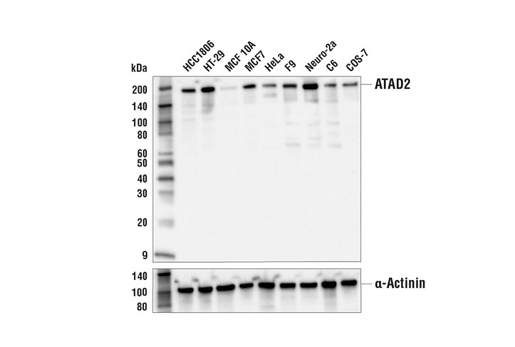
Western blot analysis of extracts from various cell lines using ATAD2 (E8Y7F) Rabbit mAb (upper) and α-Actinin (D6F6) XP® Rabbit mAb #6487 (lower).
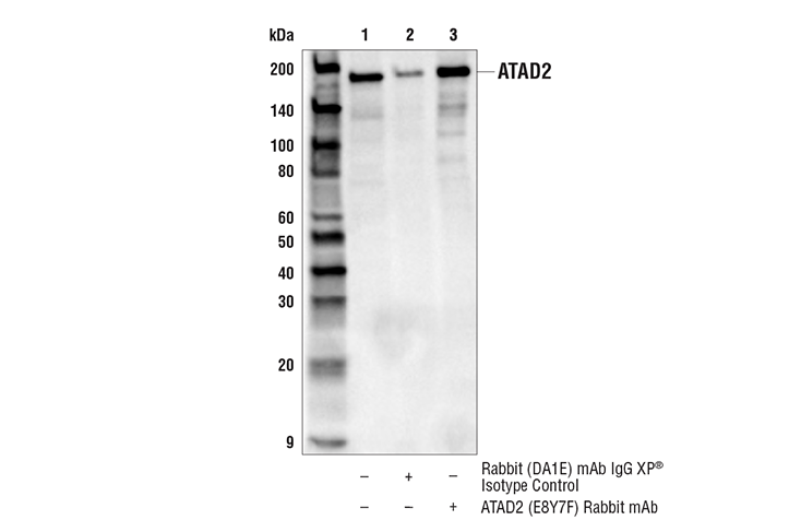
Immunoprecipitation of ATAD2 protein from Neuro-2a cell extracts. Lane 1 is 10% input, lane 2 is Rabbit (DA1E) mAb IgG XP® Isotype Control #3900, and lane 3 is ATAD2 (E8Y7F) Rabbit mAb. Western blot analysis was performed using ATAD2 (E8Y7F) Rabbit mAb. Mouse Anti-rabbit IgG (Conformation Specific) (L27A9) mAb (HRP Conjugate) #5127 was used as a secondary antibody.
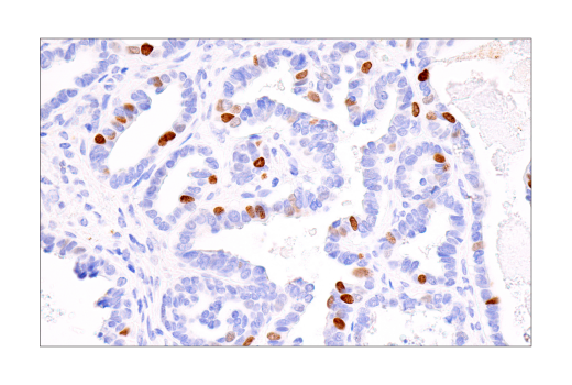
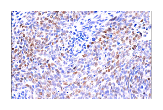
Immunohistochemical analysis of paraffin-embedded human ovarian clear cell carcinoma using ATAD2 (E8Y7F) Rabbit mAb.
Revision 1
#50563
ATAD2 (E8Y7F) Rabbit mAb
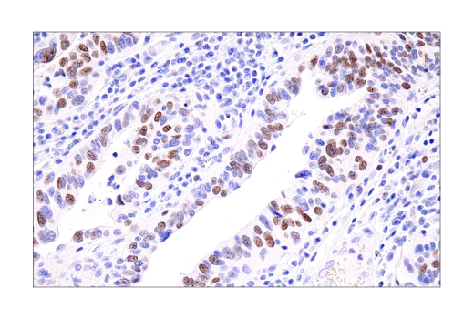
Immunohistochemical analysis of paraffin-embedded human squamous cell carcinoma of the tonsil using ATAD2 (E8Y7F) Rabbit mAb.
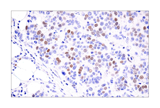
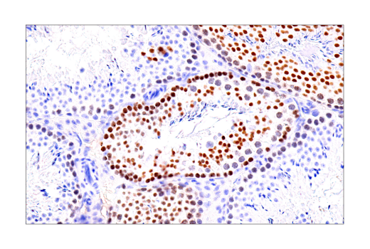
Immunohistochemical analysis of paraffin-embedded human urothelial carcinoma using ATAD2 (E8Y7F) Rabbit mAb.
Immunohistochemical analysis of paraffin-embedded human ductal breast carcinoma using ATAD2 (E8Y7F) Rabbit mAb.
Revision 1
#50563
ATAD2 (E8Y7F) Rabbit mAb
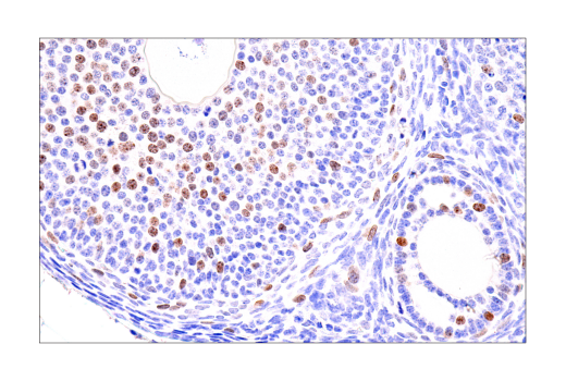
Immunohistochemical analysis of paraffin-embedded normal mouse ovary using ATAD2 (E8Y7F) Rabbit mAb.
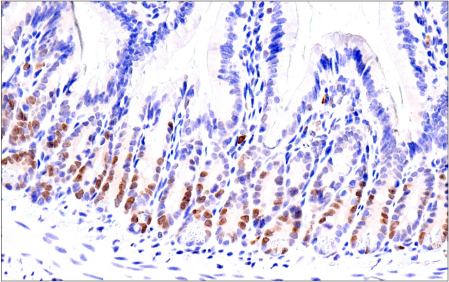
Immunohistochemical analysis of paraffin-embedded normal mouse small intestine using ATAD2 (E8Y7F) Rabbit mAb.
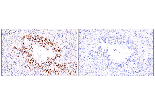
Immunohistochemical analysis of paraffin-embedded human urothelial carcinoma using ATAD2 (E8Y7F) Rabbit mAb (left) compared to concentration-matched Rabbit (DA1E) mAb IgG XP® Isotype Control #3900 (right).
Revision 1
#50563
ATAD2 (E8Y7F) Rabbit mAb
Immunohistochemical analysis of paraffin-embedded normal mouse testis using ATAD2 (E8Y7F) Rabbit mAb.
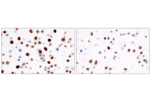
Immunohistochemical analysis of paraffin-embedded HT-29 cell pellet (left, high-expressing) or MCF 10A cell pellet (right, low-expressing) using ATAD2 (E8Y7F) Rabbit mAb.
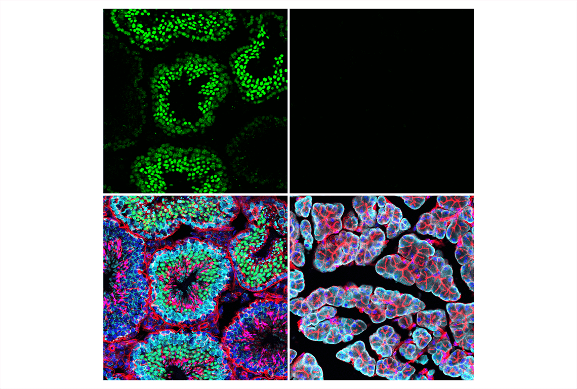
Confocal immunofluorescent analysis of mouse testis (left, positive) and pancreas (right, negative) using ATAD2 (E8Y7F) Rabbit mAb (green) and S6 Ribosomal Protein (54D2) Mouse mAb (Alexa Fluor® 647 Conjugate) #5548 (cyan pseudocolor). Actin filaments were labeled with DyLight™ 554 Phalloidin #13054 (red). Samples were mounted in ProLong® Gold Antifade Reagent with DAPI #8961 (blue).
Revision 1
#50563
ATAD2 (E8Y7F) Rabbit mAb
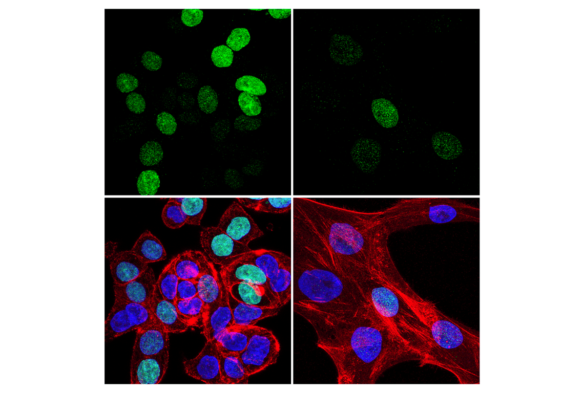
Confocal immunofluorescent analysis of HT-29 cells (left, high-expressing) and MCF 10A cells (right, low-expressing) using ATAD2 (E8Y7F) Rabbit mAb (green). Actin filaments were labeled with DyLight™ 650 Phalloidin #12956 (red). Samples were mounted in ProLong® Gold Antifade Reagent with DAPI #8961 (blue).