Revision 1
#87716
Store at -80C
877-616-CELL (2355)
877-678-TECH (8324)
3 Trask Lane | Danvers | Massachusetts | 01923 | USA
For Research Use Only. Not for Use in Diagnostic Procedures.
Applications:
W, IHC-P, IF-F, IF-IC
Reactivity:
H M R
Sensitivity:
Endogenous
MW (kDa):
29
Source/Isotype:
Rabbit IgG
UniProt ID:
#P22676
Entrez-Gene Id:
794
Product Usage Information
This formulation is ideal for use with technologies requiring specialized or custom antibody labeling, including fluorophores, metals, lanthanides, and oligonucleotides. It is not recommended for ChIP, ChIP-seq, CUT&RUN or CUT&Tag assays. If you require a carrier free formulation for chromatin profiling, please contact us. Optimal dilutions/concentrations should be determined by the end user.
Formulation
Supplied in 1X PBS, BSA and Azide Free.
For standard formulation of this product see product #92635
Storage
Specificity/Sensitivity
Calretinin (E7R6O) XP® Rabbit mAb (BSA and Azide Free) recognizes endogenous levels of total Calretinin protein.
Source / Purification
Monoclonal antibody is produced by immunizing animals with recombinant protein specific to the amino terminus of human Calretinin protein.
Background
Calretinin (29 kDa calbindin, calbindin 2) is a calcium-binding protein of the EF-hand family encoded by the CALB2 gene. It is differentially expressed from homologous family member calbindin-d28k in distinct neuronal populations of the retina, auditory system, and cerebellar granule cells (1,2), and acts as a marker for specific neuronal subsets of the subthalamic nucleus and the substantia nigra (3). Calretinin has been shown to play an important role in modulating neuronal excitability and the induction of long-term potentiation (1). Research has shown that, pathologically, Calretinin is a selective marker for epithelial mesothelioma, making it a diagnostic tool to differentiate from adenocarcinomas (4).
Species Reactivity
Species reactivity is determined by testing in at least one approved application (e.g., western blot).
Applications Key
W: Western Blotting IHC-P: Immunohistochemistry (Paraffin) IF-F: Immunofluorescence (Frozen)
Cross-Reactivity Key
H: Human M: Mouse R: Rat Hm: Hamster Mk: Monkey Vir: Virus Mi: Mink C: Chicken Dm: D. melanogaster X: Xenopus Z: Zebrafish B: Bovine Dg: Dog Pg: Pig Sc: S. cerevisiae Ce: C. elegans Hr: Horse GP: Guinea Pig Rab: Rabbit G: Goat All: All Species Expected
Trademarks and Patents
Cell Signaling Technology is a trademark of Cell Signaling Technology, Inc.
All other trademarks are the property of their respective owners. Visit cellsignal.com/trademarks for more information.
Limited Uses
Except as otherwise expressly agreed in a writing signed by a legally authorized representative of CST, the following terms apply to Products provided by CST, its affiliates or its distributors. Any Customer's terms and conditions that are in addition to, or different from, those contained herein, unless separately accepted in writing by a legally authorized representative of CST, are rejected and are of no force or effect.
Products are labeled with For Research Use Only or a similar labeling statement and have not been approved, cleared, or licensed by the FDA or other regulatory foreign or domestic entity, for any purpose. Customer shall not use any Product for any diagnostic or therapeutic purpose, or otherwise in any manner that conflicts with its labeling statement. Products sold or licensed by CST are provided for Customer as the end-user and solely for research and development uses. Any use of Product for diagnostic, prophylactic or therapeutic purposes, or any purchase of Product for resale (alone or as a component) or other commercial purpose, requires a separate license from CST. Customer shall (a) not sell, license, loan, donate or otherwise transfer or make available any Product to any third party, whether alone or in combination with other materials, or use the Products to manufacture any commercial products, (b) not copy, modify, reverse engineer, decompile, disassemble or otherwise attempt to discover the underlying structure or technology of the Products, or use the Products for the purpose of developing any products or services that would compete with CST products or services, (c) not alter or remove from the Products any trademarks, trade names, logos, patent or copyright notices or markings, (d) use the Products solely in accordance with CST Product Terms of Sale and any applicable documentation, and (e) comply with any license, terms of service or similar agreement with respect to any third party products or services used by Customer in connection with the Products.
Revision 1
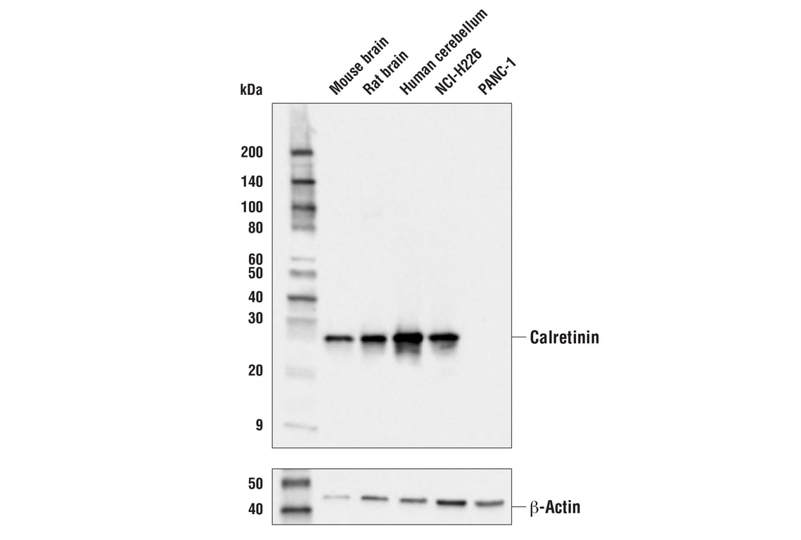
Western blot analysis of extracts from various tissues and cell lines using Calretinin (E7R6O) XP® Rabbit mAb (upper) and β-Actin (D6A8) Rabbit mAb #8457 (lower). Data were generated using the standard formulation of this product.
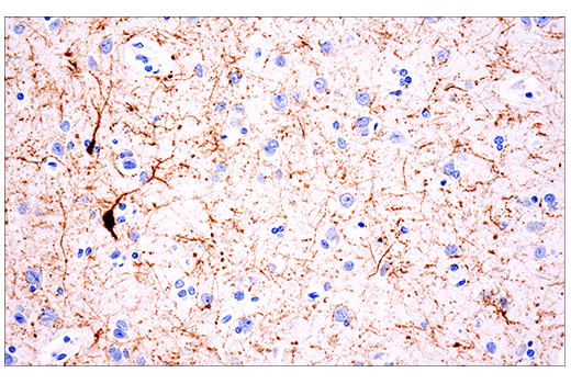
Immunohistochemical analysis of paraffin-embedded normal human brain using Calretinin (E7R6O) XP® Rabbit mAb. Data were generated using the standard formulation of this product.
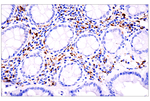
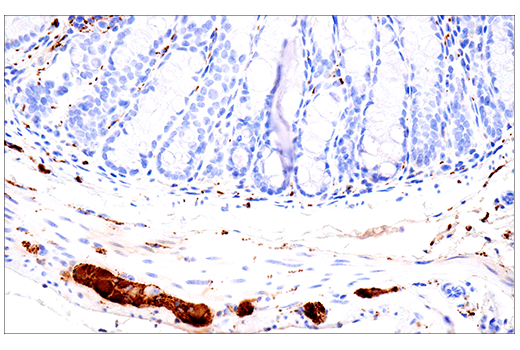
Immunohistochemical analysis of paraffin-embedded normal human colon using Calretinin (E7R6O) XP® Rabbit mAb. Data were generated using the standard formulation of this product.
Revision 1
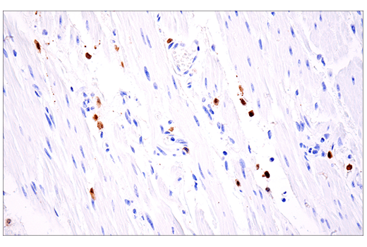
Immunohistochemical analysis of paraffin-embedded human smooth muscle adjacent to esophageal carcinoma using Calretinin (E7R6O) XP® Rabbit mAb. Data were generated using the standard formulation of this product.
Immunohistochemical analysis of paraffin-embedded human colon carcinoma using Calretinin (E7R6O) XP® Rabbit mAb. Data were generated using the standard formulation of this product.
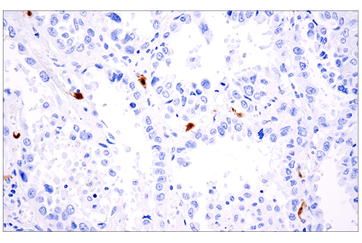
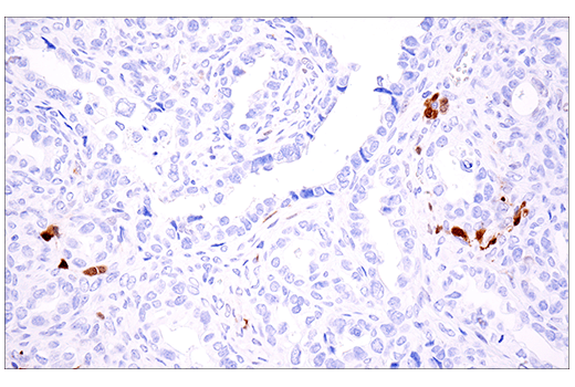
Immunohistochemical analysis of paraffin-embedded human non-small cell lung carcinoma using Calretinin (E7R6O) XP® Rabbit mAb. Data were generated using the standard formulation of this product.
Revision 1
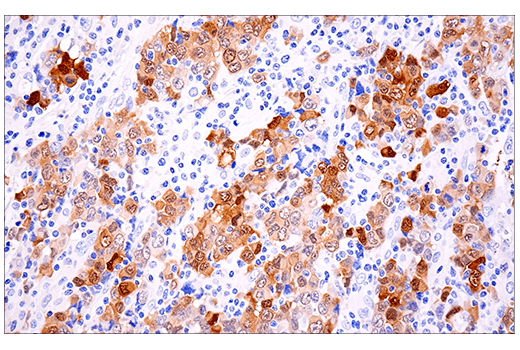
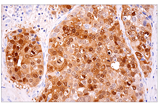
Immunohistochemical analysis of paraffin-embedded human mesothelioma using Calretinin (E7R6O) XP® Rabbit mAb. Data were generated using the standard formulation of this product.
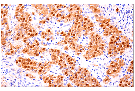
Immunohistochemical analysis of paraffin-embedded human granulosa cell tumor of the ovary using Calretinin (E7R6O) XP® Rabbit mAb. Data were generated using the standard formulation of this product.
Immunohistochemical analysis of paraffin-embedded human ductal breast carcinoma using Calretinin (E7R6O) XP® Rabbit mAb. Data were generated using the standard formulation of this product.
Revision 1
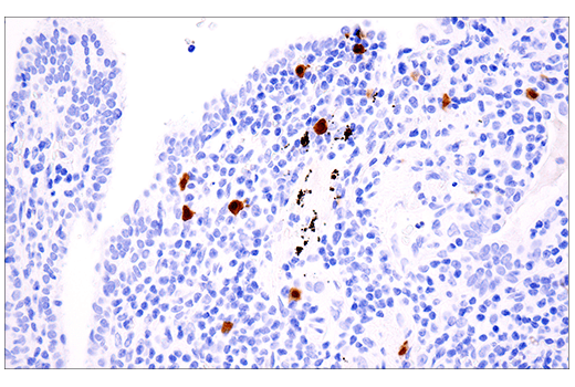
Immunohistochemical analysis of paraffin-embedded human B-cell non-Hodgkin's lymphoma using Calretinin (E7R6O) XP® Rabbit mAb. Data were generated using the standard formulation of this product.
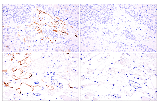
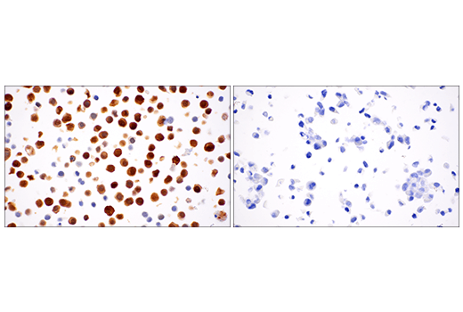
Immunohistochemical analysis of paraffin-embedded human squamous cell carcinoma of the tongue using Calretinin (E7R6O) XP® Rabbit mAb (left) compared to concentration-matched Rabbit (DA1E) mAb IgG XP® Isotype Control #3900 (right). Images depict two fields of view of the same case. Data were generated using the standard formulation of this product.
Immunohistochemical analysis of paraffin-embedded normal mouse brain using Calretinin (E7R6O) XP® Rabbit mAb. Data were generated using the standard formulation of this product.

Revision 1
Immunohistochemical analysis of paraffin-embedded normal mouse colon using Calretinin (E7R6O) XP® Rabbit mAb. Data were generated using the standard formulation of this product.
Immunohistochemical analysis of paraffin-embedded MKN-45 cell pellet (left, positive) or PANC-1 cell pellet (right, negative) using Calretinin (E7R6O) XP® Rabbit mAb. Data were generated using the standard formulation of this product.
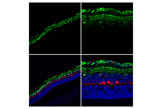
Confocal immunofluorescent analysis of mouse retina at low magnification (left) and high magnification (right) using Calretinin (E7R6O) XP® Rabbit mAb (green). After blocking free secondary antibody binding sites with Rabbit (DA1E) mAb IgG XP® Isotype Control #3900, the tissue was then labeled using Calbindin (D1I4Q) XP® Rabbit mAb (Alexa Fluor® 488 Conjugate) #65152 (red pseudocolor). Sections were mounted in ProLong® Gold Antifade Reagent with DAPI #8961 (blue). Data were generated using the standard formulation of this product.
Revision 1
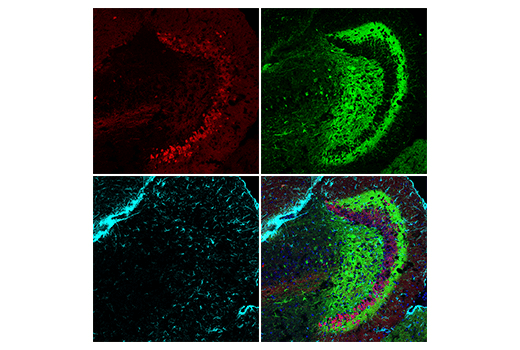
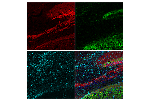
Confocal immunofluorescent analysis of mouse hippocampal formation using Calretinin (E7R6O) XP® Rabbit mAb (green) and GFAP (GA5) Mouse mAb (Alexa Fluor® 647 Conjugate) #3657 (cyan pseudocolor). After blocking free secondary antibody binding sites with Rabbit (DA1E) mAb IgG XP® Isotype Control #3900, the tissue was then labeled using Calbindin (D1I4Q) XP® Rabbit mAb (Alexa Fluor® 488 Conjugate) #65152 (red pseudocolor). Sections were mounted in ProLong® Gold Antifade Reagent with DAPI #8961 (blue). Data were generated using the standard formulation of this product.
Confocal immunofluorescent analysis of mouse dentate gyrus using Calretinin (E7R6O) XP® Rabbit mAb (green) and GFAP (GA5) Mouse mAb (Alexa Fluor® 647 Conjugate) #3657 (cyan pseudocolor). After blocking free secondary antibody binding sites with Rabbit (DA1E) mAb IgG XP® Isotype Control #3900, the tissue was then labeled using Calbindin (D1I4Q) XP® Rabbit mAb (Alexa Fluor® 488 Conjugate) #65152 (red pseudocolor). Sections were mounted in ProLong® Gold Antifade Reagent with DAPI #8961 (blue). Data were generated using the standard formulation of this product.
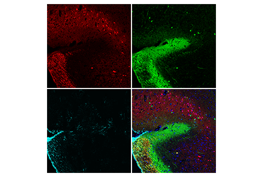
Confocal immunofluorescent analysis of mouse striatum using Calretinin (E7R6O) XP® Rabbit mAb (green) and GFAP (GA5) Mouse mAb (Alexa Fluor® 647 Conjugate) #3657 (cyan pseudocolor). After blocking free secondary antibody binding sites with Rabbit (DA1E) mAb IgG XP® Isotype Control #3900, the tissue was then labeled using Calbindin (D1I4Q) XP® Rabbit mAb (Alexa Fluor® 488 Conjugate) #65152 (red pseudocolor). Sections were mounted in ProLong® Gold Antifade Reagent with DAPI #8961 (blue). Data were generated using the standard formulation of this product.
Revision 1
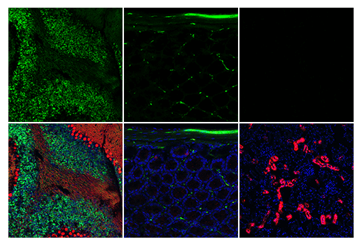
Confocal immunofluorescent analysis of mouse cerebellum (left), colon (middle), and kidney (right) using Calretinin (E7R6O) XP® Rabbit mAb (green). After blocking free secondary antibody binding sites with Rabbit (DA1E) mAb IgG XP® Isotype Control #3900, the tissues were then labeled using Calbindin (D1I4Q) XP® Rabbit mAb (Alexa Fluor® 488 Conjugate) #65152 (red pseudocolor). Sections were mounted in ProLong® Gold Antifade Reagent with DAPI #8961 (blue). Data were generated using the standard formulation of this product.
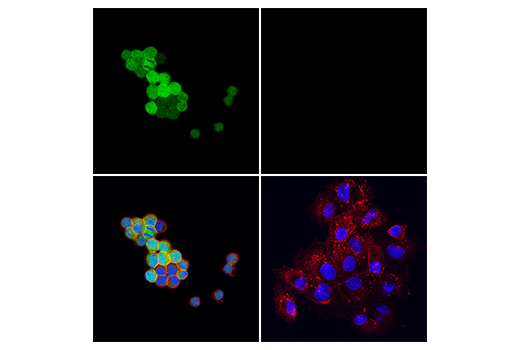
Confocal immunofluorescent analysis of MKN-45 cells (left, positive) or PANC-1 cells (right, negative) using Calretinin (E7R6O) XP® Rabbit mAb (green). Actin filaments were labeled with DyLight™ 554 Phalloidin #13054 (red). Samples were mounted in ProLong® Gold Antifade Reagent with DAPI #8961 (blue). Data were generated using the standard formulation of this product.