Revision 4
#17027
Store at -20C
877-616-CELL (2355)
877-678-TECH (8324)
3 Trask Lane | Danvers | Massachusetts | 01923 | USA
For Research Use Only. Not for Use in Diagnostic Procedures.
| Product Includes | Product # | Quantity | Mol. Wt | Isotype/Source |
|---|---|---|---|---|
| Basic FGF (E9S5A) Rabbit mAb | 98658 | 20 µl | 18, 22, 24 kDa | Rabbit IgG |
| IGF-I (E6B7O) Rabbit mAb | 73034 | 20 µl | 9, 13, 18, 20 kDa | Rabbit IgG |
| HGF β (D6S7D) XP® Rabbit mAb | 52445 | 20 µl | 35, 85 kDa | Rabbit IgG |
| TGF-β (56E4) Rabbit mAb | 3709 | 20 µl | 12, 45-60 kDa | Rabbit IgG |
| HBEGF (E5L5T) Rabbit mAb | 27450 | 20 µl | 18, 21, 27 kDa | Rabbit IgG |
| MIF (E7T1W) Rabbit mAb | 87501 | 20 µl | 12 kDa | Rabbit IgG |
| EREG (D4O5I) Rabbit mAb | 12048 | 20 µl | 17,19, 30 kDa | Rabbit IgG |
| Angiopoietin-2 (D200) Antibody | 50697 | 20 µl | 68, 70 kDa | Rabbit |
| Anti-rabbit IgG, HRP-linked Antibody | 7074 | 100 µl | Goat | |
| VEGF-A (E9X8Q) Rabbit mAb | 50661 | 20 µl | 16, 20, 23, 26 kDa | Rabbit IgG |
Please visit cellsignal.com for individual component applications, species cross-reactivity, dilutions, protocols, and additional product information.
Description
The Cancer-associated Growth Factor Antibody Sampler Kit provides an economical means of detecting selected growth factors that have been shown to influence tumor development. The kit includes enough antibodies to perform two western blot experiments with each primary antibody.
Storage
Background
The tumor microenvironment (TME) is composed of a heterogenous mixture of tumor cells, blood vessels, fibroblasts, stromal cells, infiltrating immune cells, and extracellular matrix (ECM) components, whose collective interactions play important roles in tumor development (1). Cells in the TME secrete a variety of bioactive molecules, including growth factors, cytokines, ECM proteins, and proteases (e.g., MMPs), many of which play critical roles in regulating growth and development of the tumor (2,3). Growth factors play particularly important roles in the TME, serving as cellular messengers that trigger activation or suppression of signaling pathways that govern tumor development, either directly via the tumor cells, or indirectly by way of effects on the TME. Binding of growth factors to their cognate receptors leads to activation of intracellular signaling pathways, resulting in changes in the expression of target genes that regulate cell behavior. Many growth factors (e.g., IGFs, HGFs, FGFs, HBEGF, EREG) are known to promote tumor development by way of direct effects on tumor cells; other growth factors can affect tumor development indirectly, through effects in the TME that influence tumor angiogenesis (e.g., VEGFs, angiopoietins), ECM deposition (TGF-β), or immune cell signaling (e.g., TGF-β, HBEGF, MIF) (4). The diverse and complex role played by growth factors in promoting tumorigenesis makes them important therapeutic targets in oncology, while elucidating the functions of specific growth factors in the context of tumor development remains an active area of cancer research (5).
Trademarks and Patents
Cell Signaling Technology is a trademark of Cell Signaling Technology, Inc.
XP is a registered trademark of Cell Signaling Technology, Inc.
All other trademarks are the property of their respective owners. Visit cellsignal.com/trademarks for more information.
Limited Uses
Except as otherwise expressly agreed in a writing signed by a legally authorized representative of CST, the following terms apply to Products provided by CST, its affiliates or its distributors. Any Customer's terms and conditions that are in addition to, or different from, those contained herein, unless separately accepted in writing by a legally authorized representative of CST, are rejected and are of no force or effect.
Products are labeled with For Research Use Only or a similar labeling statement and have not been approved, cleared, or licensed by the FDA or other regulatory foreign or domestic entity, for any purpose. Customer shall not use any Product for any diagnostic or therapeutic purpose, or otherwise in any manner that conflicts with its labeling statement. Products sold or licensed by CST are provided for Customer as the end-user and solely for research and development uses. Any use of Product for diagnostic, prophylactic or therapeutic purposes, or any purchase of Product for resale (alone or as a component) or other commercial purpose, requires a separate license from CST. Customer shall (a) not sell, license, loan, donate or otherwise transfer or make available any Product to any third party, whether alone or in combination with other materials, or use the Products to manufacture any commercial products, (b) not copy, modify, reverse engineer, decompile, disassemble or otherwise attempt to discover the underlying structure or technology of the Products, or use the Products for the purpose of developing any products or services that would compete with CST products or services, (c) not alter or remove from the Products any trademarks, trade names, logos, patent or copyright notices or markings, (d) use the Products solely in accordance with CST Product Terms of Sale and any applicable documentation, and (e) comply with any license, terms of service or similar agreement with respect to any third party products or services used by Customer in connection with the Products.
Revision 4
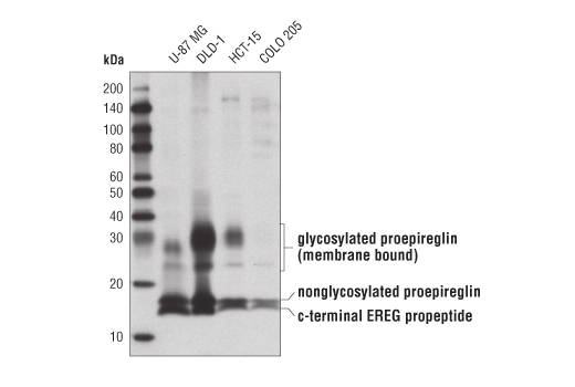
Western blot analysis of extracts from various cell lines using EREG (D4O5I) Rabbit mAb.
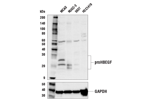
Western blot analysis of extracts from various cell lines using HBEGF (E5L5T) Rabbit mAb (upper) and GAPDH (D16H11) XP® Rabbit mAb #5174 (lower).
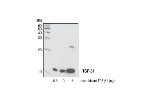
Western blot analysis of recombinant active TGF-β1 using TGF-β (56E4) Rabbit mAb.
Revision 4
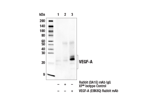
Immunoprecipitation of VEGF-A protein from U-87 MG cell extracts. Lane 1 is 10% input, lane 2 is Rabbit (DA1E) mAb IgG XP® Isotype Control #3900, and lane 3 is VEGF-A (E9X8Q) Rabbit mAb. Western blot analysis was performed using VEGF-A (E9X8Q) Rabbit mAb. Mouse Anti-rabbit IgG (Conformation Specific) (L27A9) mAb (HRP Conjugate) #5127 was used as a secondary antibody.
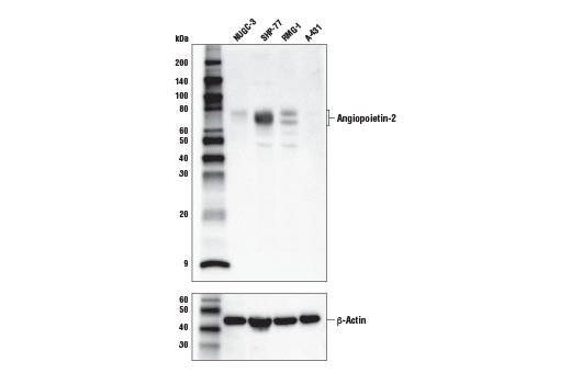
Western blot analysis of extracts from various cell lines using Angiopoietin-2 (D200) Antibody (upper) and β-Actin (D6A8) Rabbit mAb #8457 (lower). The absence of angiopoietin-2 expression in A-431 cell extracts is consistent with molecular and proteomic expression profiling data, confirming specificity of the antibody.
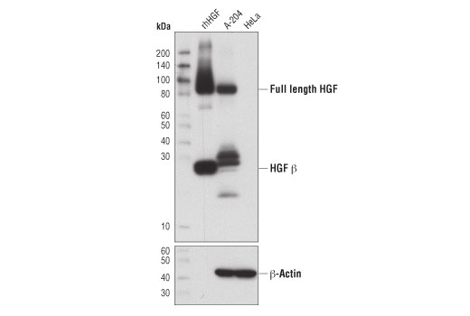
Western blot analysis of recombinant human HGF (rhHGF) and extracts from A-204 and HeLa cells using HGF β (D6S7D) Rabbit mAb (upper) and β-Actin (D6A8) XP® Rabbit mAb #8457 (lower).
Revision 4
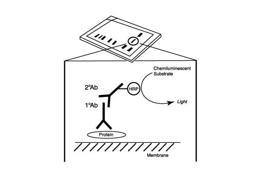
After the primary antibody is bound to the target protein, a complex with HRP-linked secondary antibody is formed. The LumiGLO® is added and emits light during enzyme catalyzed decomposition.
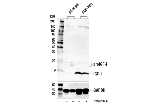
Western blot analysis of extracts from SK-N-MC and SUP-HD1 cells, untreated (-) or treated with Brefeldin A (+), using IGF-I (E6B7O) Rabbit mAb (upper) and GAPDH (D16H11) XP® Rabbit mAb #5174 (lower).
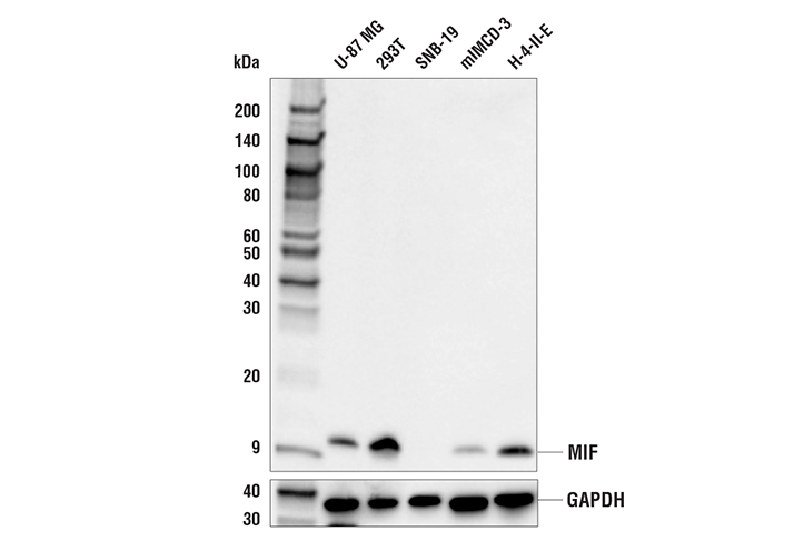
Western blot analysis of extracts from various cell lines using MIF (E7T1W) Rabbit mAb (upper) and GAPDH (D16H11) XP® Rabbit mAb #5174 (lower). The absence of MIF protein in SNB-19 cells is consistent with reported mRNA expression profiles, confirming specificity of the antibody for MIF.
Revision 4
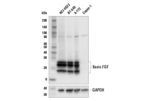
Western blot analysis of extracts from various cell lines using Basic FGF (E9S5A) Rabbit mAb (upper) and GAPDH (D16H11) XP® Rabbit mAb #5174 (lower). The absence of basic FGF protein in Capan-1 cells is consistent with mRNA expression profiles reported in public bioinformatic databases, confirming specificity of the antibody for basic FGF.
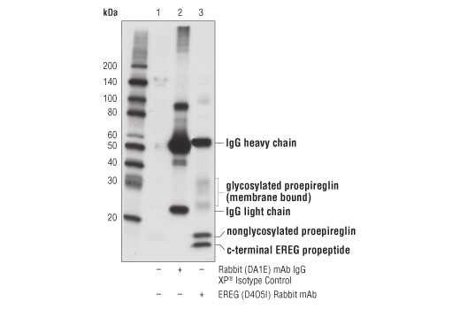
Immunoprecipitation of EREG from COLO 205 cell extracts using Rabbit (DA1E) mAb IgG XP® Isotype Control #3900 (lane 2) or EREG (D4O5I) Rabbit mAb (lane 3). Lane 1 is 10% input. Western blot analysis was performed using EREG (D4O5I) Rabbit mAb.
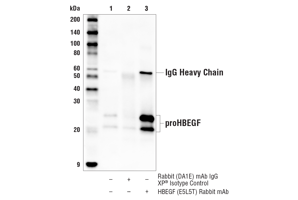
Immunoprecipitation of HBEGF/proHBEGF protein from MCAS cell extracts. Lane 1 is 10% input, lane 2 is Rabbit (DA1E) mAb IgG XP® Isotype Control #3900, and lane 3 is HBEGF (E5L5T) Rabbit mAb. Western blot analysis was performed using HBEGF (E5L5T) Rabbit mAb. Anti-rabbit IgG, HRP-linked Antibody #7074 was used as a secondary antibody.
Revision 4
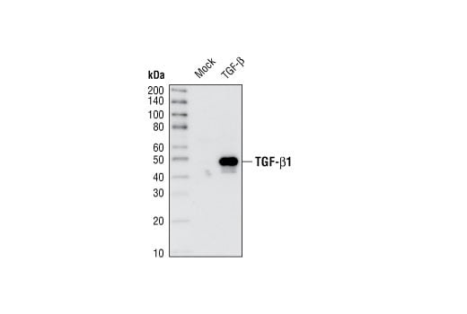
Western blot analysis of extracts of HeLa cells, mock transfected or transfected with TGF-β1 precursor, using TGF-β (56E4) Rabbit mAb.
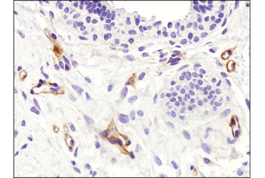
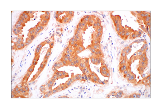
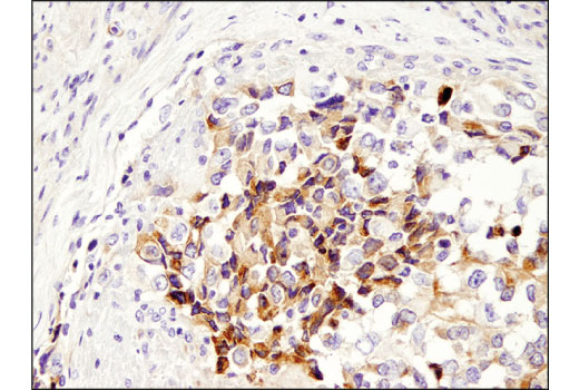
Immunohistochemical analysis of paraffin-embedded human infiltrating ductal carcinoma of the breast HGF β (D6S7D)XP® Rabbit mAb.
Immunohistochemical analysis of paraffin-embedded human ductal breast carcinoma using MIF (E7T1W) Rabbit mAb.
Revision 4
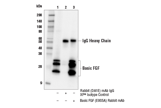
Immunoprecipitation of basic FGF protein from A-172 cell extracts. Lane 1 is 10% input, lane 2 is Rabbit (DA1E) mAb IgG XP® Isotype Control #3900, and lane 3 is Basic FGF (E9S5A) Rabbit mAb. Western blot analysis was performed using Basic FGF (E9S5A) Rabbit mAb. Anti-rabbit IgG, HRP-linked Antibody #7074 was used as a secondary antibody.
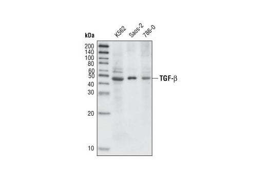
Western blot analysis of extracts from K-562, Saos-2 and 786-0 cells using TGF-β (56E4) Rabbit mAb.
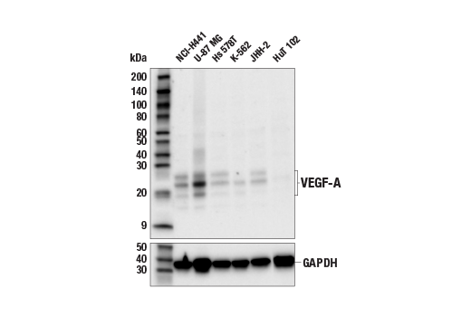
Western blot analysis of extracts from various cell lines using VEGF-A (E9X8Q) Rabbit mAb (upper) or GAPDH (D16H11) XP® Rabbit mAb #5174 (lower). Negative expression of VEGF-A protein in HuT 102 cells is consistent with the predicted expression pattern.
Revision 4
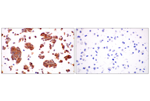
Immunohistochemical analysis of paraffin-embedded A-204 (left) and HeLa (right) cell pellets using HGF β (D6S7D) XP® Rabbit mAb.
Immunohistochemical analysis of paraffin-embedded 293T cell pellet (left, positive) or SNB-19 cell pellet (right, negative) using MIF (E7T1W) Rabbit mAb.
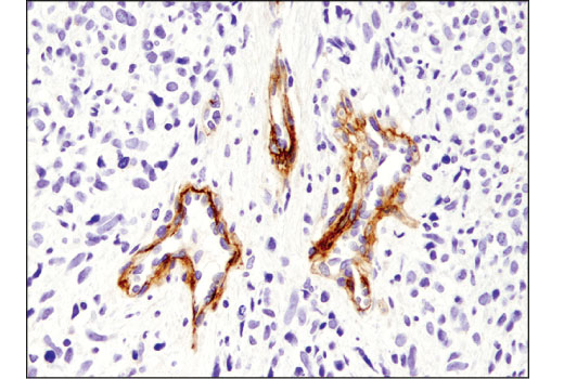
Immunohistochemical analysis of paraffin-embedded human embryonal rhabdomyosarcoma using HGF β (D6S7D) XP® Rabbit mAb.
Revision 4
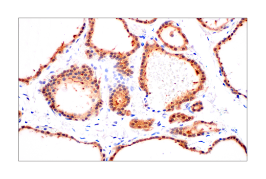
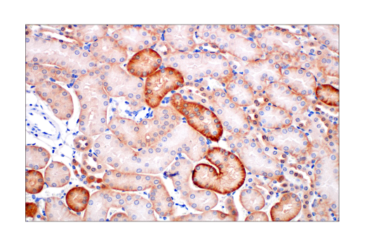
Immunohistochemical analysis of paraffin-embedded normal mouse kidney using MIF (E7T1W) Rabbit mAb.
Immunohistochemical analysis of paraffin-embedded human uterine rhabdomyosarcoma using HGF β (D6S7D) XP® Rabbit mAb.
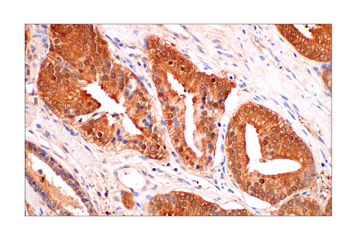
Immunohistochemical analysis of paraffin-embedded human ovarian serous carcinoma using MIF (E7T1W) Rabbit mAb.
Revision 4
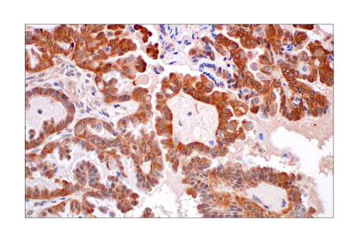
Immunohistochemical analysis of paraffin-embedded human ovarian clear cell carcinoma using MIF (E7T1W) Rabbit mAb.
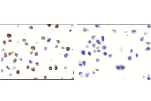
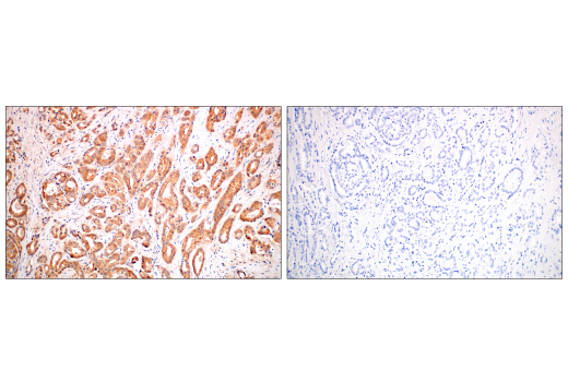
Immunohistochemical analysis of paraffin-embedded human prostate adenocarcinoma using MIF (E7T1W) Rabbit mAb (left) compared to concentration-matched Rabbit (DA1E) mAb IgG XP® Isotype Control #3900 (right).
Immunohistochemical analysis of paraffin-embedded normal human thyroid using MIF (E7T1W) Rabbit mAb.