Revision 3
#69854
Store at -20C
877-616-CELL (2355)
877-678-TECH (8324)
3 Trask Lane | Danvers | Massachusetts | 01923 | USA
For Research Use Only. Not for Use in Diagnostic Procedures.
Applications:
W, IF-F, IF-IC
Reactivity:
H M R
Sensitivity:
Endogenous
MW (kDa):
46, 43, 28
Source/Isotype:
Rabbit
UniProt ID:
#P18242
Entrez-Gene Id:
13033
Product Usage Information
| Application | Dilution |
|---|---|
| Western Blotting | 1:1000 |
| Immunofluorescence (Frozen) | 1:50 - 1:200 |
| Immunofluorescence (Immunocytochemistry) | 1:200 - 1:800 |
Storage
Specificity/Sensitivity
Cathepsin D (E179) Antibody detects endogenous levels of preprocathepsin D, procathepsin D and the heavy chain subunit of mature cathepsin D protein.
Source / Purification
Polyclonal antibodies are produced by immunizing animals with a synthetic peptide corresponding to residues surrounding Glu179 of mouse cathepsin D protein. Antibodies are purified by peptide affinity chromatography.
Background
Cathepsin D is a ubiquitously expressed lysosomal aspartyl protease involved in the normal degradation of proteins (1). It is synthesized as an inactive 43 kDa preprocathepsin D that is cleaved and glycosylated to form a 46 kDa procathepsin D and then further cleaved to produce 28 kDa and 15 kDa subunits (heavy and light chains, respectively) (2). Cathepsin D may also be secreted into the cytosol during apoptosis and contribute to cleavage of substrates implicated in the apoptotic pathway (3). Numerous studies have suggested that cathepsin D plays a role in neuronal degradation and malignant transformation, particularly in breast cancer (4-9).
Background References
- Faust, P.L. et al. (1985) Proc. Natl. Acad. Sci. USA 82, 4910-4.
- Erickson, A.H. et al. (1981) J. Biol .Chem. 256, 11224-31.
- Liaudet-Coopman, E. et al. (2006) Cancer Lett. 237, 167-79.
- Berchem, G. et al. (2002) Oncogene 21, 5951-5.
- Nomura, T. and Katunuma, N. (2005) J. Med. Invest. 52, 1-9.
- Garcia, M. et al. (1996) Stem Cells 14, 642-50.
- Nogami, M. et al. (2000) Histochem. J. 32, 505-8.
- Nakanishi, H. (2003) Ageing Res. Rev. 2, 367-81.
- Callahan, L.M. et al. Neurobiol. Aging 19, S99-105.
Species Reactivity
Species reactivity is determined by testing in at least one approved application (e.g., western blot).
Western Blot Buffer
IMPORTANT: For western blots, incubate membrane with diluted primary antibody in 5% w/v nonfat dry milk, 1X TBS, 0.1% Tween® 20 at 4°C with gentle shaking, overnight.
Applications Key
W: Western Blotting IF-F: Immunofluorescence (Frozen)
Cross-Reactivity Key
H: Human M: Mouse R: Rat Hm: Hamster Mk: Monkey Vir: Virus Mi: Mink C: Chicken Dm: D. melanogaster X: Xenopus Z: Zebrafish B: Bovine Dg: Dog Pg: Pig Sc: S. cerevisiae Ce: C. elegans Hr: Horse GP: Guinea Pig Rab: Rabbit G: Goat All: All Species Expected
Trademarks and Patents
Cell Signaling Technology is a trademark of Cell Signaling Technology, Inc.
Alexa Fluor is a registered trademark of Life Technologies Corporation.
All other trademarks are the property of their respective owners. Visit cellsignal.com/trademarks for more information.
Limited Uses
Except as otherwise expressly agreed in a writing signed by a legally authorized representative of CST, the following terms apply to Products provided by CST, its affiliates or its distributors. Any Customer's terms and conditions that are in addition to, or different from, those contained herein, unless separately accepted in writing by a legally authorized representative of CST, are rejected and are of no force or effect.
Products are labeled with For Research Use Only or a similar labeling statement and have not been approved, cleared, or licensed by the FDA or other regulatory foreign or domestic entity, for any purpose. Customer shall not use any Product for any diagnostic or therapeutic purpose, or otherwise in any manner that conflicts with its labeling statement. Products sold or licensed by CST are provided for Customer as the end-user and solely for research and development uses. Any use of Product for diagnostic, prophylactic or therapeutic purposes, or any purchase of Product for resale (alone or as a component) or other commercial purpose, requires a separate license from CST. Customer shall (a) not sell, license, loan, donate or otherwise transfer or make available any Product to any third party, whether alone or in combination with other materials, or use the Products to manufacture any commercial products, (b) not copy, modify, reverse engineer, decompile, disassemble or otherwise attempt to discover the underlying structure or technology of the Products, or use the Products for the purpose of developing any products or services that would compete with CST products or services, (c) not alter or remove from the Products any trademarks, trade names, logos, patent or copyright notices or markings, (d) use the Products solely in accordance with CST Product Terms of Sale and any applicable documentation, and (e) comply with any license, terms of service or similar agreement with respect to any third party products or services used by Customer in connection with the Products.
Revision 3
#69854
Cathepsin D (E179) Antibody
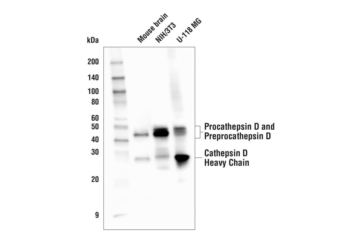
Western blot analysis of extracts from mouse brain tissue, NIH/3T3, and U-118 MG cells using Cathepsin D (E179) Antibody.
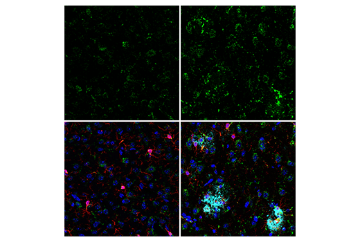
Projected confocal z-stack of mouse cortex from wild-type (left) or an amyloid mouse model of Alzheimer's disease (right) using Cathepsin D (E179) Antibody (green). After blocking free secondary antibody binding sites with Rabbit (DA1E) mAb IgG XP® Isotype Control #3900, the tissue was then labeled using Iba1/AIF-1 (E4O4W) XP® Rabbit mAb (Alexa Fluor® 555 Conjugate) #36618 (red) and β-Amyloid (D54D2) XP® Rabbit mAb (Alexa Fluor® 594 Conjugate) #35363 (cyan pseudocolor). Sections were mounted in ProLong® Gold Antifade Reagent with DAPI #8961 (blue).
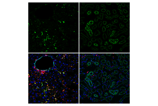
Confocal immunofluorescent analysis of mouse liver (left) and kidney (right) using Cathepsin D (E179) Antibody (green). After blocking free secondary antibody binding sites with Rabbit (DA1E) mAb IgG XP® Isotype Control #3900, the tissue was then labeled using Iba1/AIF-1 (E4O4W) XP® Rabbit mAb (Alexa Fluor® 555 Conjugate) #36618 (red) and α-Smooth Muscle Actin (D4K9N) XP® Rabbit mAb (Alexa Fluor® 647 Conjugate) #76113 (cyan pseudocolor). Sections were mounted in ProLong® Gold Antifade Reagent with DAPI #8961 (blue).