Revision 7
#93233
Store at -20C
877-616-CELL (2355)
877-678-TECH (8324)
3 Trask Lane | Danvers | Massachusetts | 01923 | USA
For Research Use Only. Not for Use in Diagnostic Procedures.
Applications:
W, IHC-Bond, IHC-P, IF-IC
Reactivity:
H
Sensitivity:
Endogenous
MW (kDa):
145
Source/Isotype:
Rabbit IgG
UniProt ID:
#P20702
Entrez-Gene Id:
3687
Product Usage Information
This product is the carrier free version of product #45581. All data were generated using the same antibody clone in the standard formulation which contains BSA and glycerol.
This formulation is ideal for use with technologies requiring specialized or custom antibody labeling, including fluorophores, metals, lanthanides, and oligonucleotides. It is not recommended for ChIP, ChIP-seq, CUT&RUN, or CUT&Tag assays. If you require a carrier-free formulation for chromatin profiling, please contact us. Optimal dilutions/concentrations should be determined by the end user.
Formulation
Supplied in 1X PBS, BSA and Azide Free.
For standard formulation of this product see product #45581.
Storage
Specificity/Sensitivity
CD11c (D3V1E) XP® Rabbit mAb (BSA and Azide Free) recognizes endogenous levels of total CD11c protein. This antibody does not cross-react with CD11b.
Source / Purification
Monoclonal antibody is produced by immunizing animals with recombinant human CD11c protein.
Background
CD11c (integrin αX, ITGAX) is a transmembrane glycoprotein that forms an α/β heterodimer with CD18 (integrin β2), which interacts with a variety of extracellular matrix molecules and cell surface proteins (1). CD11c is primarily used as a dendritic cell marker. Dendritic cells can be classified into two major types: CD11c+ conventional dendritic cells that specialize in antigen presentation, and CD11c- plasmacytoid dendritic cells that specialize in type I interferon production (2, 3). CD11c expression has also been observed on activated NK cells, subsets of B cells, monocytes, granulocytes, and some B cell malignancies including hairy cell leukemia (4-7).
Background References
- Uotila, L.M. et al. (2013) J Biol Chem 288, 33494-9.
- Kohrgruber, N. et al. (1999) J Immunol 163, 3250-9.
- Siegal, F.P. et al. (1999) Science 284, 1835-7.
- Racine, R. et al. (2008) J Immunol 181, 1375-85.
- Werfel, T. et al. (1991) J Immunol 147, 2423-7.
- Cabañas, C. et al. (1988) Hybridoma 7, 167-76.
- Kristensen, J.S. et al. (1987) Blood 70, 1063-8.
Species Reactivity
Species reactivity is determined by testing in at least one approved application (e.g., western blot).
Applications Key
W: Western Blotting IHC-Bond: IHC Leica Bond IF-IC: Immunofluorescence (Immunocytochemistry)
Cross-Reactivity Key
H: Human M: Mouse R: Rat Hm: Hamster Mk: Monkey Vir: Virus Mi: Mink C: Chicken Dm: D. melanogaster X: Xenopus Z: Zebrafish B: Bovine Dg: Dog Pg: Pig Sc: S. cerevisiae Ce: C. elegans Hr: Horse GP: Guinea Pig Rab: Rabbit G: Goat All: All Species Expected
Trademarks and Patents
Cell Signaling Technology is a trademark of Cell Signaling Technology, Inc.
SignalStain is a registered trademark of Cell Signaling Technology, Inc.
XP is a registered trademark of Cell Signaling Technology, Inc.
All other trademarks are the property of their respective owners. Visit cellsignal.com/trademarks for more information.
Limited Uses
Except as otherwise expressly agreed in a writing signed by a legally authorized representative of CST, the following terms apply to Products provided by CST, its affiliates or its distributors. Any Customer's terms and conditions that are in addition to, or different from, those contained herein, unless separately accepted in writing by a legally authorized representative of CST, are rejected and are of no force or effect.
Products are labeled with For Research Use Only or a similar labeling statement and have not been approved, cleared, or licensed by the FDA or other regulatory foreign or domestic entity, for any purpose. Customer shall not use any Product for any diagnostic or therapeutic purpose, or otherwise in any manner that conflicts with its labeling statement. Products sold or licensed by CST are provided for Customer as the end-user and solely for research and development uses. Any use of Product for diagnostic, prophylactic or therapeutic purposes, or any purchase of Product for resale (alone or as a component) or other commercial purpose, requires a separate license from CST. Customer shall (a) not sell, license, loan, donate or otherwise transfer or make available any Product to any third party, whether alone or in combination with other materials, or use the Products to manufacture any commercial products, (b) not copy, modify, reverse engineer, decompile, disassemble or otherwise attempt to discover the underlying structure or technology of the Products, or use the Products for the purpose of developing any products or services that would compete with CST products or services, (c) not alter or remove from the Products any trademarks, trade names, logos, patent or copyright notices or markings, (d) use the Products solely in accordance with CST Product Terms of Sale and any applicable documentation, and (e) comply with any license, terms of service or similar agreement with respect to any third party products or services used by Customer in connection with the Products.
Revision 7
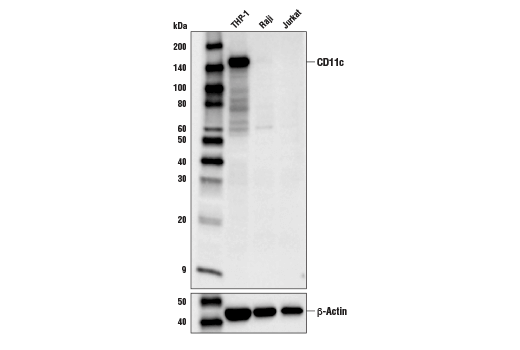
Western blot analysis of extracts from THP-1, Raji, or Jurkat cells using CD11c (D3V1E) XP® Rabbit mAb (upper) or β-Actin (D6A8) Rabbit mAb #8457 (lower). Data were generated using the standard formulation of this product.
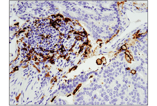
Immunohistochemical analysis of paraffin-embedded human prostate adenocarcinoma using CD11c (D3V1E) XP® Rabbit mAb performed on the Leica® BOND™ Rx. Data were generated using the standard formulation of this product.
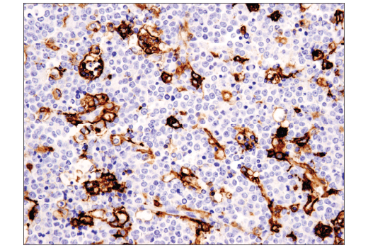
Immunohistochemical analysis of paraffin-embedded human non-Hodgkin lymphoma using CD11c (D3V1E) XP® Rabbit mAb. Data were generated using the standard formulation of this product.
Revision 7
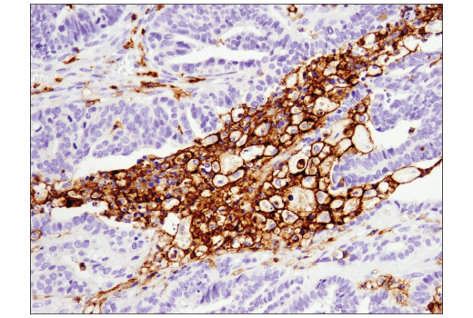
Immunohistochemical analysis of paraffin-embedded human serous papillary carcinoma of the ovary using CD11c (D3V1E) XP® Rabbit mAb. Data were generated using the standard formulation of this product.
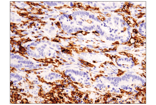
Immunohistochemical analysis of paraffin-embedded human colon carcinoma using CD11c (D3V1E) XP® Rabbit mAb. Data were generated using the standard formulation of this product.
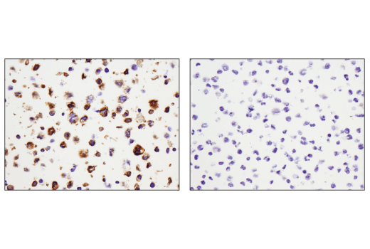
Immunohistochemical analysis of paraffin-embedded THP-1 cell pellet (left, positive) or Raji cell pellet (right, negative) using CD11c (D3V1E) XP® Rabbit mAb. Data were generated using the standard formulation of this product.
Revision 7
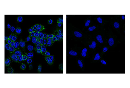
Confocal immunofluorescent analysis of THP-1 cells, differentiated with TPA (12-O-Tetradecanoylphorbol-13-Acetate) #4174 (80 nM, 24 hr) followed by 24 hr without TPA, (left, positive) or HeLa cells (right, negative), using CD11c (D3V1E) XP® Rabbit mAb #45581 (green). Samples were mounted in ProLong® Gold Antifade Reagent with DAPI #8961 (blue). Data were generated using the standard formulation of this product.