Revision 4
#29983
Store at +4C
877-616-CELL (2355)
877-678-TECH (8324)
3 Trask Lane | Danvers | Massachusetts | 01923 | USA
For Research Use Only. Not for Use in Diagnostic Procedures.
Applications:
IHC-Bond, IHC-P
Reactivity:
H
Sensitivity:
Endogenous
Source/Isotype:
Mouse IgG1
UniProt ID:
#P14209
Entrez-Gene Id:
4267
Product Usage Information
| Application | Dilution |
|---|---|
| IHC Leica Bond | 1:50 - 1:200 |
| Immunohistochemistry (Paraffin) | 1:50 - 1:200 |
Storage
Specificity/Sensitivity
CD99 (PCB1) Mouse mAb recognizes endogenous levels of CD99 protein. Non-specific staining was observed in brain by immunohistochemistry.
Source / Purification
Monoclonal antibody is produced by immunizing animals with a prokaryotic recombinant protein corresponding to 101 amino acids of the N–terminal region of human CD99 protein.
Background
CD99 is a transmembrane protein involved in many cellular functions, including cell adhesion and migration, endocytosis and exocytosis, and intracellular protein trafficking. It is highly expressed in all leukocyte lineages, Sertoli cells, granulosa cells, and pancreatic islet cells (1,2). Due to alternative splicing, there are two isoforms of CD99 that differ at the carboxy terminus. CD99 Type I (CD99wt) is the full-length form containing 185 amino acids and CD99 Type II (CD99sh) contains 161 amino acids. Their expression is differentially regulated and they may have opposite functions in different contexts (3,4). CD99 is expressed in many types of tumors and has been used for differential diagnosis of conventional Ewing sarcoma. It has been actively pursued as a therapeutic target (5,6). On the other hand, CD99 may also play a tumor suppressor role in other tumors, such as Hodgkin’s lymphomas, osteosarcomas, and pancreatic tumors (6,7).
Background References
- Muller, W.A. (2016) Immunol Rev 273, 61-75.
- Pasello, M. et al. (2018) J Cell Commun Signal 12, 55-68.
- Alberti, I. et al. (2002) FASEB J 16, 1946-8.
- Scotlandi, K. et al. (2007) Oncogene 26, 6604-18.
- Scotlandi, K. (2006) Adv Exp Med Biol 587, 13-22.
- Manara, M.C. et al. (2018) Genes (Basel) 9, 159.
- Manara, M.C. et al. (2006) Mol Biol Cell 17, 1910-21.
Species Reactivity
Species reactivity is determined by testing in at least one approved application (e.g., western blot).
Applications Key
IHC-Bond: IHC Leica Bond
Cross-Reactivity Key
H: Human M: Mouse R: Rat Hm: Hamster Mk: Monkey Vir: Virus Mi: Mink C: Chicken Dm: D. melanogaster X: Xenopus Z: Zebrafish B: Bovine Dg: Dog Pg: Pig Sc: S. cerevisiae Ce: C. elegans Hr: Horse GP: Guinea Pig Rab: Rabbit G: Goat All: All Species Expected
Trademarks and Patents
Cell Signaling Technology is a trademark of Cell Signaling Technology, Inc.
SignalStain is a registered trademark of Cell Signaling Technology, Inc.
All other trademarks are the property of their respective owners. Visit cellsignal.com/trademarks for more information.
Limited Uses
Except as otherwise expressly agreed in a writing signed by a legally authorized representative of CST, the following terms apply to Products provided by CST, its affiliates or its distributors. Any Customer's terms and conditions that are in addition to, or different from, those contained herein, unless separately accepted in writing by a legally authorized representative of CST, are rejected and are of no force or effect.
Products are labeled with For Research Use Only or a similar labeling statement and have not been approved, cleared, or licensed by the FDA or other regulatory foreign or domestic entity, for any purpose. Customer shall not use any Product for any diagnostic or therapeutic purpose, or otherwise in any manner that conflicts with its labeling statement. Products sold or licensed by CST are provided for Customer as the end-user and solely for research and development uses. Any use of Product for diagnostic, prophylactic or therapeutic purposes, or any purchase of Product for resale (alone or as a component) or other commercial purpose, requires a separate license from CST. Customer shall (a) not sell, license, loan, donate or otherwise transfer or make available any Product to any third party, whether alone or in combination with other materials, or use the Products to manufacture any commercial products, (b) not copy, modify, reverse engineer, decompile, disassemble or otherwise attempt to discover the underlying structure or technology of the Products, or use the Products for the purpose of developing any products or services that would compete with CST products or services, (c) not alter or remove from the Products any trademarks, trade names, logos, patent or copyright notices or markings, (d) use the Products solely in accordance with CST Product Terms of Sale and any applicable documentation, and (e) comply with any license, terms of service or similar agreement with respect to any third party products or services used by Customer in connection with the Products.
Revision 4
#29983
CD99 (PCB1) Mouse mAb
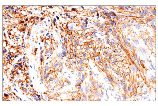
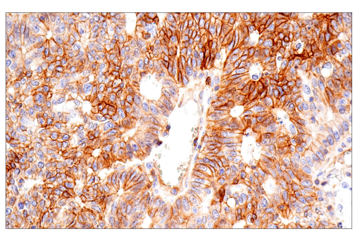
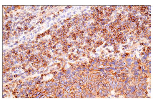
Immunohistochemical analysis of paraffin-embedded human lung sarcoma using CD99 (PCB1) Mouse mAb performed on the Leica® BOND™ Rx.
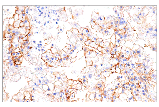
Immunohistochemical analysis of paraffin-embedded human renal cell carcinoma using CD99 (PCB1) Mouse mAb performed on the Leica® BOND™ Rx.
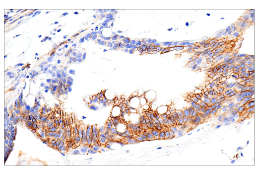
Immunohistochemical analysis of paraffin-embedded human colon carcinoma using CD99 (PCB1) Mouse mAb performed on the Leica® BOND™ Rx.
Revision 4
#29983
CD99 (PCB1) Mouse mAb
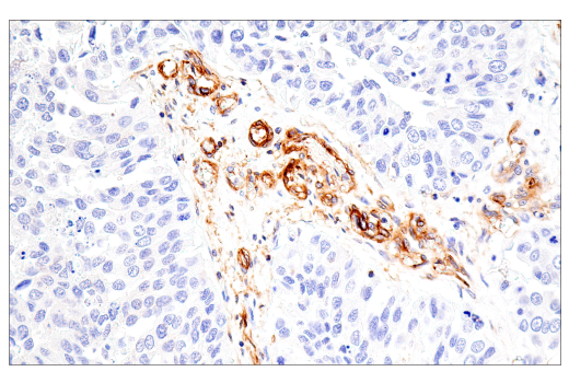
Immunohistochemical analysis of paraffin-embedded human prostate carcinoma using CD99 (PCB1) Mouse mAb performed on the Leica® BOND™ Rx.
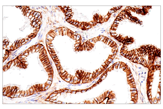
Immunohistochemical analysis of paraffin-embedded human colon carcinoma using CD99 (PCB1) Mouse mAb.
Immunohistochemical analysis of paraffin-embedded human urothelial carcinoma using CD99 (PCB1) Mouse mAb.
Revision 4
#29983
CD99 (PCB1) Mouse mAb
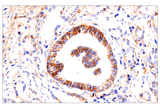
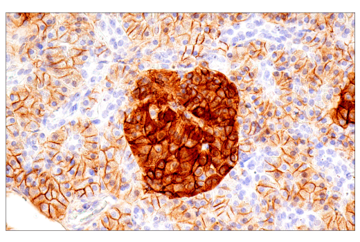
Immunohistochemical analysis of paraffin-embedded human renal cell carcinoma using CD99 (PCB1) Mouse mAb.
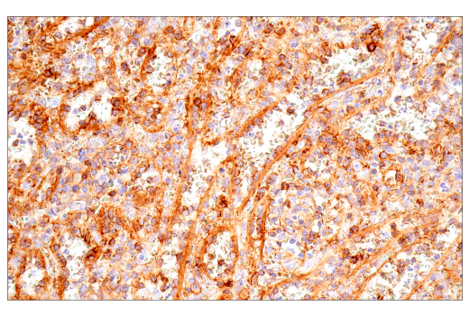
Immunohistochemical analysis of paraffin-embedded human endometrioid adenocarcinoma using CD99 (PCB1) Mouse mAb.
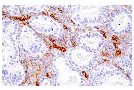
Immunohistochemical analysis of paraffin-embedded human small cell carcinoma of the salivary gland using CD99 (PCB1) Mouse mAb.
Revision 4
#29983
CD99 (PCB1) Mouse mAb
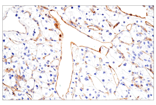
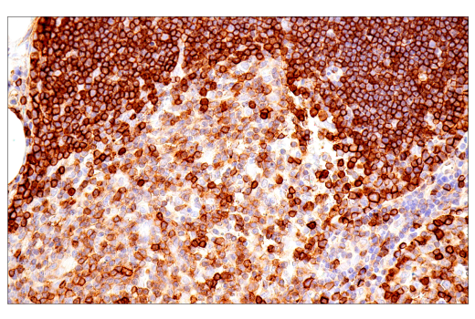
Immunohistochemical analysis of paraffin-embedded normal human thymus using CD99 (PCB1) Mouse mAb.
Immunohistochemical analysis of paraffin-embedded normal human placenta using CD99 (PCB1) Mouse mAb.
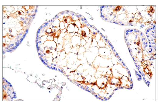
Immunohistochemical analysis of paraffin-embedded normal human prostate using CD99 (PCB1) Mouse mAb.
Revision 4
#29983
CD99 (PCB1) Mouse mAb
Immunohistochemical analysis of paraffin-embedded normal human pancreas using CD99 (PCB1) Mouse mAb.
Immunohistochemical analysis of paraffin-embedded normal human spleen using CD99 (PCB1) Mouse mAb.
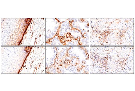
Immunohistochemical analysis of paraffin-embedded normal human esophagus (left), lung adenocarcinoma (middle), or urothelial carcinoma (right) using CD99 (PCB1) Mouse mAb (top) or a CD99 Rabbit mAb (bottom). These two antibodies detect unique, non-overlapping epitopes on human CD99. The similar staining patterns obtained with both antibodies help to confirm the specificity of the staining.
Revision 4
#29983
CD99 (PCB1) Mouse mAb
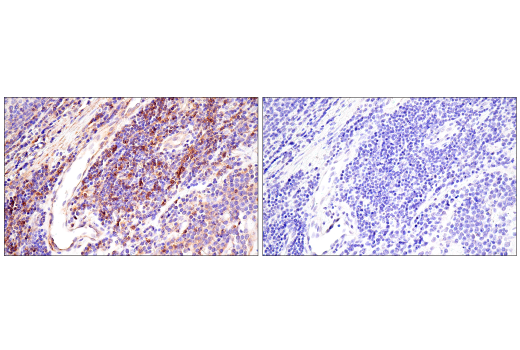
Immunohistochemical analysis of paraffin-embedded human B-cell non-Hodgkin lymphoma using CD99 (PCB1) Mouse mAb (left) compared to concentration-matched Mouse (G3A1) mAb IgG1 Isotype Control #5415 (right).
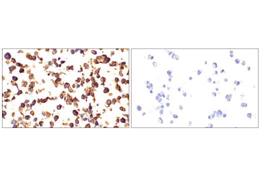
Immunohistochemical analysis of paraffin-embedded A-673 cell pellet (left, positive) or SCLC-21H cell pellet (right, negative) using CD99 (PCB1) Mouse mAb.