Revision 6
#92904
Store at -20C
877-616-CELL (2355)
877-678-TECH (8324)
3 Trask Lane | Danvers | Massachusetts | 01923 | USA
For Research Use Only. Not for Use in Diagnostic Procedures.
Applications:
W, IHC-P, IF-F, IF-IC, FC-FP
Reactivity:
H M
Sensitivity:
Endogenous
MW (kDa):
60
Source/Isotype:
Rabbit IgG
UniProt ID:
#P31146
Entrez-Gene Id:
11151
Product Usage Information
| Application | Dilution |
|---|---|
| Western Blotting | 1:1000 |
| Immunohistochemistry (Paraffin) | 1:200 |
| Immunofluorescence (Frozen) | 1:100 |
| Immunofluorescence (Immunocytochemistry) | 1:100 |
| Flow Cytometry (Fixed/Permeabilized) | 1:50 |
Storage
Specificity/Sensitivity
Coronin 1A (D6K5B) XP® Rabbit mAb recognizes endogenous levels of total coronin 1A protein. Based on the sequence of the immunogenic peptide, this antibody is not expected to cross-react with other coronin protein family members.
Source / Purification
Monoclonal antibody is produced by immunizing animals with a synthetic peptide corresponding to residues surrounding Val200 of human coronin 1A protein.
Background
The coronin family of actin-binding proteins regulates a variety of cellular functions, including migration, phagocytosis, and cytokinesis. Coronin 1A is highly expressed in lymphocytes, and is required for appropriate immune regulation in mice and humans. Researchers are investigating coronin 1A as a potential therapeutic target for autoimmune diseases and lymphoid cancers (1,2). Coronin 1A affects bone resorption through its regulation of lysosome fusion and secretion of cathepsin K in osteoclasts (3). In the nervous system, coronin 1A has been shown to regulate GPCR signaling and neurite outgrowth (4,5).
Species Reactivity
Species reactivity is determined by testing in at least one approved application (e.g., western blot).
Western Blot Buffer
IMPORTANT: For western blots, incubate membrane with diluted primary antibody in 5% w/v BSA, 1X TBS, 0.1% Tween® 20 at 4°C with gentle shaking, overnight.
Applications Key
W: Western Blotting IHC-P: Immunohistochemistry (Paraffin) IF-F: Immunofluorescence (Frozen) FC-FP: Flow Cytometry (Fixed/Permeabilized)
Cross-Reactivity Key
H: Human M: Mouse R: Rat Hm: Hamster Mk: Monkey Vir: Virus Mi: Mink C: Chicken Dm: D. melanogaster X: Xenopus Z: Zebrafish B: Bovine Dg: Dog Pg: Pig Sc: S. cerevisiae Ce: C. elegans Hr: Horse GP: Guinea Pig Rab: Rabbit G: Goat All: All Species Expected
Trademarks and Patents
Cell Signaling Technology is a trademark of Cell Signaling Technology, Inc.
Alexa Fluor is a registered trademark of Life Technologies Corporation.
All other trademarks are the property of their respective owners. Visit cellsignal.com/trademarks for more information.
Limited Uses
Except as otherwise expressly agreed in a writing signed by a legally authorized representative of CST, the following terms apply to Products provided by CST, its affiliates or its distributors. Any Customer's terms and conditions that are in addition to, or different from, those contained herein, unless separately accepted in writing by a legally authorized representative of CST, are rejected and are of no force or effect.
Products are labeled with For Research Use Only or a similar labeling statement and have not been approved, cleared, or licensed by the FDA or other regulatory foreign or domestic entity, for any purpose. Customer shall not use any Product for any diagnostic or therapeutic purpose, or otherwise in any manner that conflicts with its labeling statement. Products sold or licensed by CST are provided for Customer as the end-user and solely for research and development uses. Any use of Product for diagnostic, prophylactic or therapeutic purposes, or any purchase of Product for resale (alone or as a component) or other commercial purpose, requires a separate license from CST. Customer shall (a) not sell, license, loan, donate or otherwise transfer or make available any Product to any third party, whether alone or in combination with other materials, or use the Products to manufacture any commercial products, (b) not copy, modify, reverse engineer, decompile, disassemble or otherwise attempt to discover the underlying structure or technology of the Products, or use the Products for the purpose of developing any products or services that would compete with CST products or services, (c) not alter or remove from the Products any trademarks, trade names, logos, patent or copyright notices or markings, (d) use the Products solely in accordance with CST Product Terms of Sale and any applicable documentation, and (e) comply with any license, terms of service or similar agreement with respect to any third party products or services used by Customer in connection with the Products.
Revision 6
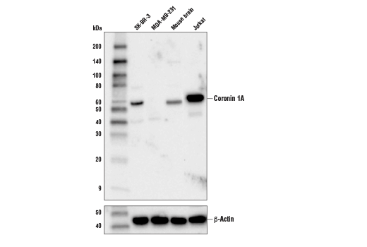
Western blot analysis of extracts from various cells and tissues using Coronin 1A (D6K5B) XP® Rabbit mAb (upper) or β-Actin (D6A8) Rabbit mAb #8457 (lower).
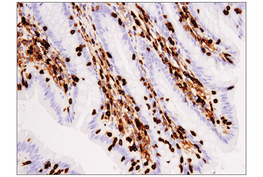
Immunohistochemical analysis of paraffin-embedded human appendix using Coronin 1A (D6K5B) XP® Rabbit mAb.
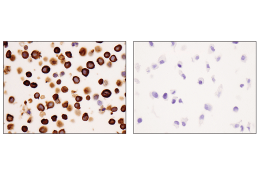
Immunohistochemical analysis of paraffin-embedded Jurkat cell pellet (left, positive) or MDA-MB-231 cell pellet (right, negative) using Coronin 1A (D6K5B) XP® Rabbit mAb.
Revision 6
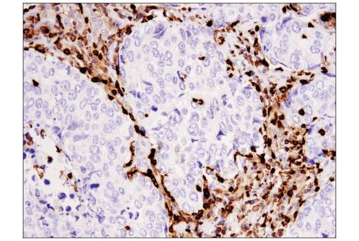
Immunohistochemical analysis of paraffin-embedded human squamous cell carcinoma of the lung using Coronin 1A (D6K5B) XP® Rabbit mAb.
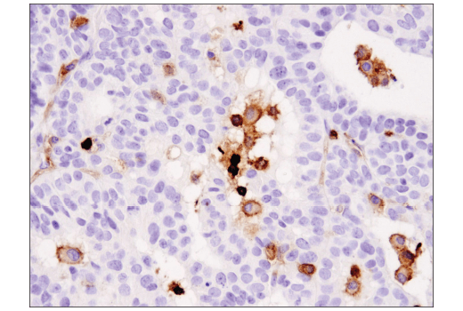
Immunohistochemical analysis of paraffin-embedded human endometrioid adenocarcinoma using Coronin 1A (D6K5B) XP® Rabbit mAb.
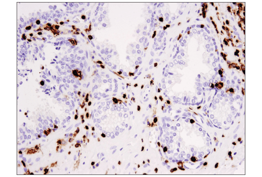
Immunohistochemical analysis of paraffin-embedded human prostate carcinoma using Coronin 1A (D6K5B) XP® Rabbit mAb.
Revision 6
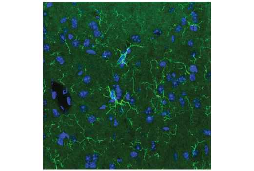
Confocal immunofluorescent analysis of mouse brain using Coronin 1A (D6K5B) XP® Rabbit mAb (green). Samples were mounted in ProLong® Gold Antifade Reagent with DAPI #8961 (blue). Citrate antigen retrieval is required to visualize Coronin 1A expression in microglia. This processing step may induce low-level, non-specific fluorescence.
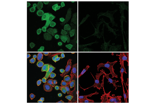
Confocal immunofluorescent analysis of SK-BR-3 cells (left, positive) and MDA-MB-231 cells (right, negative) using Coronin 1A (D6K5B) XP® Rabbit mAb (green). Actin filaments were labeled with DyLight™ 554 Phalloidin #13054 (red). Samples were mounted in ProLong® Gold Antifade Reagent with DAPI #8961 (blue).
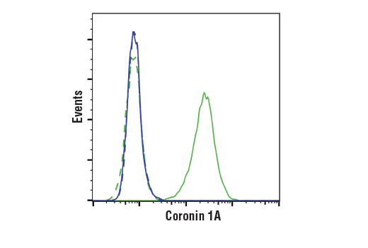
Flow cytometric analysis of MDA-MB-231 cells (blue) and Jurkat cells (green) using Coronin 1A (D6K5B) XP® Rabbit mAb (solid lines) or a concentration-matched Rabbit (DA1E) mAb IgG XP® Isotype Control #3900 (dashed lines). Anti-rabbit IgG (H+L), F(ab')2 Fragment (Alexa Fluor® 488 Conjugate) #4412 was used as a secondary antibody.