Revision 1
#97361
Store at -20C
877-616-CELL (2355)
877-678-TECH (8324)
3 Trask Lane | Danvers | Massachusetts | 01923 | USA
For Research Use Only. Not for Use in Diagnostic Procedures.
Applications:
W, IP, IF-IC, FC-FP
Reactivity:
H
Sensitivity:
Endogenous
MW (kDa):
88
Source/Isotype:
Rabbit IgG
UniProt ID:
#P50416
Entrez-Gene Id:
1374
Product Usage Information
| Application | Dilution |
|---|---|
| Western Blotting | 1:1000 |
| Immunoprecipitation | 1:50 |
| Immunofluorescence (Immunocytochemistry) | 1:400 - 1:1600 |
| Flow Cytometry (Fixed/Permeabilized) | 1:200 - 1:800 |
Storage
Specificity/Sensitivity
CPT1A (E3Y1V) Rabbit mAb recognizes endogenous levels of total CPT1A protein. This antibody does not cross-react with CPT1B, CPT1C, or CPT2.
Source / Purification
Monoclonal antibody is produced by immunizing animals with a synthetic peptide corresponding to residues surrounding Leu696 of human CPT1A protein.
Background
Carnitine palmitoyltransferase-1 (CPT1), localized to the mitochondrial outer membrane, translocates fatty acids across the mitochondrial membranes and catalyzes the rate-limiting step of β-oxidation (1,2). There are three isoforms of this enzyme: CPT1A (liver), CPT1B (muscle), and CPT1C (brain) (1,2). Deficiency of CPT1A results in an autosomal recessive mitochondrial fatty acid oxidation disorder (3). Studies have shown that physiological high blood glucose and insulin levels inhibit CPT1B activity in human muscle and therefore divert long-chain fatty acids toward storage in human muscle as triglycerides (4). Furthermore, mice deficient in CPT1C show less food intake and reduced body weight (5). These findings suggest that CPT1 may play a role in metabolic syndromes.
Species Reactivity
Species reactivity is determined by testing in at least one approved application (e.g., western blot).
Western Blot Buffer
IMPORTANT: For western blots, incubate membrane with diluted primary antibody in 5% w/v BSA, 1X TBS, 0.1% Tween® 20 at 4°C with gentle shaking, overnight.
Applications Key
W: Western Blotting IP: Immunoprecipitation IF-IC: Immunofluorescence (Immunocytochemistry) FC-FP: Flow Cytometry (Fixed/Permeabilized)
Cross-Reactivity Key
H: Human M: Mouse R: Rat Hm: Hamster Mk: Monkey Vir: Virus Mi: Mink C: Chicken Dm: D. melanogaster X: Xenopus Z: Zebrafish B: Bovine Dg: Dog Pg: Pig Sc: S. cerevisiae Ce: C. elegans Hr: Horse GP: Guinea Pig Rab: Rabbit G: Goat All: All Species Expected
Trademarks and Patents
Cell Signaling Technology is a trademark of Cell Signaling Technology, Inc.
Alexa Fluor is a registered trademark of Life Technologies Corporation.
All other trademarks are the property of their respective owners. Visit cellsignal.com/trademarks for more information.
Limited Uses
Except as otherwise expressly agreed in a writing signed by a legally authorized representative of CST, the following terms apply to Products provided by CST, its affiliates or its distributors. Any Customer's terms and conditions that are in addition to, or different from, those contained herein, unless separately accepted in writing by a legally authorized representative of CST, are rejected and are of no force or effect.
Products are labeled with For Research Use Only or a similar labeling statement and have not been approved, cleared, or licensed by the FDA or other regulatory foreign or domestic entity, for any purpose. Customer shall not use any Product for any diagnostic or therapeutic purpose, or otherwise in any manner that conflicts with its labeling statement. Products sold or licensed by CST are provided for Customer as the end-user and solely for research and development uses. Any use of Product for diagnostic, prophylactic or therapeutic purposes, or any purchase of Product for resale (alone or as a component) or other commercial purpose, requires a separate license from CST. Customer shall (a) not sell, license, loan, donate or otherwise transfer or make available any Product to any third party, whether alone or in combination with other materials, or use the Products to manufacture any commercial products, (b) not copy, modify, reverse engineer, decompile, disassemble or otherwise attempt to discover the underlying structure or technology of the Products, or use the Products for the purpose of developing any products or services that would compete with CST products or services, (c) not alter or remove from the Products any trademarks, trade names, logos, patent or copyright notices or markings, (d) use the Products solely in accordance with CST Product Terms of Sale and any applicable documentation, and (e) comply with any license, terms of service or similar agreement with respect to any third party products or services used by Customer in connection with the Products.
Revision 1
#97361
CPT1A (E3Y1V) Rabbit mAb
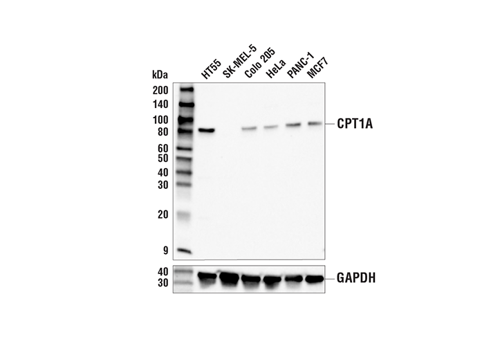
Western blot analysis of extracts from various cell lines using CPT1A (E3Y1V) Rabbit mAb (upper) and GAPDH (D16H11) XP® Rabbit mAb #5174 (lower). The absence of detected CPT1A expression in SK-MEL-5 cell extracts is consistent with RNAseq expression profiling data, confirming specificity of the antibody for CPT1A.
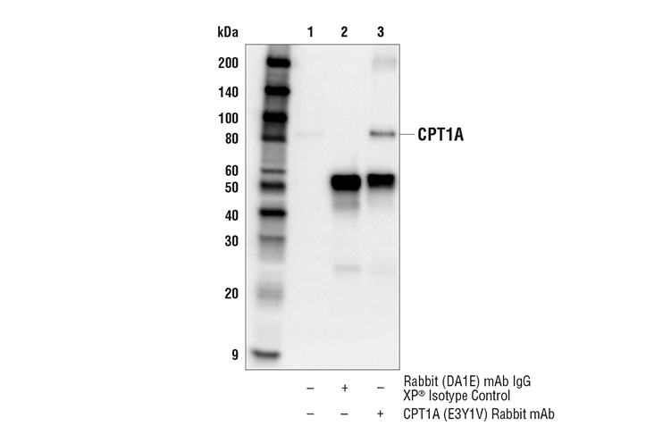
Immunoprecipitation of CPT1A protein from HeLa cell extracts. Lane 1 is 10% input, lane 2 is Rabbit (DA1E) mAb IgG XP® Isotype Control #3900, and lane 3 is CPT1A (E3Y1V) Rabbit mAb. Western blot analysis was performed using CPT1A (E3Y1V) Rabbit mAb. Anti-rabbit IgG, HRP-linked Antibody #7074 was used as the secondary antibody.
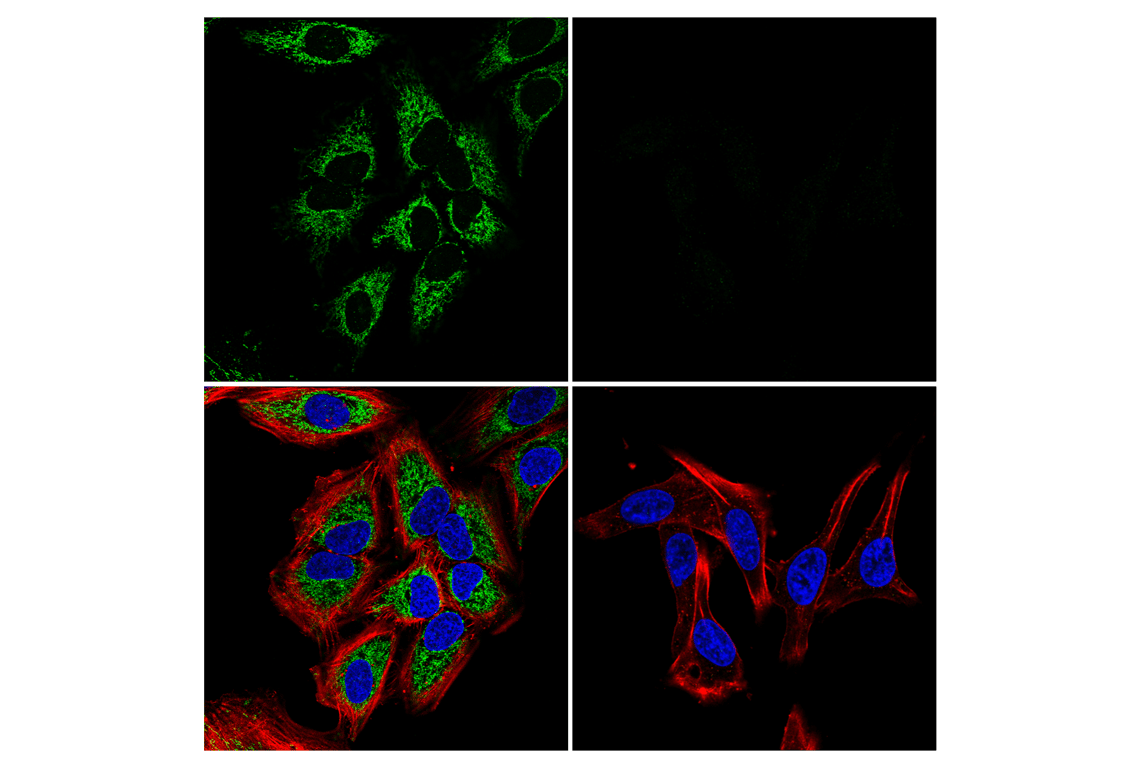
Confocal immunofluorescent analysis of HeLa cells (left, positive) or SK-MEL-5 cells (right, negative) using CPT1A (E3Y1V) Rabbit mAb (green). Actin filaments were labeled with DyLight™ 650 Phalloidin #12956 (red). Samples were mounted in ProLong® Gold Antifade Reagent with DAPI #8961 (blue).
Revision 1
#97361
CPT1A (E3Y1V) Rabbit mAb
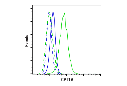
Flow cytometric analysis of SK-MEL-5 cells (blue, negative) and HeLa cells (green, positive) using CPT1A (E3Y1V) Rabbit mAb (solid lines) or a concentration-matched Rabbit (DA1E) mAb IgG XP® Isotype Control #3900 (dashed lines). Anti-rabbit IgG (H+L), F(ab')2 Fragment (Alexa Fluor® 488 Conjugate) #4412 was used as a secondary antibody.