Revision 1
#96615
Store at -20C
877-616-CELL (2355)
877-678-TECH (8324)
3 Trask Lane | Danvers | Massachusetts | 01923 | USA
For Research Use Only. Not for Use in Diagnostic Procedures.
Applications:
W, IHC-P
Reactivity:
H
Sensitivity:
Endogenous
MW (kDa):
220
Source/Isotype:
Rabbit IgG
UniProt ID:
#P17927
Entrez-Gene Id:
1378
Product Usage Information
| Application | Dilution |
|---|---|
| Western Blotting | 1:1000 |
| Immunohistochemistry (Paraffin) | 1:800 - 1:3200 |
Storage
Specificity/Sensitivity
CR1/CD35 (E8B1Z) Rabbit mAb recognizes endogenous levels of total CR1/CD35 protein. Non-specific staining was observed in skeletal muscle by immunohistochemistry.
Source / Purification
Monoclonal antibody is produced by immunizing animals with recombinant protein specific to the extracellular domain of human CR1/CD35 protein.
Background
Complement receptor type 1 (CR1/CD35) is a type I transmembrane glycoprotein that is expressed on the surface of B cells, neutrophils, monocytes, and renal podocytes (1,2). As a component of the host innate immune system, CR1/CD35 expressed on neutrophils and monocytes binds to ligands coated with the complement opsonins, C3b and C4b, which facilitates phagocytosis and production of proinflammatory cytokines (3,4). CR1/CD35 also participates in negative regulation of the complement cascade through its ability to promote dissociation of C3 and C5 convertases (4,5) and by serving as one of multiple cofactors for factor-I-mediated cleavage and inactivation of C3b and C4b (6).
Background References
- Fearon, D.T. (1980) J Exp Med 152, 20-30.
- Tedder, T.F. et al. (1983) J Immunol 130, 1668-73.
- Krych-Goldberg, M. and Atkinson, J.P. (2001) Immunol Rev 180, 112-22.
- Bacle, F. et al. (1990) J Immunol 144, 147-52.
- Hourcade, D.E. et al. (2002) J Biol Chem 277, 1107-12.
- Masaki, T. et al. (1992) J Biochem 111, 573-8.
Species Reactivity
Species reactivity is determined by testing in at least one approved application (e.g., western blot).
Western Blot Buffer
IMPORTANT: For western blots, incubate membrane with diluted primary antibody in 5% w/v BSA, 1X TBS, 0.1% Tween® 20 at 4°C with gentle shaking, overnight.
Applications Key
W: Western Blotting IHC-P: Immunohistochemistry (Paraffin)
Cross-Reactivity Key
H: Human M: Mouse R: Rat Hm: Hamster Mk: Monkey Vir: Virus Mi: Mink C: Chicken Dm: D. melanogaster X: Xenopus Z: Zebrafish B: Bovine Dg: Dog Pg: Pig Sc: S. cerevisiae Ce: C. elegans Hr: Horse GP: Guinea Pig Rab: Rabbit G: Goat All: All Species Expected
Trademarks and Patents
Cell Signaling Technology is a trademark of Cell Signaling Technology, Inc.
All other trademarks are the property of their respective owners. Visit cellsignal.com/trademarks for more information.
Limited Uses
Except as otherwise expressly agreed in a writing signed by a legally authorized representative of CST, the following terms apply to Products provided by CST, its affiliates or its distributors. Any Customer's terms and conditions that are in addition to, or different from, those contained herein, unless separately accepted in writing by a legally authorized representative of CST, are rejected and are of no force or effect.
Products are labeled with For Research Use Only or a similar labeling statement and have not been approved, cleared, or licensed by the FDA or other regulatory foreign or domestic entity, for any purpose. Customer shall not use any Product for any diagnostic or therapeutic purpose, or otherwise in any manner that conflicts with its labeling statement. Products sold or licensed by CST are provided for Customer as the end-user and solely for research and development uses. Any use of Product for diagnostic, prophylactic or therapeutic purposes, or any purchase of Product for resale (alone or as a component) or other commercial purpose, requires a separate license from CST. Customer shall (a) not sell, license, loan, donate or otherwise transfer or make available any Product to any third party, whether alone or in combination with other materials, or use the Products to manufacture any commercial products, (b) not copy, modify, reverse engineer, decompile, disassemble or otherwise attempt to discover the underlying structure or technology of the Products, or use the Products for the purpose of developing any products or services that would compete with CST products or services, (c) not alter or remove from the Products any trademarks, trade names, logos, patent or copyright notices or markings, (d) use the Products solely in accordance with CST Product Terms of Sale and any applicable documentation, and (e) comply with any license, terms of service or similar agreement with respect to any third party products or services used by Customer in connection with the Products.
Revision 1
#96615
CR1/CD35 (E8B1Z) Rabbit mAb
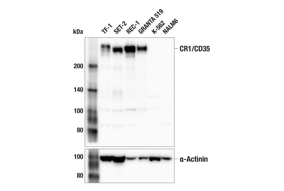
Western blot analysis of extracts from various cell lines using CR1/CD35 (E8B1Z) Rabbit mAb (upper) or α-Actinin (D6F6) XP® Rabbit mAb #6487 (lower). Negative expression of CR1/CD35 protein in K-562 and NALM6 cells is consistent with the predicted expression pattern.
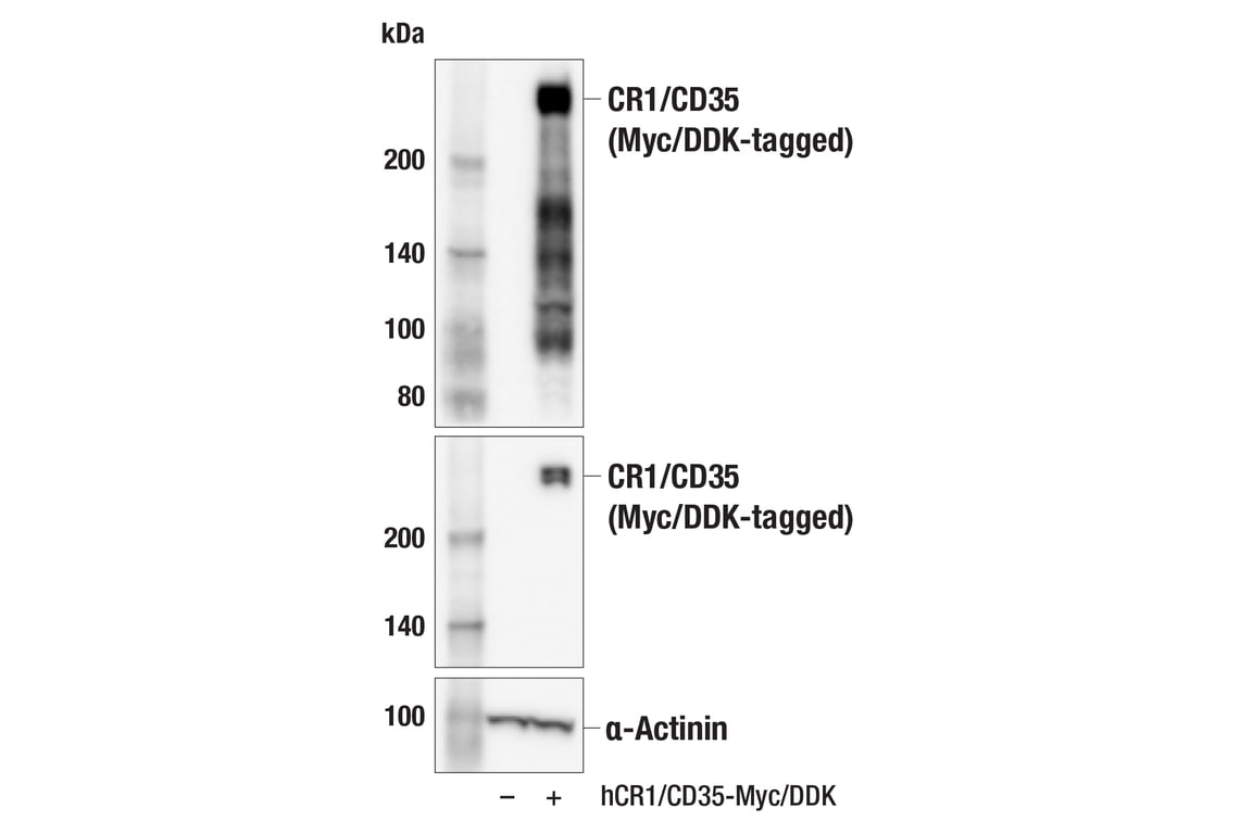
Western blot analysis of extracts from 293T cells, mock transfected (-) or transfected with a construct expressing Myc/DDK-tagged full-length human CR1/CD35 (hCR1/CD35-Myc/DDK; +), using CR1/CD35 (E8B1Z) Rabbit mAb (upper), DYKDDDDK Tag (D6W5B) Rabbit mAb #14793 (middle), or α-Actinin (D6F6) XP® Rabbit mAb #6487 (lower).
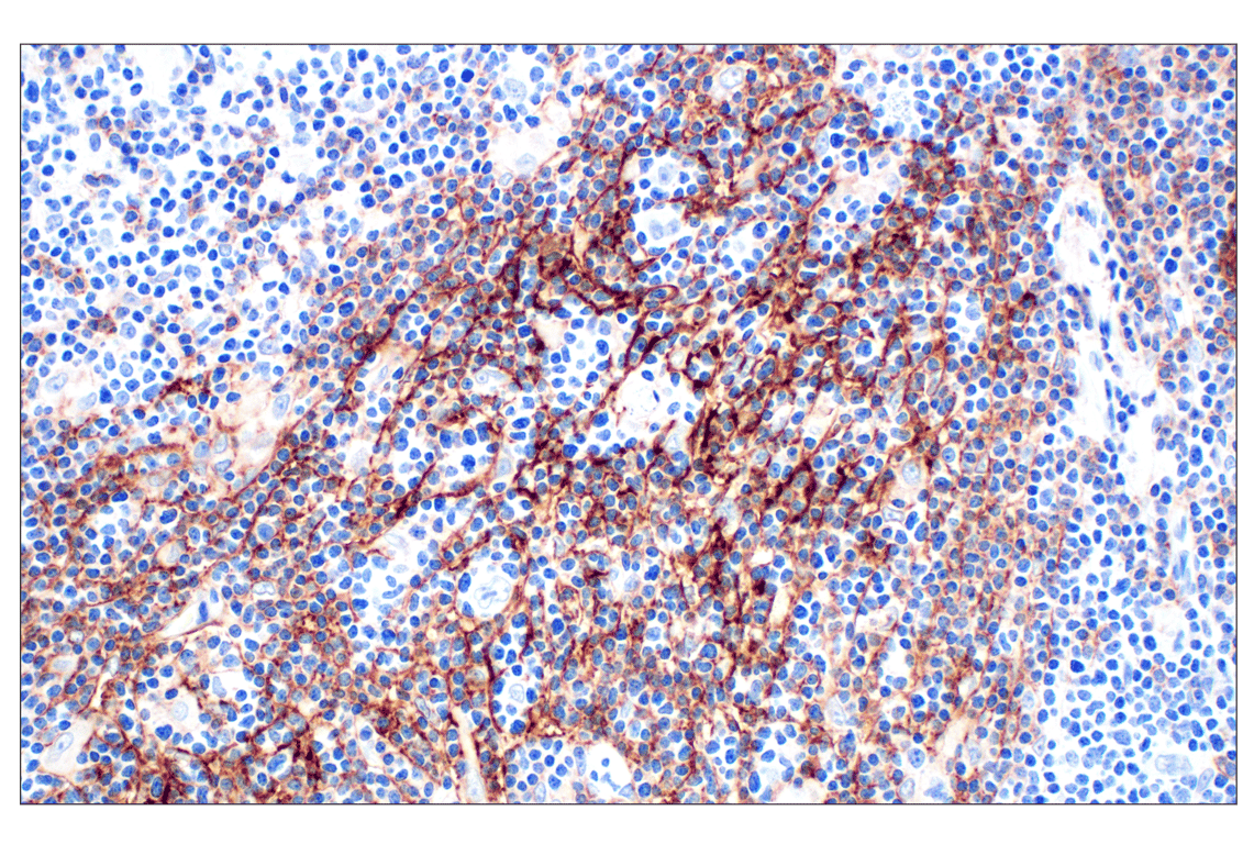
Immunohistochemical analysis of paraffin-embedded human Hodgkin lymphoma using CR1/CD35 (E8B1Z) Rabbit mAb.
Revision 1
#96615
CR1/CD35 (E8B1Z) Rabbit mAb
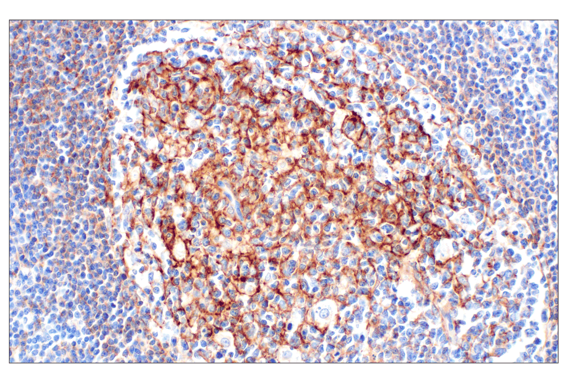
Immunohistochemical analysis of paraffin-embedded human tonsil using CR1/CD35 (E8B1Z) Rabbit mAb.
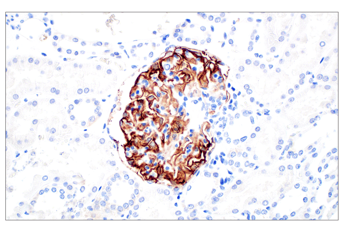
Immunohistochemical analysis of paraffin-embedded normal human kidney using CR1/CD35 (E8B1Z) Rabbit mAb.
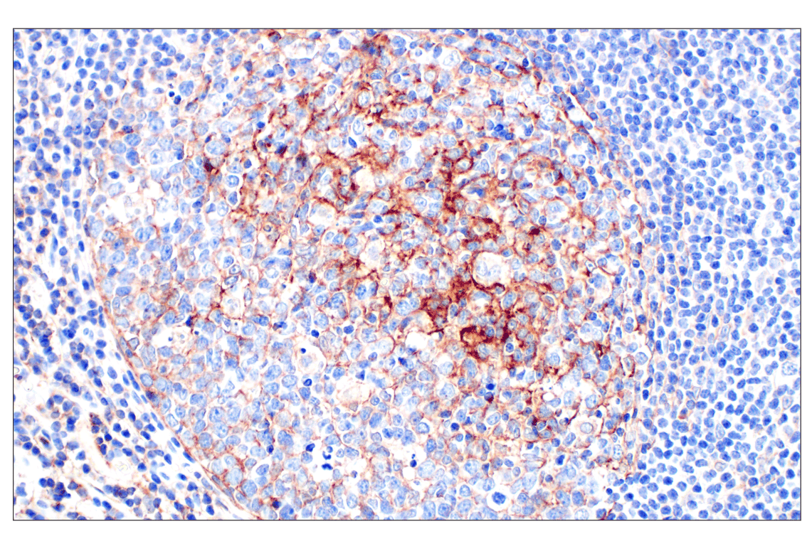
Immunohistochemical analysis of paraffin-embedded Peyer's patch within normal human small intestine using CR1/CD35 (E8B1Z) Rabbit mAb.
Revision 1
#96615
CR1/CD35 (E8B1Z) Rabbit mAb
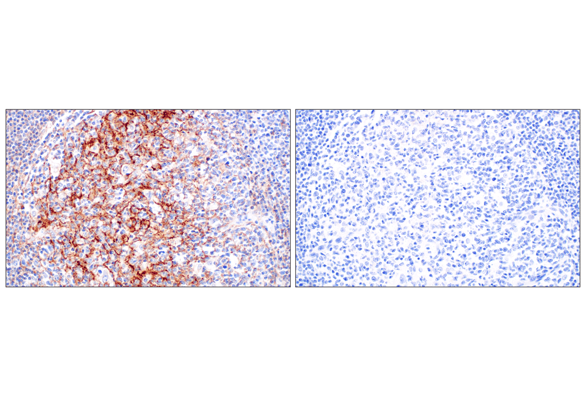
Immunohistochemical analysis of paraffin-embedded human tonsil using CR1/CD35 (E8B1Z) Rabbit mAb (left) compared to concentration-matched Rabbit (DA1E) mAb IgG XP® Isotype Control #3900 (right).
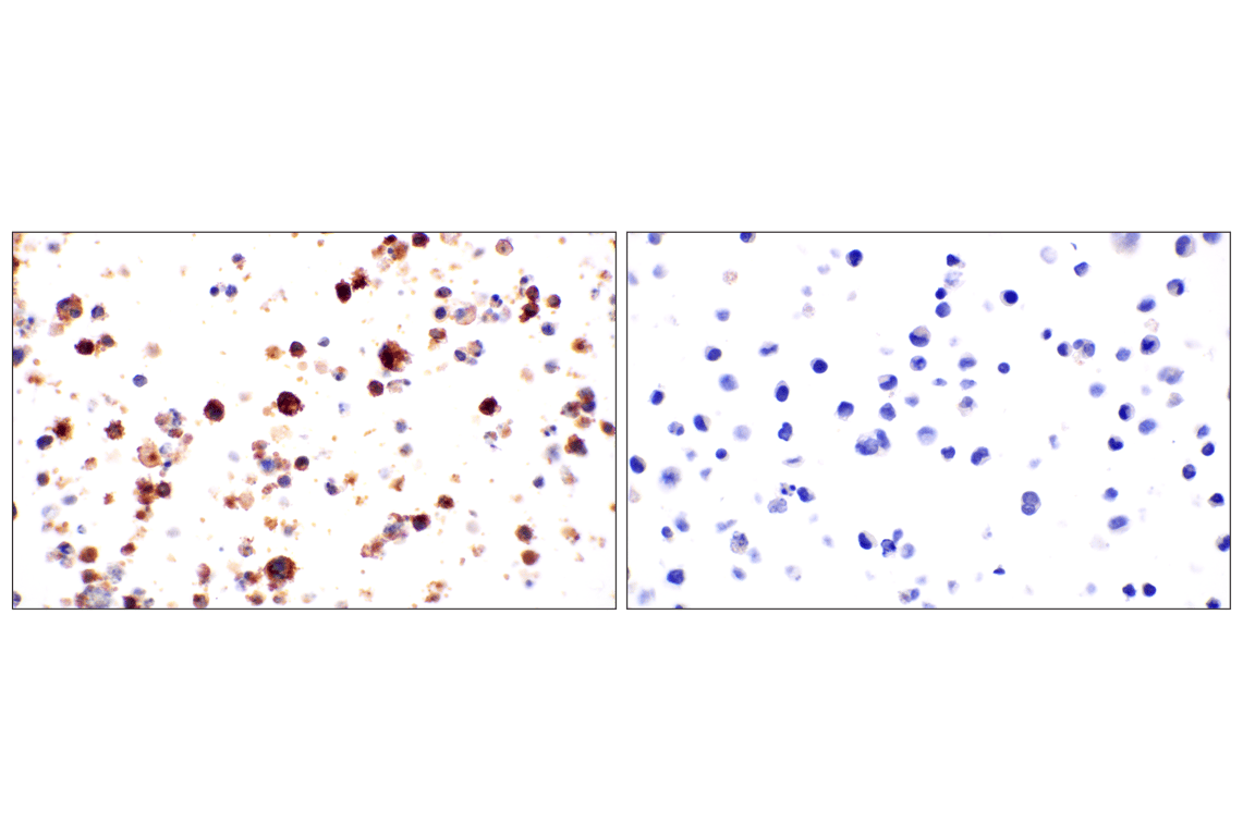
Immunohistochemical analysis of paraffin-embedded TF-1 cell pellet (left, positive) or K-562 cell pellet (right, negative) using CR1/CD35 (E8B1Z) Rabbit mAb.