WB, IP, IHC-P
H
Endogenous
55
Rabbit IgG
#P14635
891
Product Information
Product Usage Information
| Application | Dilution |
|---|---|
| Western Blotting | 1:1000 |
| Immunoprecipitation | 1:100 |
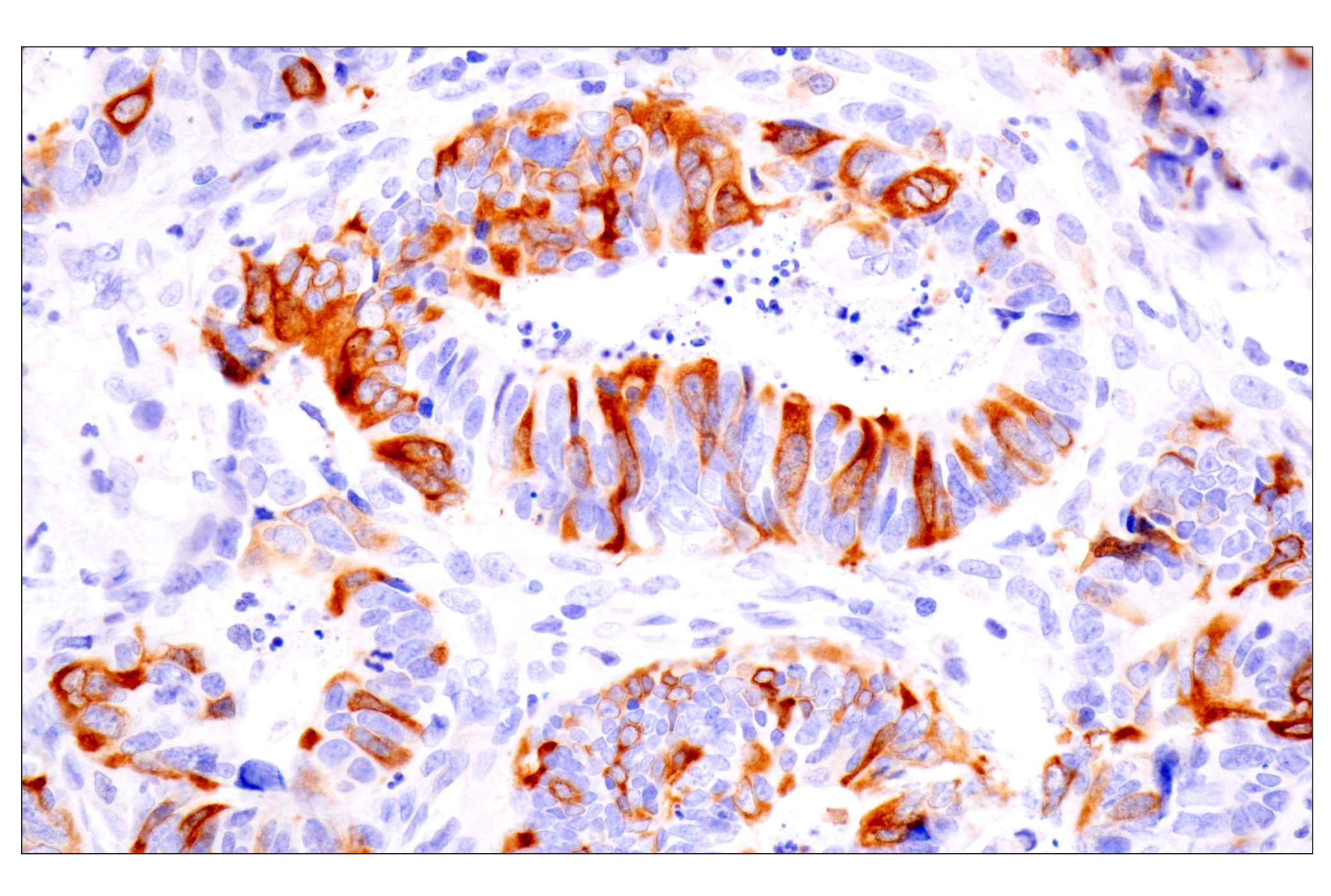
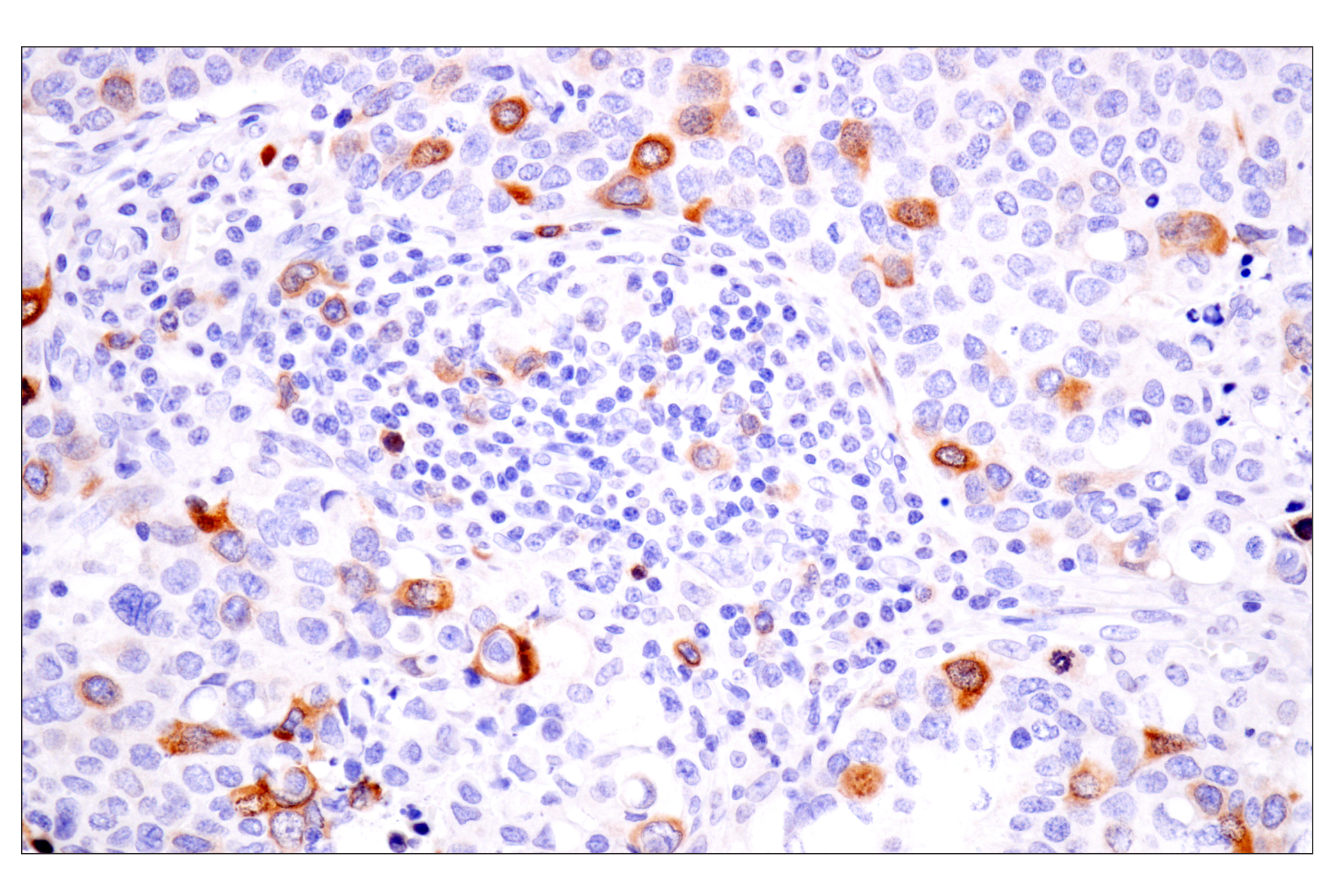
| Immunohistochemistry (Paraffin) | 1:50 - 1:200 |
Storage
For a carrier free (BSA and azide free) version of this product see product #67873.
Specificity / Sensitivity
Species Reactivity:
Human
Source / Purification
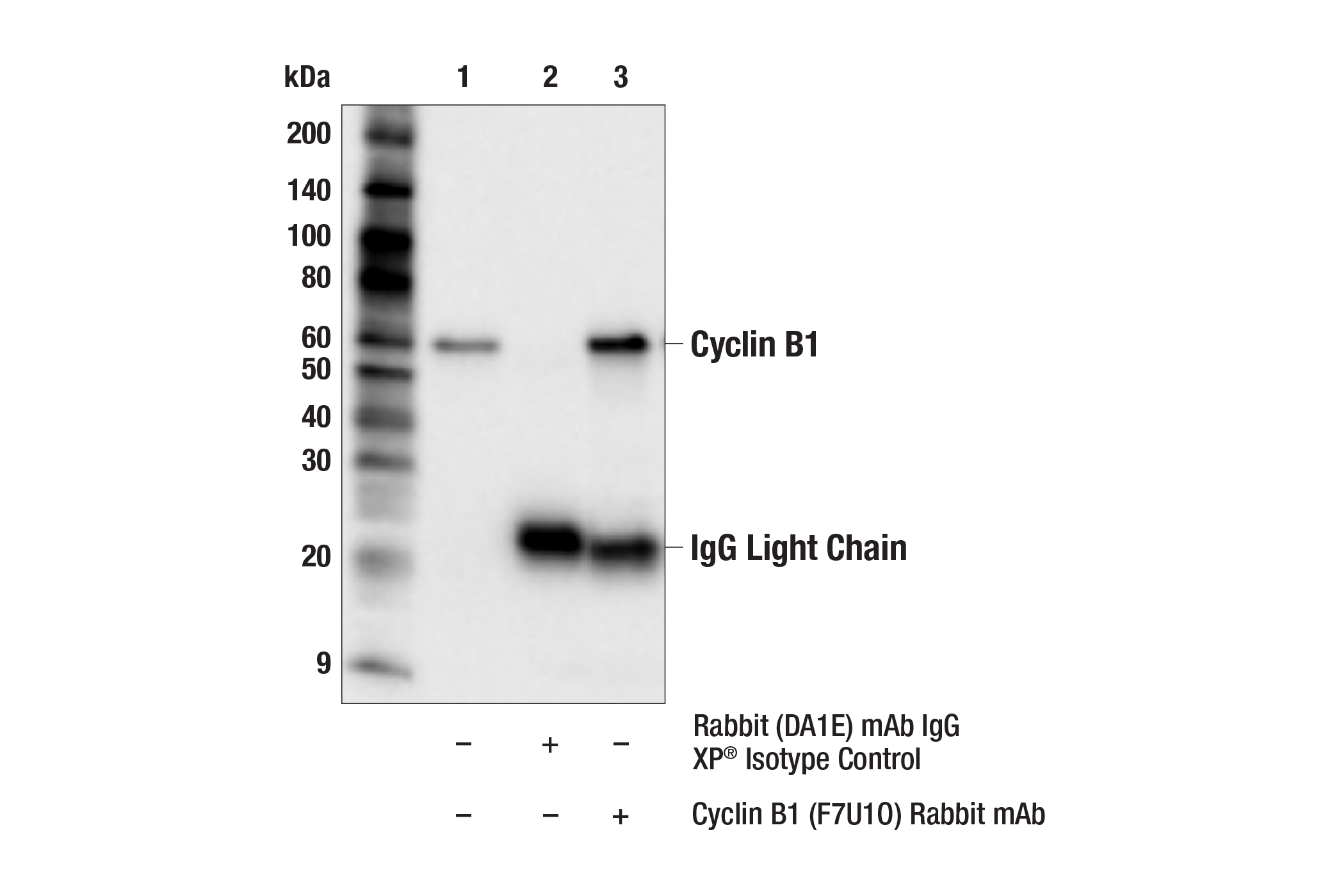
Monoclonal antibody is produced by immunizing animals with a synthetic peptide corresponding to residues surrounding Asn275 of human cyclin B1 protein.
Background
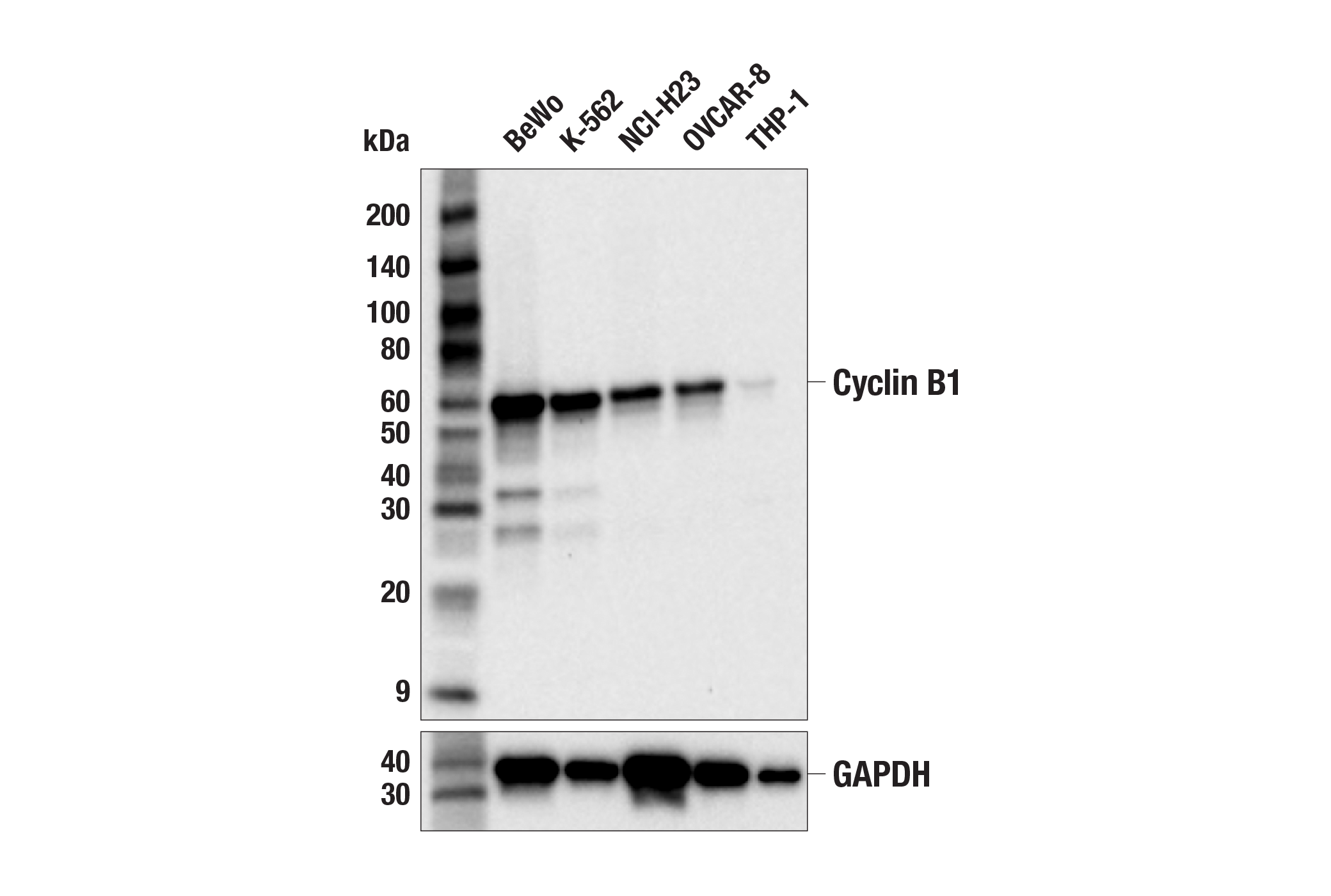
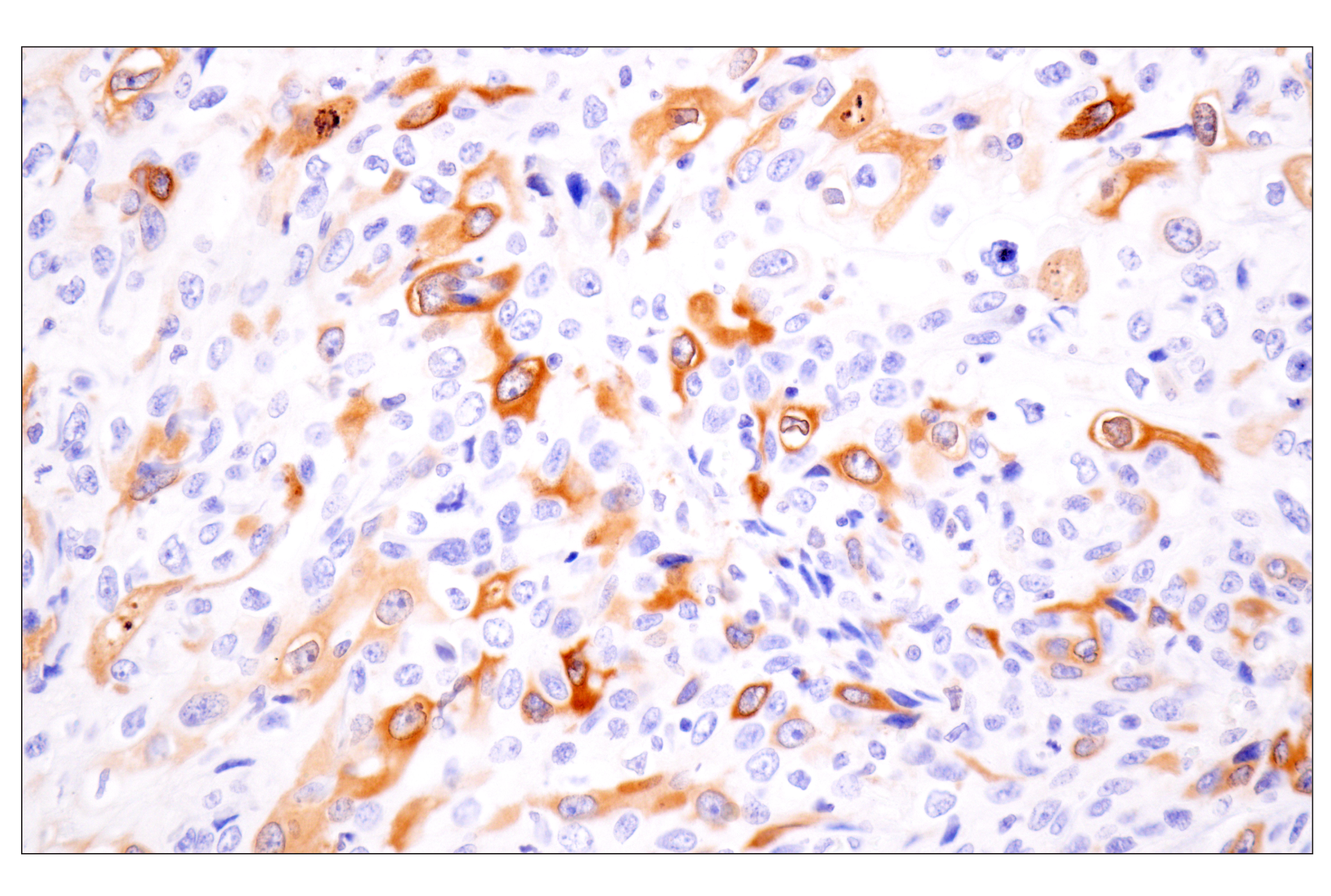
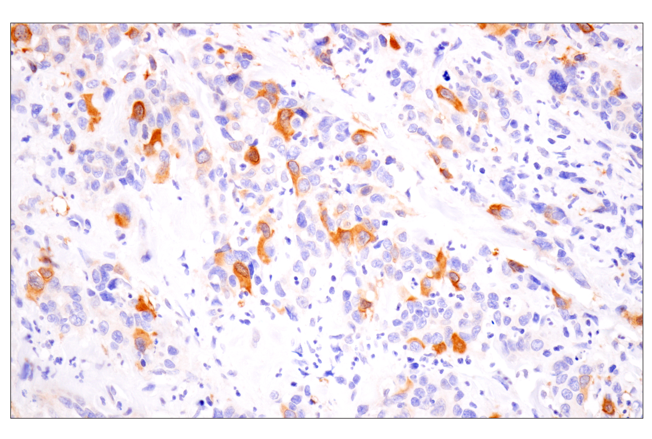
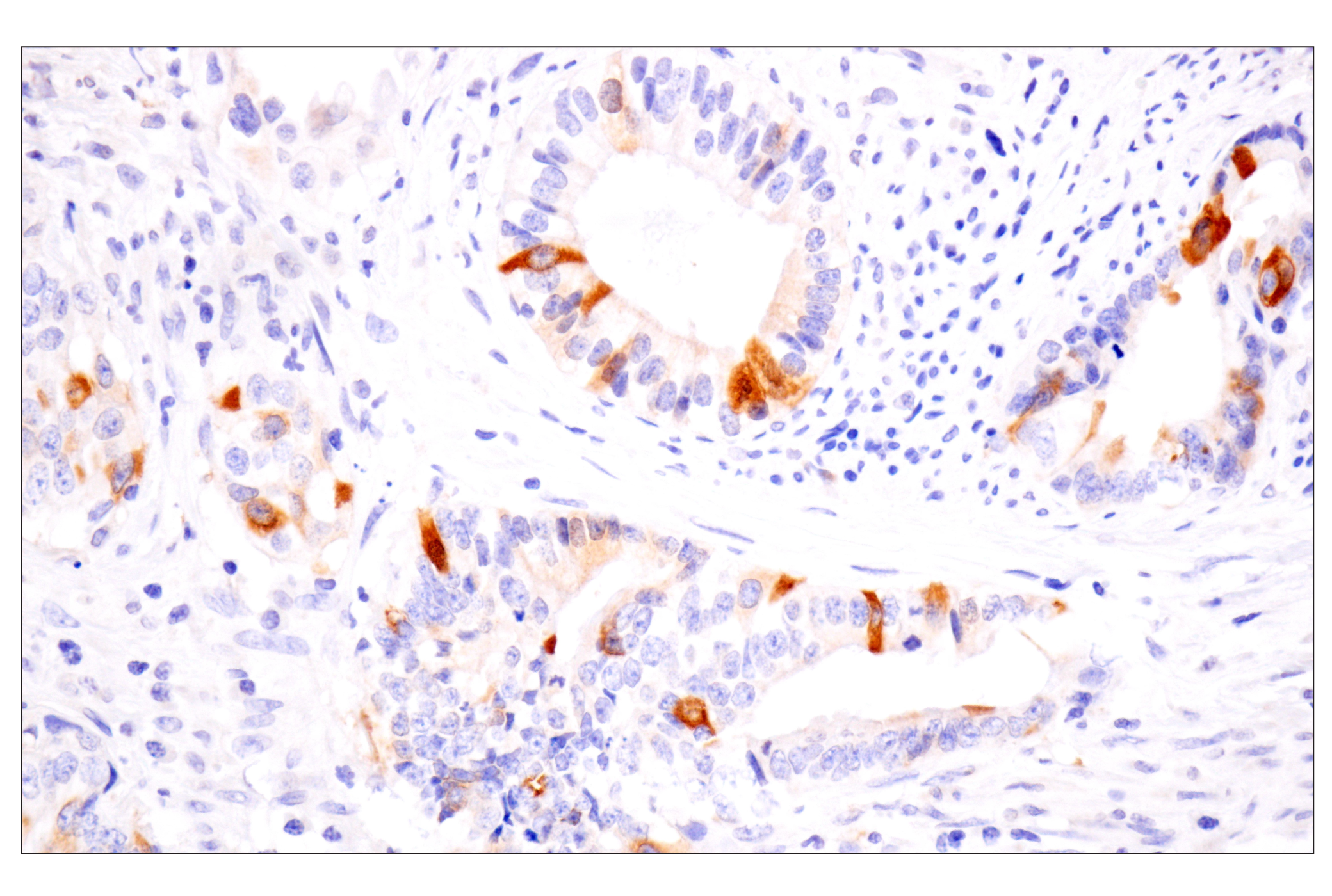
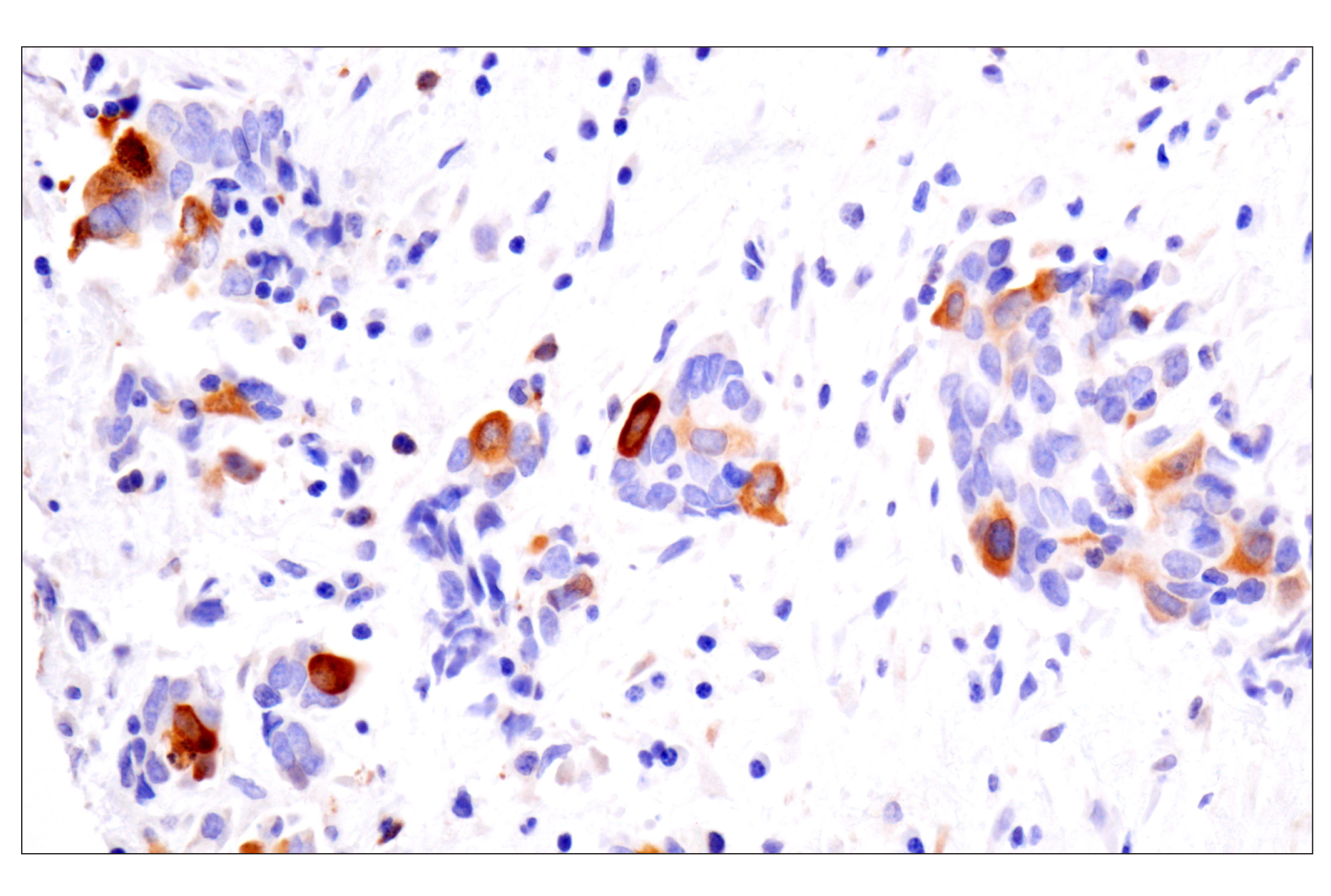
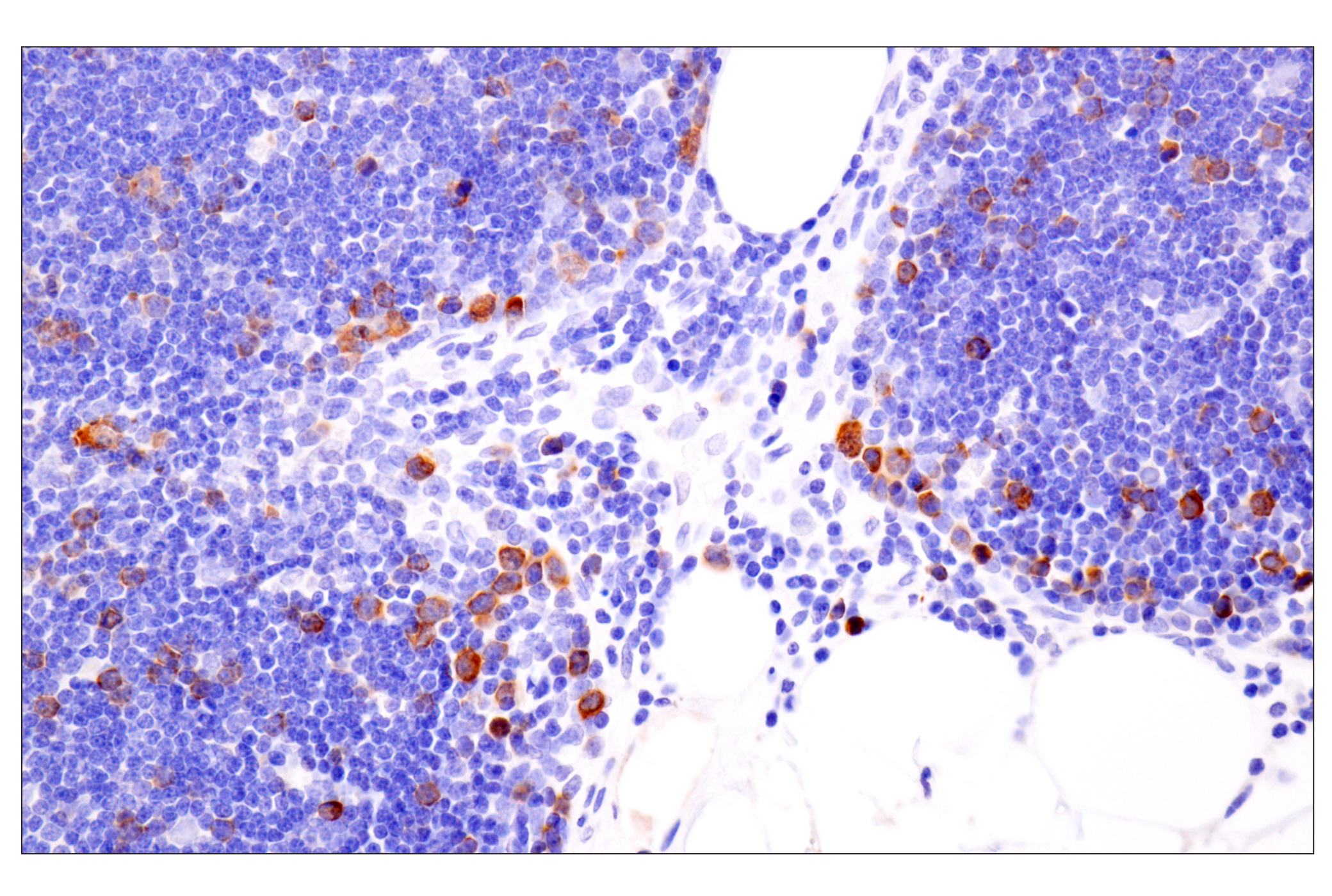
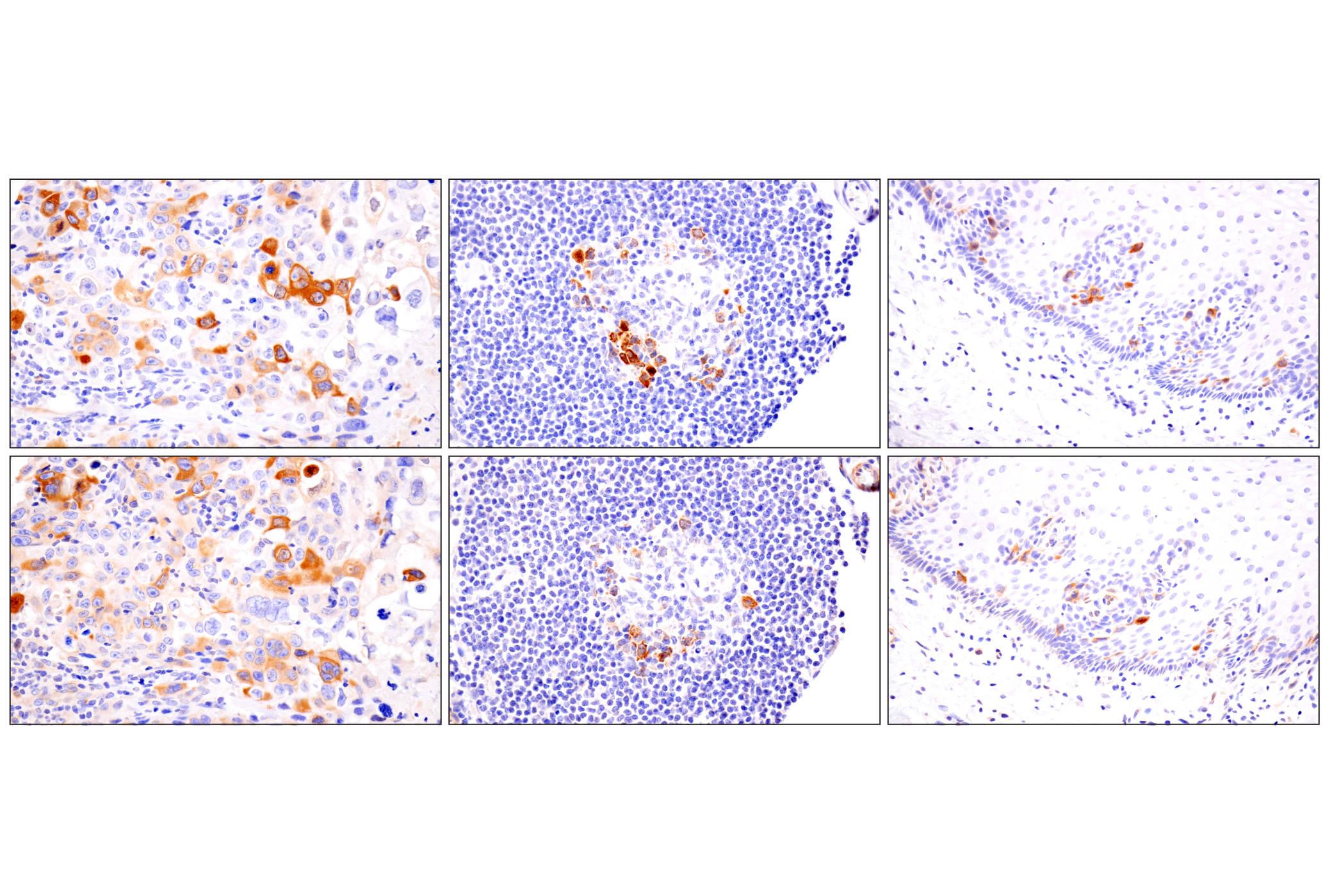
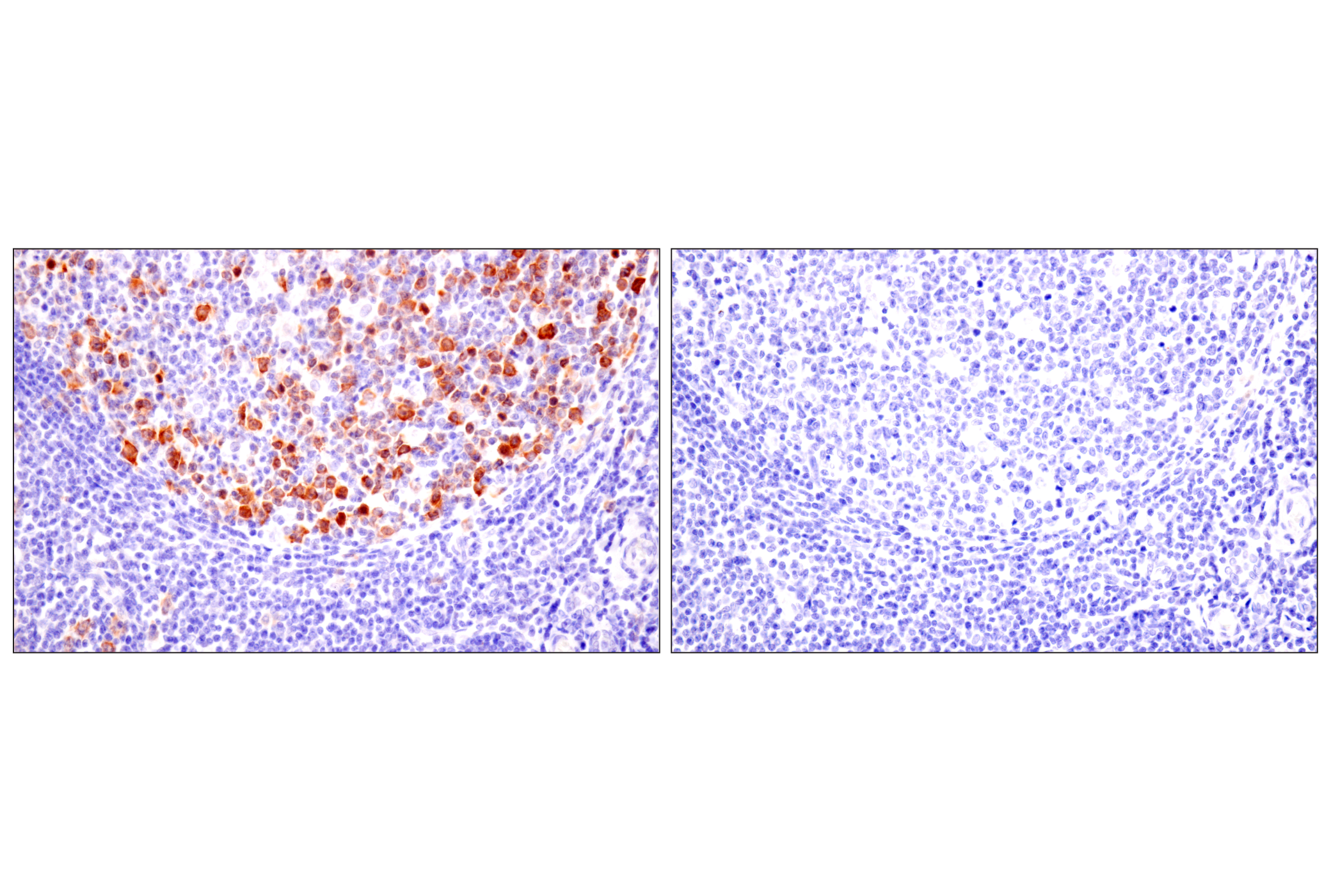
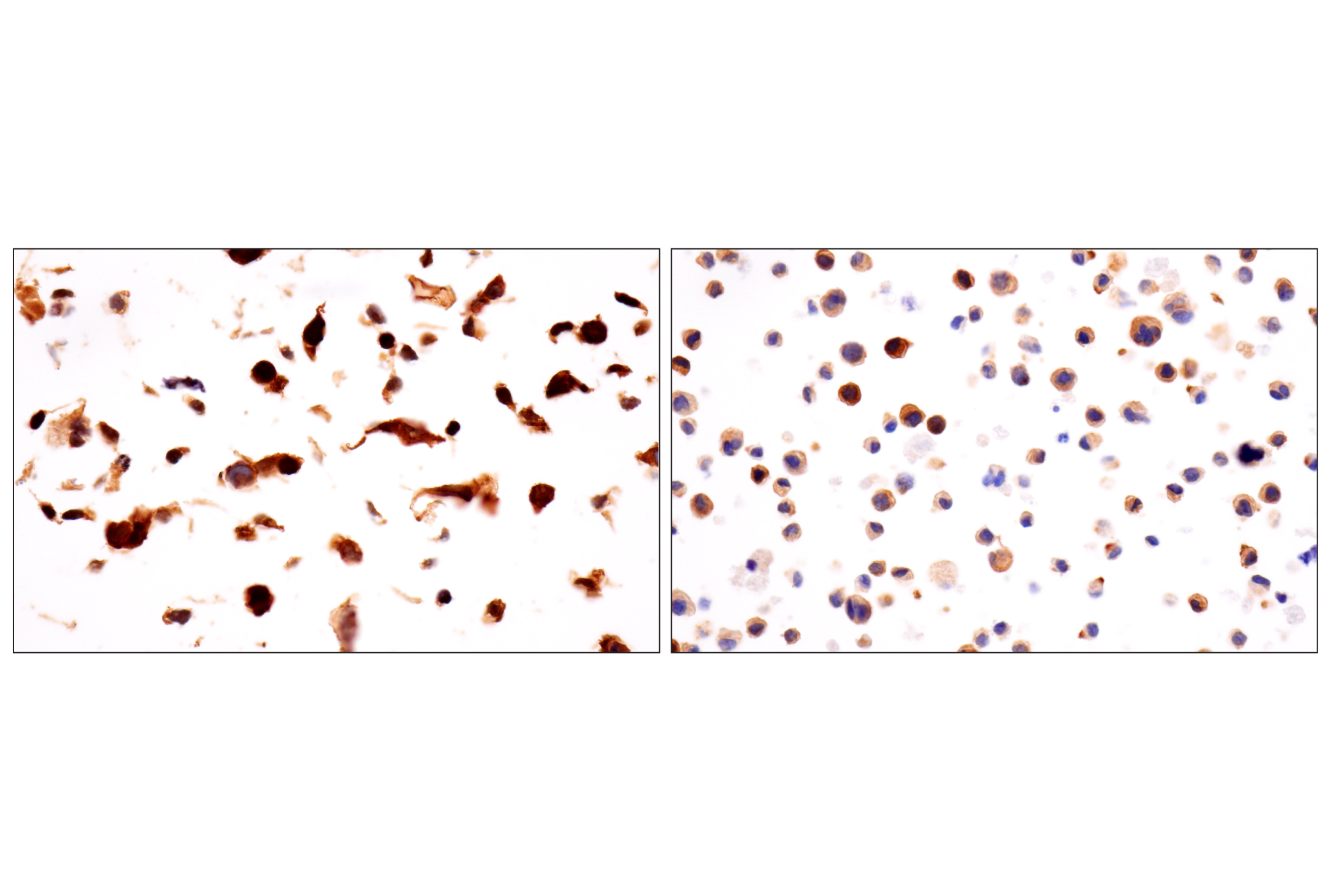
Cyclins are a family of proteins that activate specific cyclin-dependent kinases required for progression through the cell cycle. The entry of all eukaryotic cells into mitosis is regulated by activation of cdc2/cdk1 at the G2/M transition. This activation is a multi-step process that begins with the binding of the regulatory subunit, cyclin B1, to cdc2/cdk1 to form the mitosis-promoting factor (MPF). MPF remains in the inactive state until phosphorylation of cdc2/cdk1 at Thr161 by cdk activating kinase (CAK) (1,2) and dephosphorylation of cdc2/cdk1 at Thr14/Tyr15 by cdc25C (3-5). Five cyclin B1 phosphorylation sites (Ser116, 126, 128, 133, and 147) are located in the cytoplasmic retention signal (CRS) domain and are thought to regulate the translocation of cyclin B1 to the nucleus at the G2/M checkpoint, promoting nuclear accumulation and initiation of mitosis (6-9). While MPF itself can phosphorylate Ser126 and Ser128, polo-like kinase 1 (PLK1) phosphorylates cyclin B1 preferentially at Ser133 and possibly at Ser147 (6,10). At the end of mitosis, cyclin B1 is targeted for degradation by the anaphase-promoting complex (APC), allowing for cell cycle progression (11). Research studies have shown that cyclin B1 is overexpressed in breast, prostate, and non-small cell lung cancers (12-14).
- Lorca, T. et al. (1992) EMBO J 11, 2381-90.
- Harper, J.W. and Elledge, S.J. (1998) Genes Dev 12, 285-9.
- Norbury, C. et al. (1991) EMBO J 10, 3321-9.
- McGowan, C.H. and Russell, P. (1993) EMBO J 12, 75-85.
- Atherton-Fessler, S. et al. (1994) Mol Biol Cell 5, 989-1001.
- Toyoshima-Morimoto, F. et al. (2001) Nature 410, 215-20.
- Li, J. et al. (1997) Proc Natl Acad Sci U S A 94, 502-7.
- Takizawa, C.G. and Morgan, D.O. (2000) Curr Opin Cell Biol 12, 658-65.
- Santos, S.D. et al. (2012) Cell 149, 1500-13.
- Jackman, M. et al. (2003) Nat Cell Biol 5, 143-8.
- Gong, D. and Ferrell, J.E. (2010) Mol Biol Cell 21, 3149-61.
- Mashal, R.D. et al. (1996) Cancer Res 56, 4159-63.
- Kawamoto, H. et al. (1997) Am J Pathol 150, 15-23.
- Soria, J.C. et al. (2000) Cancer Res 60, 4000-4.
Species Reactivity
Species reactivity is determined by testing in at least one approved application (e.g., western blot).
Western Blot Buffer
IMPORTANT: For western blots, incubate membrane with diluted primary antibody in 5% w/v BSA, 1X TBS, 0.1% Tween® 20 at 4°C with gentle shaking, overnight.
Applications Key
WB: Western Blotting IP: Immunoprecipitation IHC-P: Immunohistochemistry (Paraffin)
Cross-Reactivity Key
H: human M: mouse R: rat Hm: hamster Mk: monkey Vir: virus Mi: mink C: chicken Dm: D. melanogaster X: Xenopus Z: zebrafish B: bovine Dg: dog Pg: pig Sc: S. cerevisiae Ce: C. elegans Hr: horse GP: Guinea Pig Rab: rabbit All: all species expected
Trademarks and Patents
Limited Uses
Except as otherwise expressly agreed in a writing signed by a legally authorized representative of CST, the following terms apply to Products provided by CST, its affiliates or its distributors. Any Customer's terms and conditions that are in addition to, or different from, those contained herein, unless separately accepted in writing by a legally authorized representative of CST, are rejected and are of no force or effect.
Products are labeled with For Research Use Only or a similar labeling statement and have not been approved, cleared, or licensed by the FDA or other regulatory foreign or domestic entity, for any purpose. Customer shall not use any Product for any diagnostic or therapeutic purpose, or otherwise in any manner that conflicts with its labeling statement. Products sold or licensed by CST are provided for Customer as the end-user and solely for research and development uses. Any use of Product for diagnostic, prophylactic or therapeutic purposes, or any purchase of Product for resale (alone or as a component) or other commercial purpose, requires a separate license from CST. Customer shall (a) not sell, license, loan, donate or otherwise transfer or make available any Product to any third party, whether alone or in combination with other materials, or use the Products to manufacture any commercial products, (b) not copy, modify, reverse engineer, decompile, disassemble or otherwise attempt to discover the underlying structure or technology of the Products, or use the Products for the purpose of developing any products or services that would compete with CST products or services, (c) not alter or remove from the Products any trademarks, trade names, logos, patent or copyright notices or markings, (d) use the Products solely in accordance with CST Product Terms of Sale and any applicable documentation, and (e) comply with any license, terms of service or similar agreement with respect to any third party products or services used by Customer in connection with the Products.