Revision 1
#83458
Store at -20C
877-616-CELL (2355)
877-678-TECH (8324)
3 Trask Lane | Danvers | Massachusetts | 01923 | USA
For Research Use Only. Not for Use in Diagnostic Procedures.
Applications:
W, IF-IC
Reactivity:
H
Sensitivity:
Endogenous
MW (kDa):
75-100
Source/Isotype:
Rabbit IgG
UniProt ID:
#Q16610
Entrez-Gene Id:
1893
Product Usage Information
| Application | Dilution |
|---|---|
| Western Blotting | 1:1000 |
| Immunofluorescence (Immunocytochemistry) | 1:800 - 1:3200 |
Storage
Specificity/Sensitivity
ECM1 (E3B9A) Rabbit mAb recognizes endogenous levels of total ECM1 protein.
Source / Purification
Monoclonal antibody is produced by immunizing animals with recombinant protein specific to the amino terminus of human ECM1 protein.
Background
Extracellular matrix gene 1 (ECM1) is a secreted glycoprotein found primarily in the extracellular matrix. It acts as both a multifunctional binding core and as a scaffolding protein that interacts with a variety of extracellular and structural proteins, including perlecan, fibulin-1C/1D, and MMP-9. (1). Research studies have shown that ECM1 is involved in the maintenance of tissue integrity and homeostasis, T cell immune responses, and angiogenesis (2-5). Its importance in maintaining tissue integrity is demonstrated by reports showing that mutations in the gene encoding ECM1 lead to skin lipoid proteinosis (1). Notably, ECM1 expression is upregulated in a variety of cancers, and during metastasis, suggesting a role for ECM1 in promoting tumor cell proliferation and invasion (6). In this context, research studies have shown that ECM1 can activate various signaling pathways, such as integrin/FAK, MMP-9/galectin-3, and S100A4/RhoA (7-9). In hepatic tissues, it was furthermore shown that ECM1 contributes to maintaining the latency of TGF-β in the extracellular matrix, thereby preventing spontaneous TGF-β activation and liver fibrosis (10,11).
Background References
- Chan, I. et al. (2007) Exp Dermatol 16, 881-90.
- Sercu, S. et al. (2008) J Invest Dermatol 128, 1397-408.
- Kong, L. et al. (2010) Matrix Biol 29, 276-86.
- Han, Z. et al. (2001) FASEB J 15, 988-94.
- He, L. et al. (2018) Proc Natl Acad Sci U S A 115, 8621-8626.
- Wang, L. et al. (2003) Cancer Lett 200, 57-67.
- Gan, L. et al. (2018) Oncogene 37, 744-755.
- Lee, K.M. et al. (2014) Breast Cancer Res 16, 479.
- Gómez-Contreras, P. et al. (2017) Clin Exp Metastasis 34, 37-49.
- Fan, W. et al. (2019) Gastroenterology 157, 1352-1367.e13.
- Li, Y. et al. (2022) JHEP Rep 4, 100397.
Species Reactivity
Species reactivity is determined by testing in at least one approved application (e.g., western blot).
Western Blot Buffer
IMPORTANT: For western blots, incubate membrane with diluted primary antibody in 5% w/v BSA, 1X TBS, 0.1% Tween® 20 at 4°C with gentle shaking, overnight.
Applications Key
W: Western Blotting IF-IC: Immunofluorescence (Immunocytochemistry)
Cross-Reactivity Key
H: Human M: Mouse R: Rat Hm: Hamster Mk: Monkey Vir: Virus Mi: Mink C: Chicken Dm: D. melanogaster X: Xenopus Z: Zebrafish B: Bovine Dg: Dog Pg: Pig Sc: S. cerevisiae Ce: C. elegans Hr: Horse GP: Guinea Pig Rab: Rabbit G: Goat All: All Species Expected
Trademarks and Patents
Cell Signaling Technology is a trademark of Cell Signaling Technology, Inc.
XP is a registered trademark of Cell Signaling Technology, Inc.
All other trademarks are the property of their respective owners. Visit cellsignal.com/trademarks for more information.
Limited Uses
Except as otherwise expressly agreed in a writing signed by a legally authorized representative of CST, the following terms apply to Products provided by CST, its affiliates or its distributors. Any Customer's terms and conditions that are in addition to, or different from, those contained herein, unless separately accepted in writing by a legally authorized representative of CST, are rejected and are of no force or effect.
Products are labeled with For Research Use Only or a similar labeling statement and have not been approved, cleared, or licensed by the FDA or other regulatory foreign or domestic entity, for any purpose. Customer shall not use any Product for any diagnostic or therapeutic purpose, or otherwise in any manner that conflicts with its labeling statement. Products sold or licensed by CST are provided for Customer as the end-user and solely for research and development uses. Any use of Product for diagnostic, prophylactic or therapeutic purposes, or any purchase of Product for resale (alone or as a component) or other commercial purpose, requires a separate license from CST. Customer shall (a) not sell, license, loan, donate or otherwise transfer or make available any Product to any third party, whether alone or in combination with other materials, or use the Products to manufacture any commercial products, (b) not copy, modify, reverse engineer, decompile, disassemble or otherwise attempt to discover the underlying structure or technology of the Products, or use the Products for the purpose of developing any products or services that would compete with CST products or services, (c) not alter or remove from the Products any trademarks, trade names, logos, patent or copyright notices or markings, (d) use the Products solely in accordance with CST Product Terms of Sale and any applicable documentation, and (e) comply with any license, terms of service or similar agreement with respect to any third party products or services used by Customer in connection with the Products.
Revision 1
#83458
ECM1 (E3B9A) Rabbit mAb
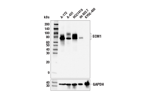
Western blot analysis of extracts from various cell lines using ECM1 (E3B9A) Rabbit mAb (upper) or GAPDH (D16H11) XP® Rabbit mAb #5174 (lower). Negative expression of ECM1 protein in KYSE-450 cells is consistent with the predicted expression pattern.
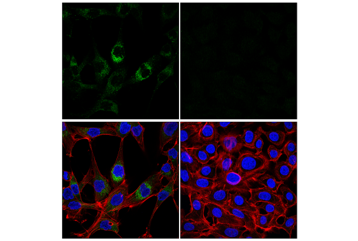
Confocal immunofluorescent analysis of A-172 cells (left, positive) and KYSE-450 (right, negative) using ECM1 (E3B9A) Rabbit mAb (green), DyLight™ 650 Phalloidin #12956 (red), and DAPI #4083 (blue).