Revision 3
#96380
Store at -20C
877-616-CELL (2355)
877-678-TECH (8324)
3 Trask Lane | Danvers | Massachusetts | 01923 | USA
For Research Use Only. Not for Use in Diagnostic Procedures.
Applications:
W, IP, IHC-P
Reactivity:
H
Sensitivity:
Endogenous
MW (kDa):
95-110
Source/Isotype:
Rabbit IgG
UniProt ID:
#Q9ULC0
Entrez-Gene Id:
51705
Product Usage Information
| Application | Dilution |
|---|---|
| Western Blotting | 1:1000 |
| Immunoprecipitation | 1:50 |
| Immunohistochemistry (Paraffin) | 1:1000 - 1:4000 |
Storage
For a carrier free (BSA and azide free) version of this product see product #89278.
Specificity/Sensitivity
EMCN (E3Z4D) Rabbit mAb recognizes endogenous levels of total EMCN protein.
Source / Purification
Monoclonal antibody is produced by immunizing animals with recombinant protein specific to the extracellular domain of human EMCN protein.
Background
Endomucin (EMCN) is a type I transmembrane O-sialoglycoprotein expressed in endothelium in venules and capillaries, but not arteries (1,2). EMCN prevents leukocyte adhesion in non-inflamed tissues, and downregulation of EMCN during inflammation facilitates migration of inflammatory cells into target tissues (3). Notch activation in endothelial cells downregulates EMCN and promotes migration of neutrophils, modulating acute inflammation in hepatic ischemia/reperfusion injury (4). EMCN deficiency in endothelial cells drives tumor lung metastasis by providing a premetastatic niche for cancer cell colonization, and is a potential prognostic biomarker for highly metastatic cancers (5). Combined analysis of EMCN/MUC15 has been suggested as a prognostic signature for gastric cancer (6).
Background References
- Kuhn, A. et al. (2002) J Invest Dermatol 119, 1388-93.
- dela Paz, N.G. and D'Amore, P.A. (2009) Cell Tissue Res 335, 5-16.
- Zahr, A. et al. (2016) Nat Commun 7, 10363.
- Zhang, P. et al. (2020) Sci China Life Sci 63, 375-387.
- Zhang, G. et al. (2022) J Transl Med 20, 446.
- Dai, W. et al. (2020) Front Mol Biosci 7, 19.
Species Reactivity
Species reactivity is determined by testing in at least one approved application (e.g., western blot).
Western Blot Buffer
IMPORTANT: For western blots, incubate membrane with diluted primary antibody in 5% w/v BSA, 1X TBS, 0.1% Tween® 20 at 4°C with gentle shaking, overnight.
Applications Key
W: Western Blotting IP: Immunoprecipitation IHC-P: Immunohistochemistry (Paraffin)
Cross-Reactivity Key
H: Human M: Mouse R: Rat Hm: Hamster Mk: Monkey Vir: Virus Mi: Mink C: Chicken Dm: D. melanogaster X: Xenopus Z: Zebrafish B: Bovine Dg: Dog Pg: Pig Sc: S. cerevisiae Ce: C. elegans Hr: Horse GP: Guinea Pig Rab: Rabbit G: Goat All: All Species Expected
Trademarks and Patents
Cell Signaling Technology is a trademark of Cell Signaling Technology, Inc.
All other trademarks are the property of their respective owners. Visit cellsignal.com/trademarks for more information.
Limited Uses
Except as otherwise expressly agreed in a writing signed by a legally authorized representative of CST, the following terms apply to Products provided by CST, its affiliates or its distributors. Any Customer's terms and conditions that are in addition to, or different from, those contained herein, unless separately accepted in writing by a legally authorized representative of CST, are rejected and are of no force or effect.
Products are labeled with For Research Use Only or a similar labeling statement and have not been approved, cleared, or licensed by the FDA or other regulatory foreign or domestic entity, for any purpose. Customer shall not use any Product for any diagnostic or therapeutic purpose, or otherwise in any manner that conflicts with its labeling statement. Products sold or licensed by CST are provided for Customer as the end-user and solely for research and development uses. Any use of Product for diagnostic, prophylactic or therapeutic purposes, or any purchase of Product for resale (alone or as a component) or other commercial purpose, requires a separate license from CST. Customer shall (a) not sell, license, loan, donate or otherwise transfer or make available any Product to any third party, whether alone or in combination with other materials, or use the Products to manufacture any commercial products, (b) not copy, modify, reverse engineer, decompile, disassemble or otherwise attempt to discover the underlying structure or technology of the Products, or use the Products for the purpose of developing any products or services that would compete with CST products or services, (c) not alter or remove from the Products any trademarks, trade names, logos, patent or copyright notices or markings, (d) use the Products solely in accordance with CST Product Terms of Sale and any applicable documentation, and (e) comply with any license, terms of service or similar agreement with respect to any third party products or services used by Customer in connection with the Products.
Revision 3
#96380
EMCN (E3Z4D) Rabbit mAb
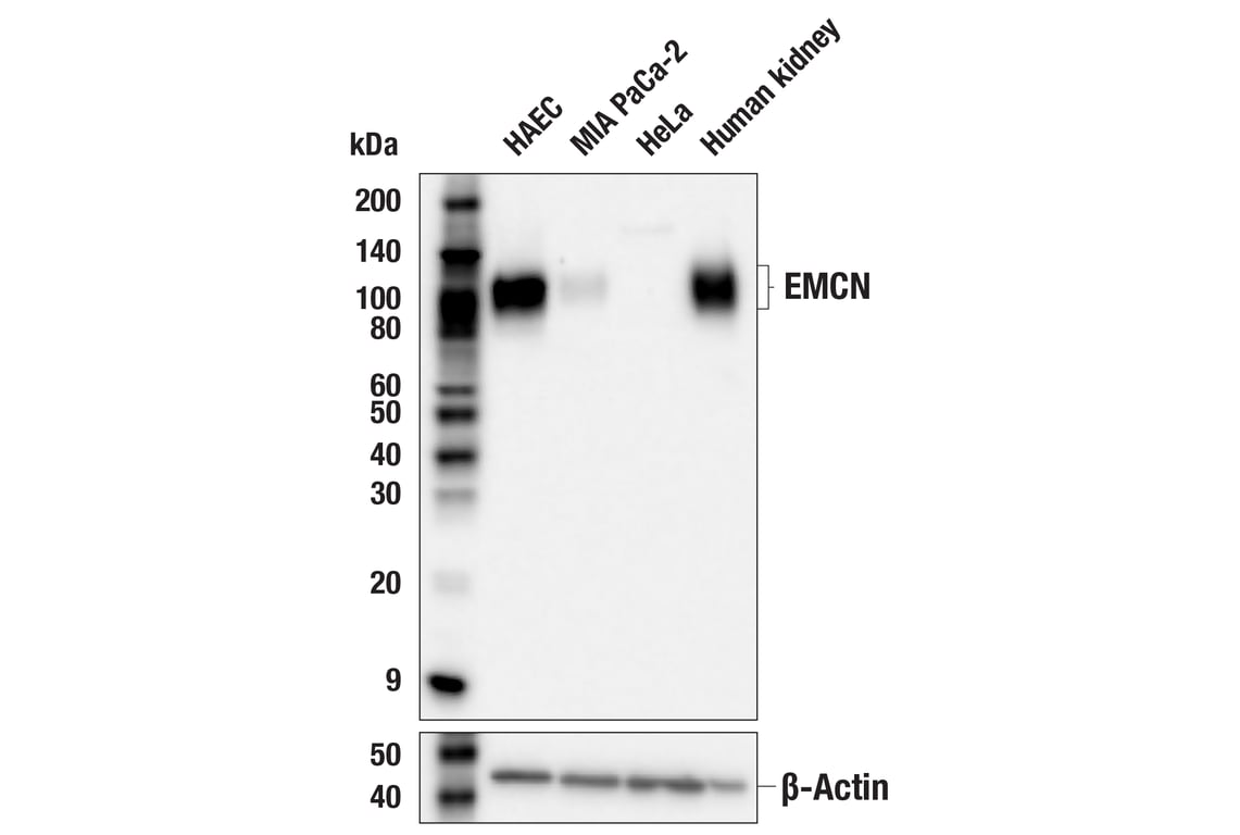
Western blot analysis of extracts from various cells and human kidney tissue using EMCN (E3Z4D) Rabbit mAb (upper) or β-Actin (D6A8) Rabbit mAb #8457 (lower). Negative expression of EMCN protein in HeLa cells is consistent with the predicted expression pattern.
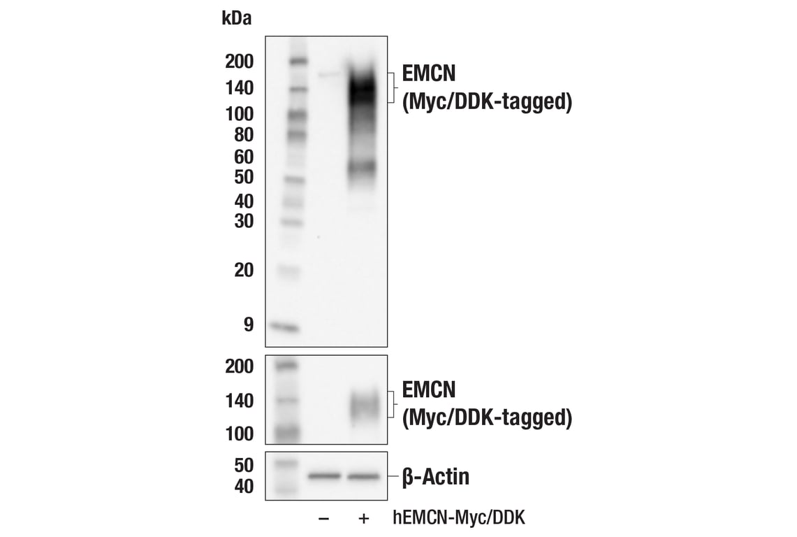
Western blot analysis of extracts from 293T cells, mock transfected (-) or transfected with a construct expressing Myc/DDK-tagged full-length human EMCN (hEMCN-Myc/DDK; +), using EMCN (E3Z4D) Rabbit mAb (upper), Myc-Tag (71D10) Rabbit mAb #2278 (middle), or β-Actin (D6A8) Rabbit mAb #8457 (lower).
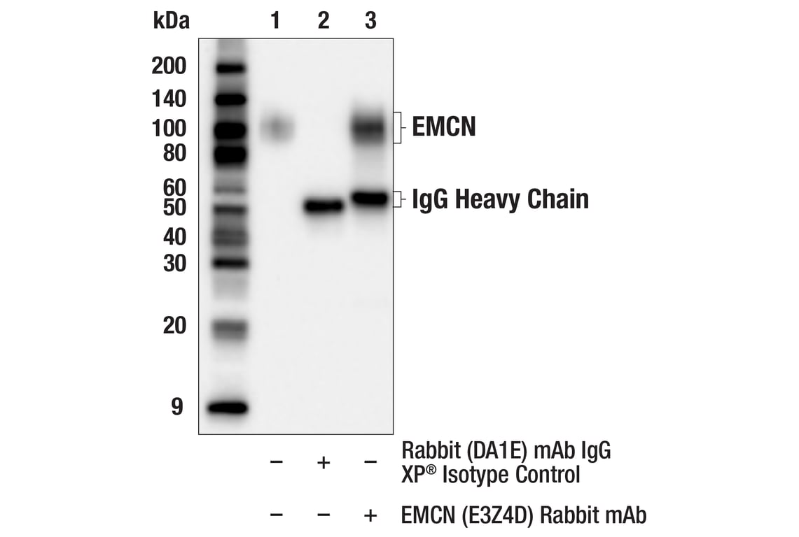
Immunoprecipitation of EMCN protein from MIA PaCa-2 cell extracts. Lane 1 is 10% input, lane 2 is Rabbit (DA1E) mAb IgG XP® Isotype Control #3900, and lane 3 is EMCN (E3Z4D) Rabbit mAb. Western blot analysis was performed using EMCN (E3Z4D) Rabbit mAb. Anti-rabbit IgG, HRP-linked Antibody #7074 was used as a secondary antibody.
Revision 3
#96380
EMCN (E3Z4D) Rabbit mAb
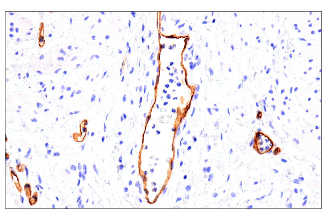
Immunohistochemical analysis of paraffin-embedded human renal cell carcinoma using EMCN (E3Z4D) Rabbit mAb.
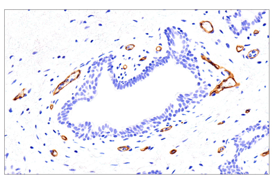
Immunohistochemical analysis of paraffin-embedded human papillary thyroid carcinoma using EMCN (E3Z4D) Rabbit mAb.
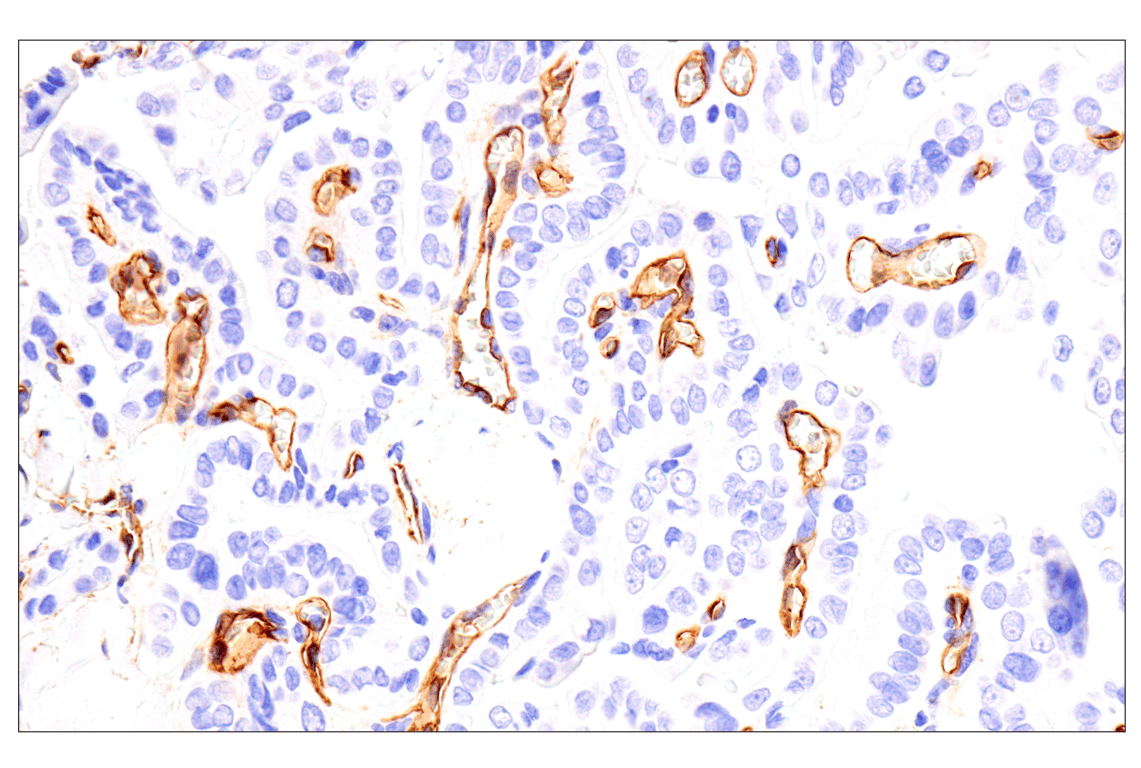
Immunohistochemical analysis of paraffin-embedded human prostate adenocarcinoma using EMCN (E3Z4D) Rabbit mAb.
Revision 3
#96380
EMCN (E3Z4D) Rabbit mAb
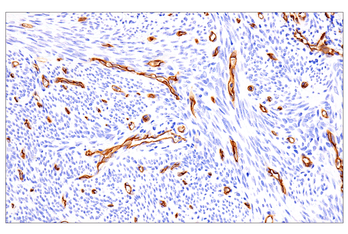
Immunohistochemical analysis of paraffin-embedded human serous papillary carcinoma of the ovary using EMCN (E3Z4D) Rabbit mAb.
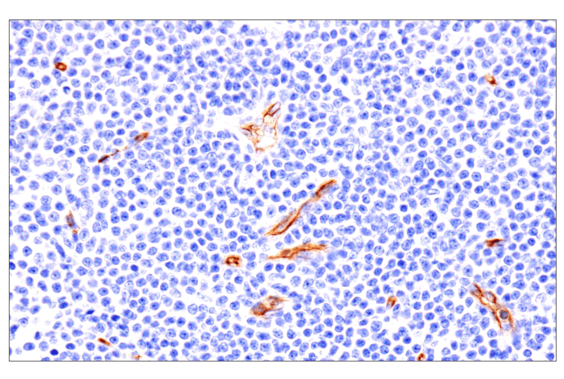
Immunohistochemical analysis of paraffin-embedded human B-cell non-Hodgkin lymphoma using EMCN (E3Z4D) Rabbit mAb.
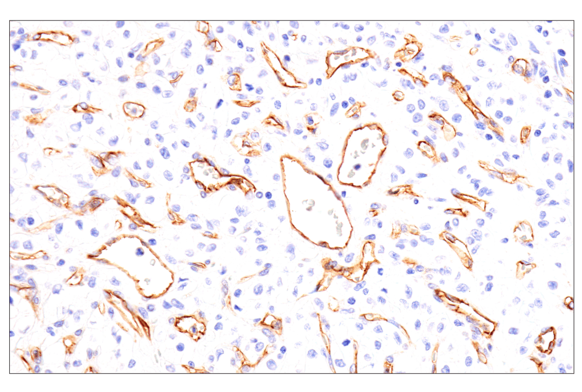
Immunohistochemical analysis of paraffin-embedded human hepatocellular carcinoma using EMCN (E3Z4D) Rabbit mAb.
Revision 3
#96380
EMCN (E3Z4D) Rabbit mAb
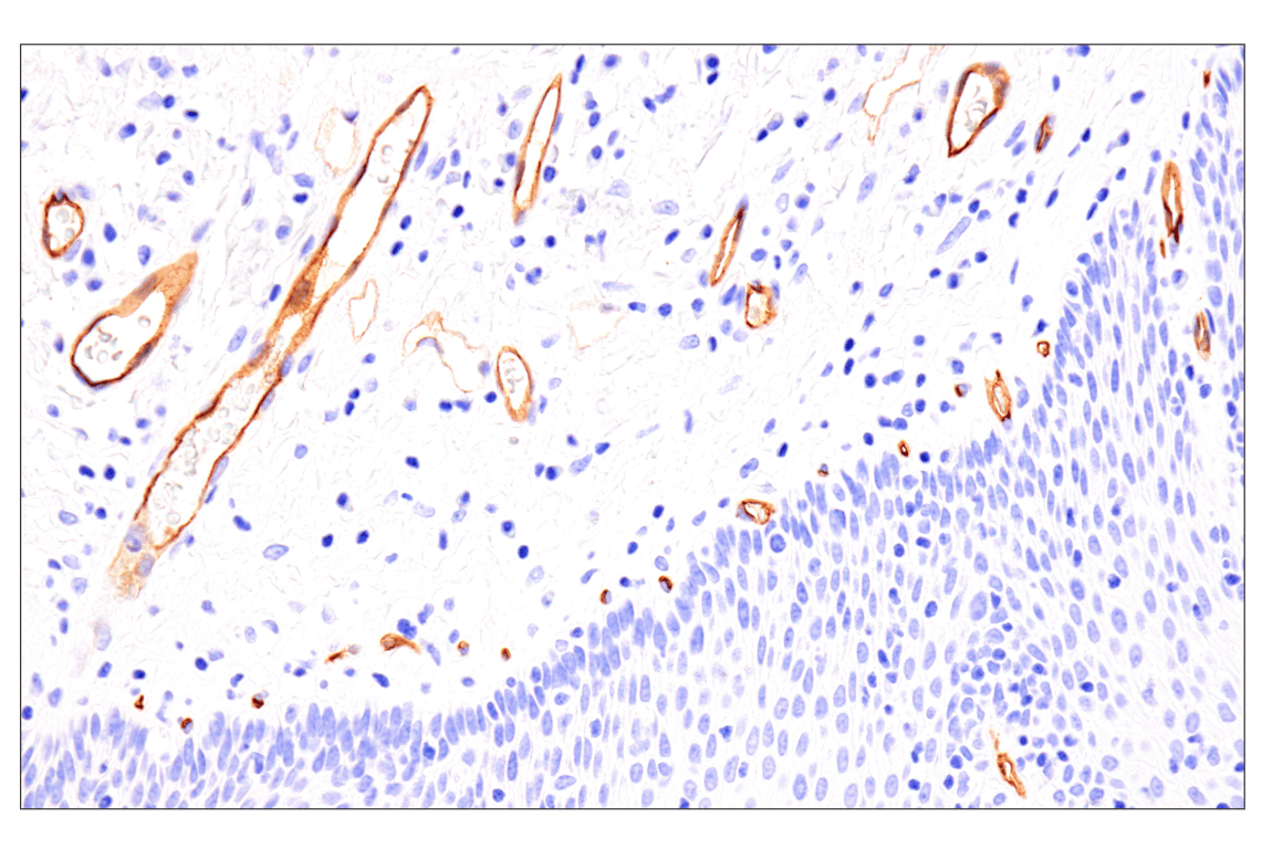
Immunohistochemical analysis of paraffin-embedded human esophageal adenocarcinoma using EMCN (E3Z4D) Rabbit mAb.
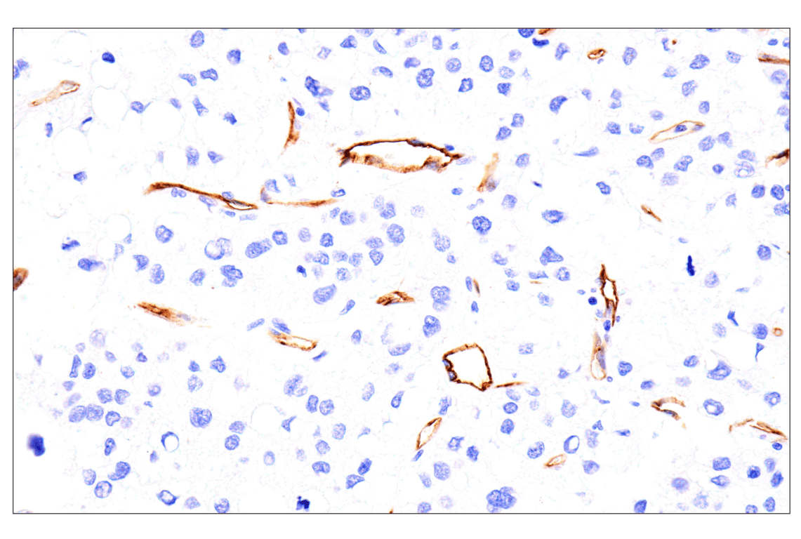
Immunohistochemical analysis of paraffin-embedded normal human kidney using EMCN (E3Z4D) Rabbit mAb.
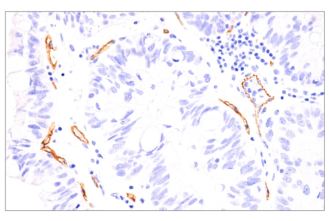
Immunohistochemical analysis of paraffin-embedded normal human uterus using EMCN (E3Z4D) Rabbit mAb.
Revision 3
#96380
EMCN (E3Z4D) Rabbit mAb
Immunohistochemical analysis of paraffin-embedded normal human lung using EMCN (E3Z4D) Rabbit mAb.

Immunohistochemical analysis of paraffin-embedded normal human esophagus using EMCN (E3Z4D) Rabbit mAb.
Immunohistochemical analysis of paraffin-embedded normal human breast using EMCN (E3Z4D) Rabbit mAb.

Revision 3
#96380
EMCN (E3Z4D) Rabbit mAb
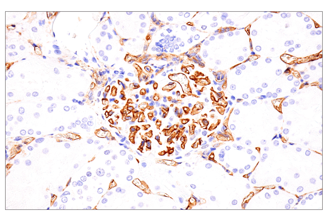
Immunohistochemical analysis of paraffin-embedded normal human adrenal gland using EMCN (E3Z4D) Rabbit mAb.
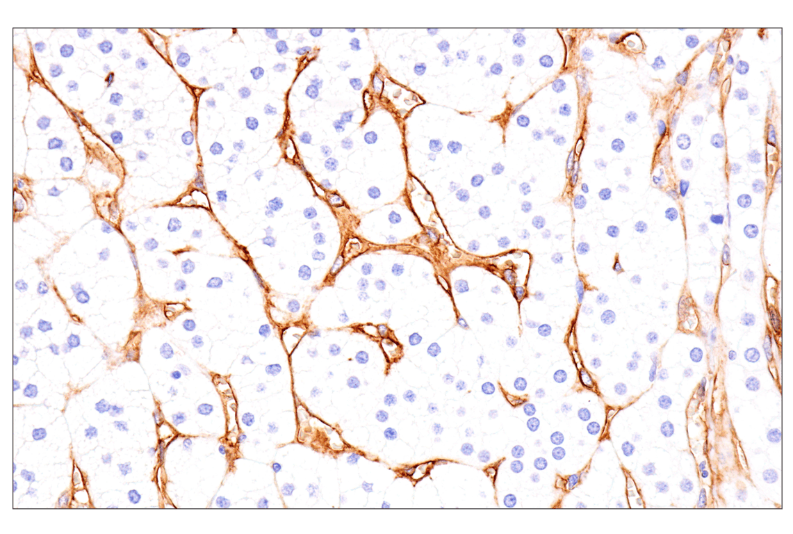
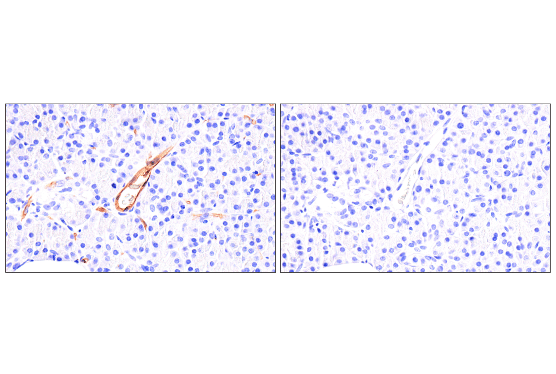
Immunohistochemical analysis of paraffin-embedded normal human pancreas using EMCN (E3Z4D) Rabbit mAb (left) compared to concentration-matched Rabbit (DA1E) mAb IgG XP® Isotype Control #3900 (right).
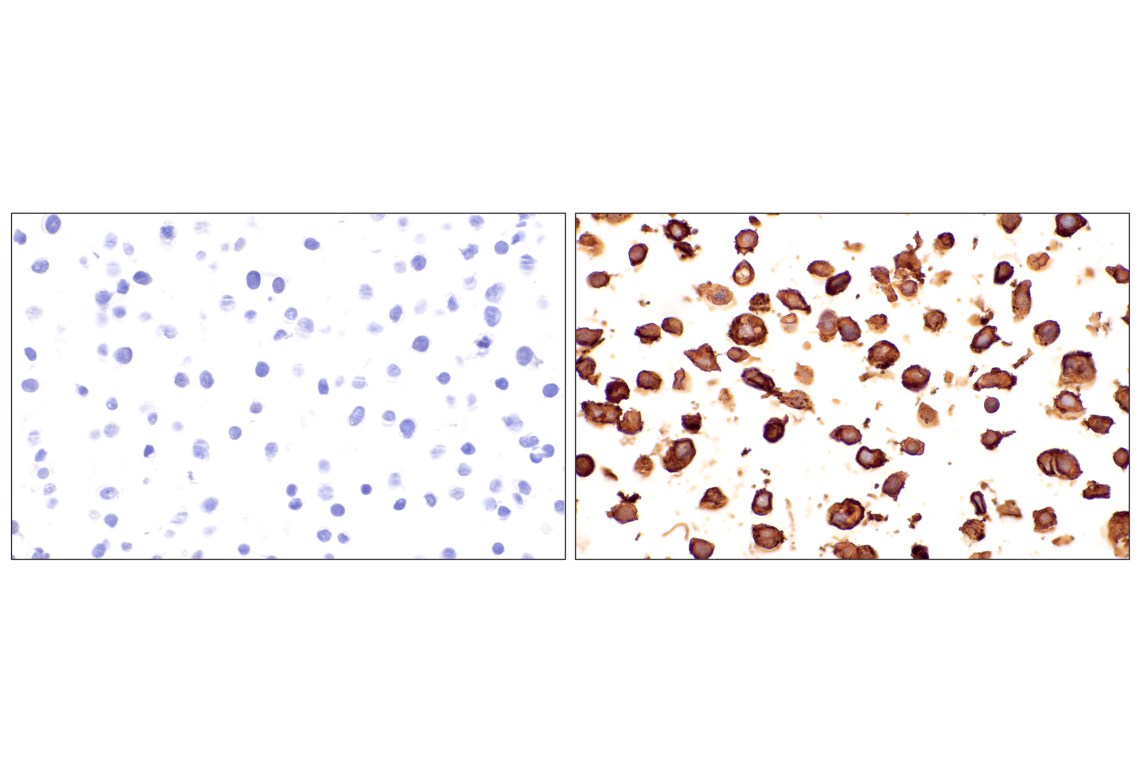
Immunohistochemical analysis of paraffin-embedded HAEC cell pellet (left, positive) or HeLa cell pellet (right, negative) using EMCN (E3Z4D) Rabbit mAb.

Revision 3
#96380
EMCN (E3Z4D) Rabbit mAb
Immunohistochemical analysis of paraffin-embedded 293T cell pellet, untransfected (left) or EMCN-transfected (right), using EMCN (E3Z4D) Rabbit mAb.