Revision 1
#82484
Store at -20C
ER-phagy Cargo Receptor Antibody Sampler Kit
1 Kit
(4 x 20 microliters)
877-616-CELL (2355)
877-678-TECH (8324)
3 Trask Lane | Danvers | Massachusetts | 01923 | USA
For Research Use Only. Not for Use in Diagnostic Procedures.
| Product Includes | Product # | Quantity | Mol. Wt | Isotype/Source |
|---|---|---|---|---|
| FAM134B (E8Y9R) Rabbit mAb | 83414 | 20 µl | 70 kDa | Rabbit IgG |
| CCPG1 (E3C5G) Rabbit mAb | 80158 | 20 µl | 105-120 kDa | Rabbit IgG |
| TEX264 (E2J3J) Rabbit mAb | 44830 | 20 µl | 45 kDa | Rabbit IgG |
| ATL3 (F5T5J) Rabbit mAb | 19901 | 20 µl | 60 kDa | Rabbit IgG |
| Anti-rabbit IgG, HRP-linked Antibody | 7074 | 100 µl | Goat |
Please visit cellsignal.com for individual component applications, species cross-reactivity, dilutions, protocols, and additional product information.
Description
The ER-phagy Cargo Receptor Antibody Sampler Kit provides an economical means of detecting proteins functioning as ER-phagy cargo receptors. The kit includes enough antibodies to perform two western blot experiments with each primary antibody.
Storage
Background
The endoplasmic reticulum (ER) is a large multifaceted organelle that functions in protein folding and processing, calcium storage, and steroid and lipid biogenesis. It is composed of a heterogeneous and continuous network of flattened sacs, or sheets, and tubules that extend from the nuclear envelope to the plasma membrane. It can also be divided into rough ER and smooth peripheral ER; rough ER is predominantly within perinuclear sheets and decorated with ribosomes, and smooth peripheral ER is involved in metabolic activities such as lipid and steroid synthesis. Tubular ER extends throughout the cytoplasm and provides contact points for signaling to other organelles including providing lipids for phagophore formation needed for autophagy. The ER structure is regulated in a dynamic fashion to maintain homeostasis and adjust to cellular stress. Defects in this process may contribute to pathological conditions including metabolic and neurological disorders, cancer, and defense against infectious diseases.
ER-phagy is one of the key processes that regulates ER morphology and functions in the fragmentation and removal of ER segments. Accumulation of unfolded proteins or exposure to conditions such as metabolic or oxidative stress leads to compensatory ER stress programs that remodel the ER structure and activate signaling pathways to cope with those challenges. Activation of ER stress pathways, such as the unfolded protein response (UPR), increases ER membrane to help manage increased demand. Once the stress conditions subside, ER-phagy can eliminate unneeded ER fragments (reviewed in 1-3). Over the last several years, there have been advances in our understanding of ER-phagy. A significant advancement is the discovery of several ER-resident autophagy receptors, including FAM134B, CCPG1, ATL3, TEX264, SEC62, and RTN3L. These cargo receptors have distinct modes of regulation, expression patterns, and localization within the ER. Each of these proteins contains at least one LIR or GIM, which facilitates binding to LC3 or GABARAP family members on the autophagosome. These autophagy receptors contribute to ER-phagy at specific sites on the ER and in response to different stimuli, including nutrient deprivation, ER stress, and changes in calcium. Loss of these proteins is associated with damaged ER expansion, sustained activation of ER stress pathways, and pathophysiological regulation.
Trademarks and Patents
Cell Signaling Technology is a trademark of Cell Signaling Technology, Inc.
All other trademarks are the property of their respective owners. Visit cellsignal.com/trademarks for more information.
Limited Uses
Except as otherwise expressly agreed in a writing signed by a legally authorized representative of CST, the following terms apply to Products provided by CST, its affiliates or its distributors. Any Customer's terms and conditions that are in addition to, or different from, those contained herein, unless separately accepted in writing by a legally authorized representative of CST, are rejected and are of no force or effect.
Products are labeled with For Research Use Only or a similar labeling statement and have not been approved, cleared, or licensed by the FDA or other regulatory foreign or domestic entity, for any purpose. Customer shall not use any Product for any diagnostic or therapeutic purpose, or otherwise in any manner that conflicts with its labeling statement. Products sold or licensed by CST are provided for Customer as the end-user and solely for research and development uses. Any use of Product for diagnostic, prophylactic or therapeutic purposes, or any purchase of Product for resale (alone or as a component) or other commercial purpose, requires a separate license from CST. Customer shall (a) not sell, license, loan, donate or otherwise transfer or make available any Product to any third party, whether alone or in combination with other materials, or use the Products to manufacture any commercial products, (b) not copy, modify, reverse engineer, decompile, disassemble or otherwise attempt to discover the underlying structure or technology of the Products, or use the Products for the purpose of developing any products or services that would compete with CST products or services, (c) not alter or remove from the Products any trademarks, trade names, logos, patent or copyright notices or markings, (d) use the Products solely in accordance with CST Product Terms of Sale and any applicable documentation, and (e) comply with any license, terms of service or similar agreement with respect to any third party products or services used by Customer in connection with the Products.
Revision 1
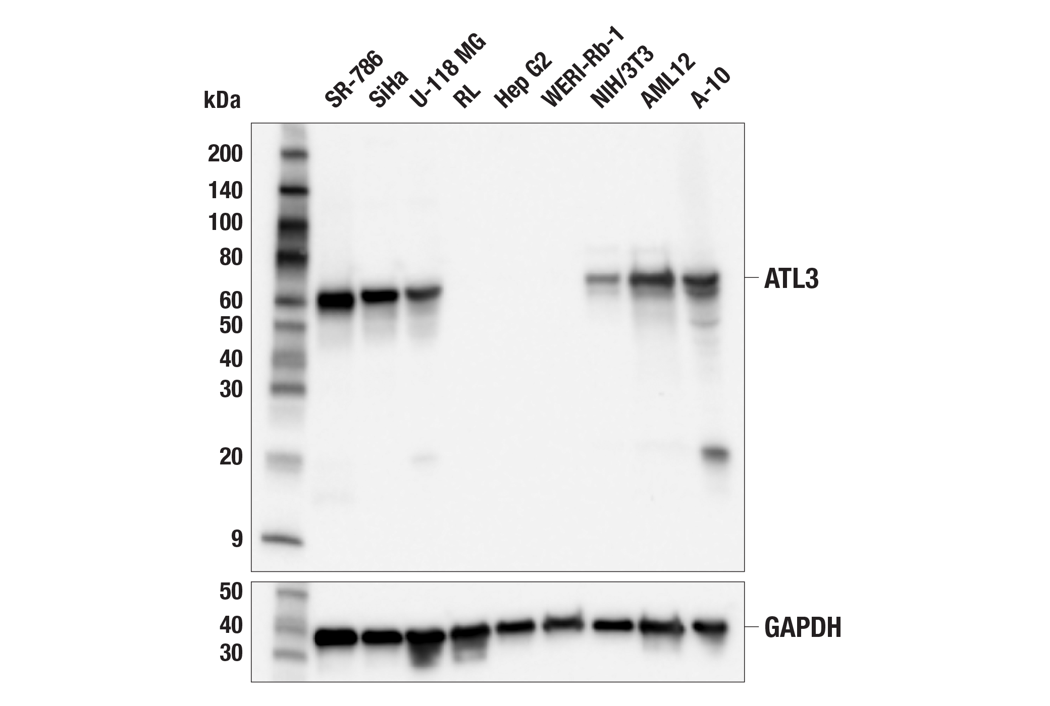
Western blot analysis of extracts from various cell lines using ATL3 (F5T5J) Rabbit mAb (upper) or GAPDH (D16H11) XP® Rabbit mAb #5174 (lower). Negative expression of ATL3 protein in RL, Hep G2, and WERI-Rb-1 cells is consistent with the predicted expression pattern.
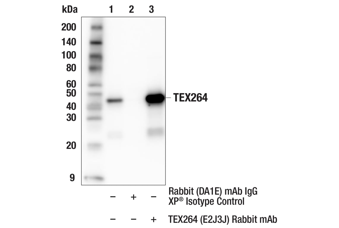
Immunoprecipitation of TEX264 protein from OCI-AML3 cell extracts. Lane 1 is 10% input, lane 2 is Rabbit (DA1E) mAb IgG XP® Isotype Control #3900, and lane 3 is TEX264 (E2J3J) Rabbit mAb. Western blot analysis was performed using TEX264 (E2J3J) Rabbit mAb. Mouse Anti-rabbit IgG (Conformation Specific) (L27A9) mAb (HRP Conjugate) #5127 was used as a secondary antibody.
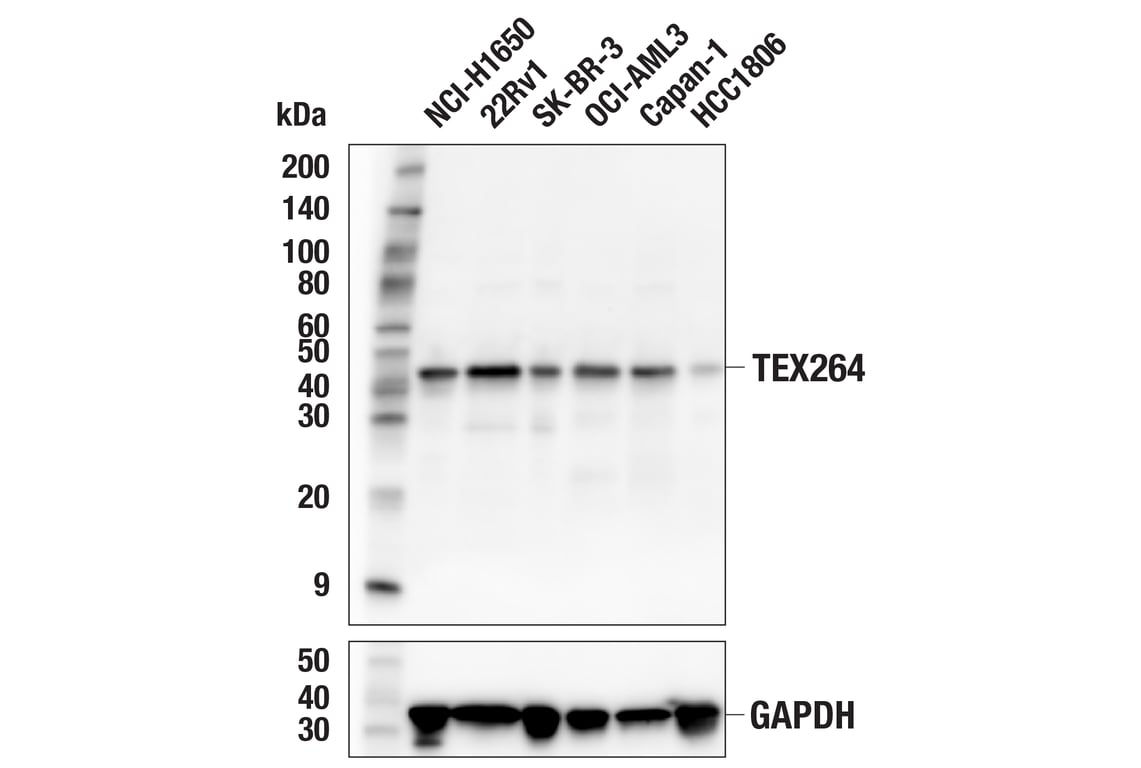
Western blot analysis of extracts from various cell lines using TEX264 (E2J3J) Rabbit mAb (upper) or GAPDH (D16H11) XP® Rabbit mAb #5174 (lower). Low expression of TEX264 protein in HCC1806 cells is consistent with the predicted expression pattern.
Revision 1
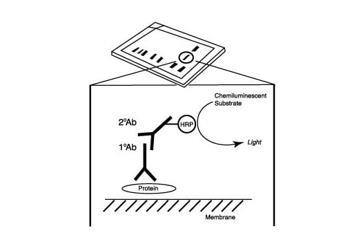
After the primary antibody is bound to the target protein, a complex with HRP-linked secondary antibody is formed. The LumiGLO® is added and emits light during enzyme catalyzed decomposition.
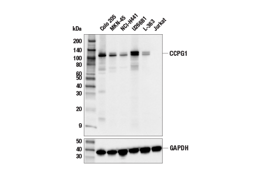
Western blot analysis of extracts from various cell lines using CCPG1 (E3C5G) Rabbit mAb (upper) or GAPDH (D16H11) XP® Rabbit mAb #5174 (lower). Absence of signal in Jurkat cells is predicted by RNAseq and confirms the specificity of the antibody.
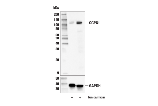
Western blot analysis of extracts from SNU-475 cells, untreated (-) or treated with Tunicamycin #12819 (5 μg/mL, 24 hr; +), using CCPG1 (E3C5G) Rabbit mAb (upper) or GAPDH (D16H11) XP® Rabbit mAb #5174 (lower).
Revision 1
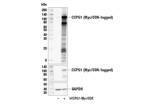
Western blot analysis of extracts from 293T cells, mock transfected (-) or transfected with a construct expressing Myc/DDK-tagged full-length human CCPG1 protein (hCCPG1-Myc/DDK; +), using CCPG1 (E3C5G) Rabbit mAb (upper), Myc-Tag (71D10) Rabbit mAb #2278 (middle), or GAPDH (D16H11) XP® Rabbit mAb #5174 (lower).
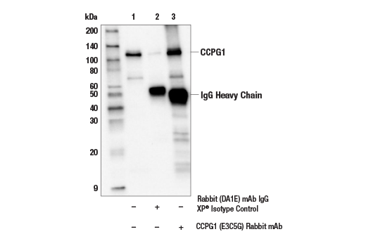
Immunoprecipitation of CCPG1 protein from Colo 205 extracts. Lane 1 is 10% input, lane 2 is Rabbit (DA1E) mAb IgG XP® Isotype Control #3900, and lane 3 is CCPG1 (E3C5G) Rabbit mAb. Western blot analysis was performed using CCPG1 (E3C5G) Rabbit mAb. Anti-rabbit IgG, HRP-linked Antibody #7074 was used as the secondary antibody.
Western blot analysis of extracts from various cell lines using FAM134B (E8Y9R) Rabbit mAb (upper) or GAPDH (D16H11) XP® Rabbit mAb #5174 (lower). The absence of signal for FAM134B in Hep G2 cells is consistent with RNAseq data and confirms the specificity of the antibody for FAM134B.
Revision 1
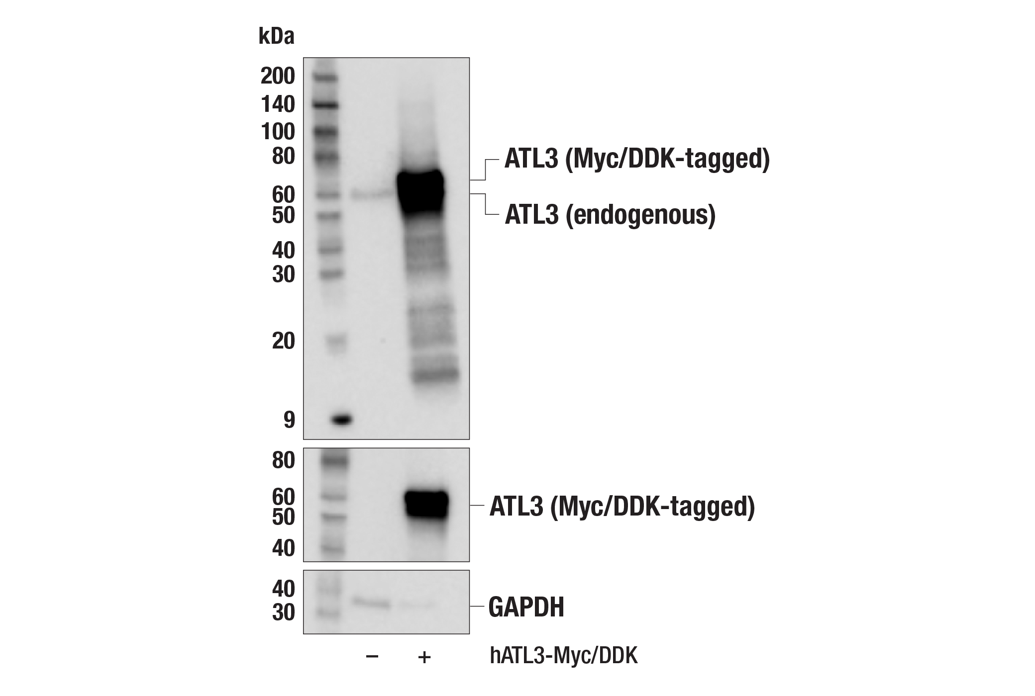
Western blot analysis of extracts from 293T cells, mock transfected (-) or transfected with a construct expressing Myc/DDK-tagged full-length human ATL3 protein (hATL3-Myc/DDK; +), using ATL3 (F5T5J) Rabbit mAb (upper), Myc-Tag (71D10) Rabbit mAb #2278 (middle), or GAPDH (D16H11) XP® Rabbit mAb #5174 (lower).
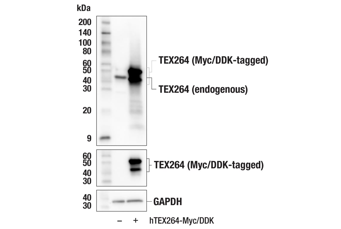
Western blot analysis of extracts from 293T cells, mock transfected (-) or transiently transfected with plasmid expressing Myc/DDK-tagged human TEX264 protein (hTEX264-Myc/DDK; +), using TEX264 (E2J3J) Rabbit mAb (upper), Myc-Tag (71D10) Rabbit mAb #2278 (middle), or GAPDH (D16H11) XP® Rabbit mAb #5174 (lower).
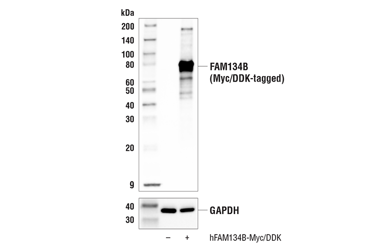
Western blot analysis of extracts from 293T cells, mock transfected (-) or transfected with a construct expressing Myc/DDK-tagged full-length human FAM134B protein (hFAM134B-Myc/DDK; +), using FAM134B (E8Y9R) Rabbit mAb (upper) or GAPDH (D16H11) XP® Rabbit mAb #5174 (lower).
Revision 1
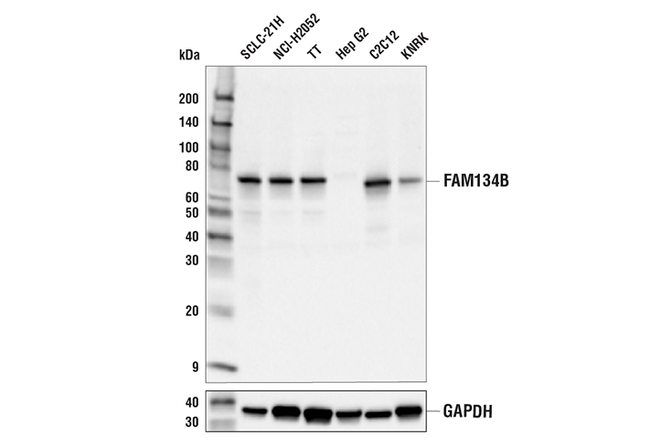
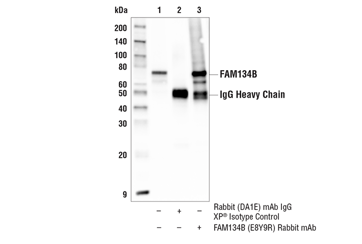
Immunoprecipitation of FAM134B protein from SCLC-21H cell extracts. Lane 1 is 10% input, lane 2 is Rabbit (DA1E) mAb IgG XP® Isotype Control #3900, and lane 3 is FAM134B (E8Y9R) Rabbit mAb. Western blot analysis was performed using FAM134B (E8Y9R) Rabbit mAb. Anti-rabbit IgG, HRP-linked Antibody #7074 was used as a secondary antibody.
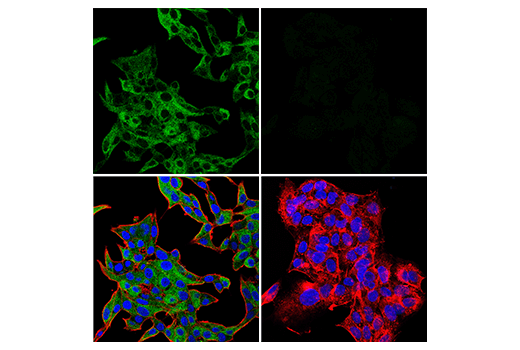
Confocal immunofluorescent analysis of TT cells (left, positive) and Hep G2 cells (right, negative) using FAM134B (E8Y9R) Rabbit mAb (green) and β-Actin (8H10D10) Mouse mAb #3700 (red). Samples were mounted in ProLong® Gold Antifade Reagent with DAPI #8961 (blue).