Revision 2
#48888
Store at -20C
877-616-CELL (2355)
877-678-TECH (8324)
3 Trask Lane | Danvers | Massachusetts | 01923 | USA
For Research Use Only. Not for Use in Diagnostic Procedures.
Applications:
W, IHC-P, IF-IC, FC-FP
Reactivity:
H M
Sensitivity:
Endogenous
MW (kDa):
55
Source/Isotype:
Rabbit IgG
UniProt ID:
#Q92908
Entrez-Gene Id:
2627
Product Usage Information
This formulation is ideal for use with technologies requiring specialized or custom antibody labeling, including fluorophores, metals, lanthanides, and oligonucleotides. It is not recommended for ChIP, ChIP-seq, CUT&RUN or CUT&Tag assays. If you require a carrier free formulation for chromatin profiling, please contact us. Optimal dilutions/concentrations should be determined by the end user.
Formulation
Supplied in 1X PBS, BSA and Azide Free.
For standard formulation of this product see product #5851
Storage
Specificity/Sensitivity
GATA-6 (D61E4) XP® Rabbit mAb (BSA and Azide Free) recognizes endogenous levels of total GATA-6 protein. Non-specific cytoplasmic staining of limited immune cells in mouse small intestine, thymus and spleen was observed by immunohistochemistry.
Species predicted to react based on 100% sequence homology
Mouse, Rat, Dog, Pig
Source / Purification
Monoclonal antibody is produced by immunizing animals with a synthetic peptide corresponding to residues near the amino terminus of human GATA-6 protein.
Background
GATA proteins comprise a group of transcription factors that are related by the presence of conserved zinc finger DNA-binding domains, which bind directly to the nucleotide sequence core element GATA (1-3). There are six vertebrate GATA proteins, designated GATA-1 to GATA-6 (3).
GATA-6 plays a critical role in endoderm development (4). It is essential for development of the heart, gut, and other organs (5,6). Knock out of GATA-6 is embryonic lethal due to defects in formation of the heart tube and a failure to develop extraembryonic endoderm (4). Loss of expression, or loss of nuclear localization of GATA-6 is apparent in a large number of ovarian tumors (7).
Background References
- Ko, L.J. and Engel, J.D. (1993) Mol Cell Biol 13, 4011-22.
- Merika, M. and Orkin, S.H. (1993) Mol Cell Biol 13, 3999-4010.
- Lowry, J.A. and Atchley, W.R. (2000) J Mol Evol 50, 103-15.
- Cai, K.Q. et al. (2008) Dev Dyn 237, 2820-9.
- Charron, F. and Nemer, M. (1999) Semin Cell Dev Biol 10, 85-91.
- Haveri, H. et al. (2008) BMC Gastroenterol 8, 9.
- Caslini, C. et al. (2006) Oncogene 25, 5446-61.
Species Reactivity
Species reactivity is determined by testing in at least one approved application (e.g., western blot).
Applications Key
W: Western Blotting IHC-P: Immunohistochemistry (Paraffin) IF-IC: Immunofluorescence (Immunocytochemistry) FC-FP: Flow Cytometry (Fixed/Permeabilized)
Cross-Reactivity Key
H: Human M: Mouse R: Rat Hm: Hamster Mk: Monkey Vir: Virus Mi: Mink C: Chicken Dm: D. melanogaster X: Xenopus Z: Zebrafish B: Bovine Dg: Dog Pg: Pig Sc: S. cerevisiae Ce: C. elegans Hr: Horse GP: Guinea Pig Rab: Rabbit G: Goat All: All Species Expected
Trademarks and Patents
Cell Signaling Technology is a trademark of Cell Signaling Technology, Inc.
XP is a registered trademark of Cell Signaling Technology, Inc.
All other trademarks are the property of their respective owners. Visit cellsignal.com/trademarks for more information.
Limited Uses
Except as otherwise expressly agreed in a writing signed by a legally authorized representative of CST, the following terms apply to Products provided by CST, its affiliates or its distributors. Any Customer's terms and conditions that are in addition to, or different from, those contained herein, unless separately accepted in writing by a legally authorized representative of CST, are rejected and are of no force or effect.
Products are labeled with For Research Use Only or a similar labeling statement and have not been approved, cleared, or licensed by the FDA or other regulatory foreign or domestic entity, for any purpose. Customer shall not use any Product for any diagnostic or therapeutic purpose, or otherwise in any manner that conflicts with its labeling statement. Products sold or licensed by CST are provided for Customer as the end-user and solely for research and development uses. Any use of Product for diagnostic, prophylactic or therapeutic purposes, or any purchase of Product for resale (alone or as a component) or other commercial purpose, requires a separate license from CST. Customer shall (a) not sell, license, loan, donate or otherwise transfer or make available any Product to any third party, whether alone or in combination with other materials, or use the Products to manufacture any commercial products, (b) not copy, modify, reverse engineer, decompile, disassemble or otherwise attempt to discover the underlying structure or technology of the Products, or use the Products for the purpose of developing any products or services that would compete with CST products or services, (c) not alter or remove from the Products any trademarks, trade names, logos, patent or copyright notices or markings, (d) use the Products solely in accordance with CST Product Terms of Sale and any applicable documentation, and (e) comply with any license, terms of service or similar agreement with respect to any third party products or services used by Customer in connection with the Products.
Revision 2
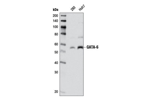
Western blot analysis of extracts from Huh7 and 293 cells using GATA-6 (D61E4) XP® Rabbit mAb. Data were generated using the standard formulation of this product.
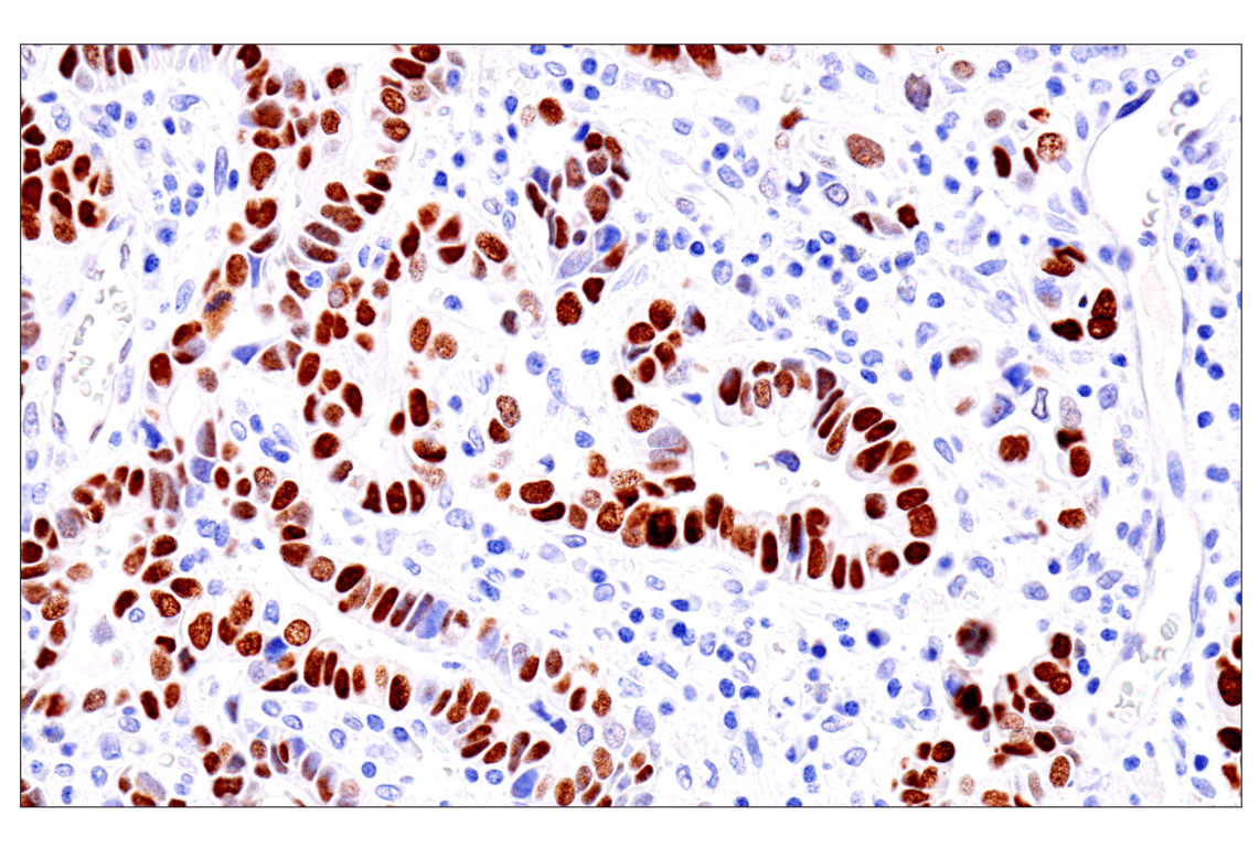
Immunohistochemical analysis of paraffin-embedded human esophageal carcinoma using GATA-6 (D61E4) XP® Rabbit mAb. Data were generated using the standard formulation of this product.
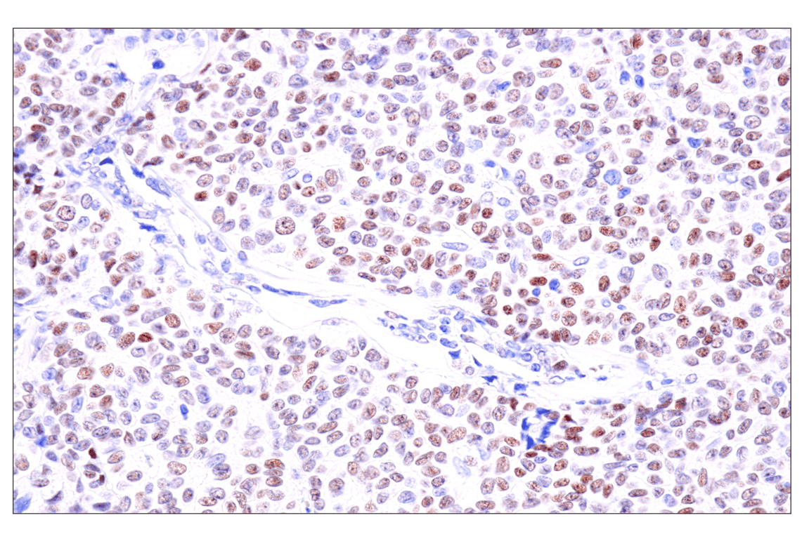
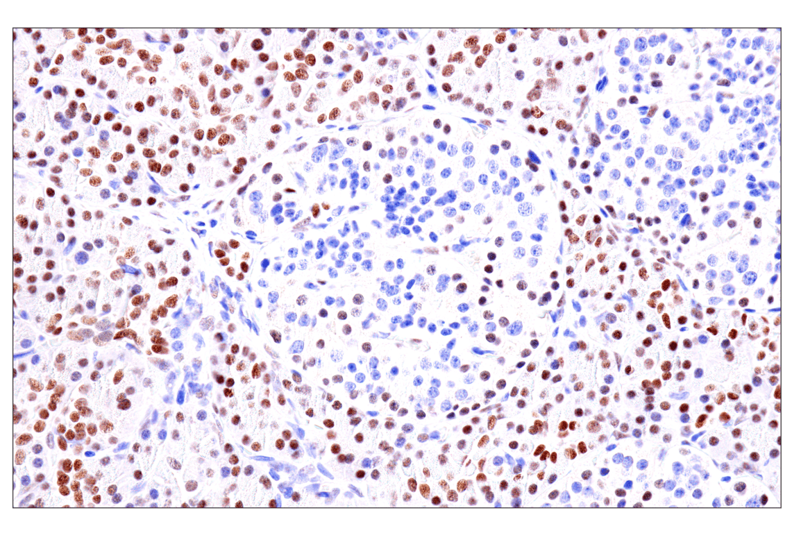
Immunohistochemical analysis of paraffin-embedded human granulosa cell tumor of the ovary using GATA-6 (D61E4) XP® Rabbit mAb. Data were generated using the standard formulation of this product.
Revision 2
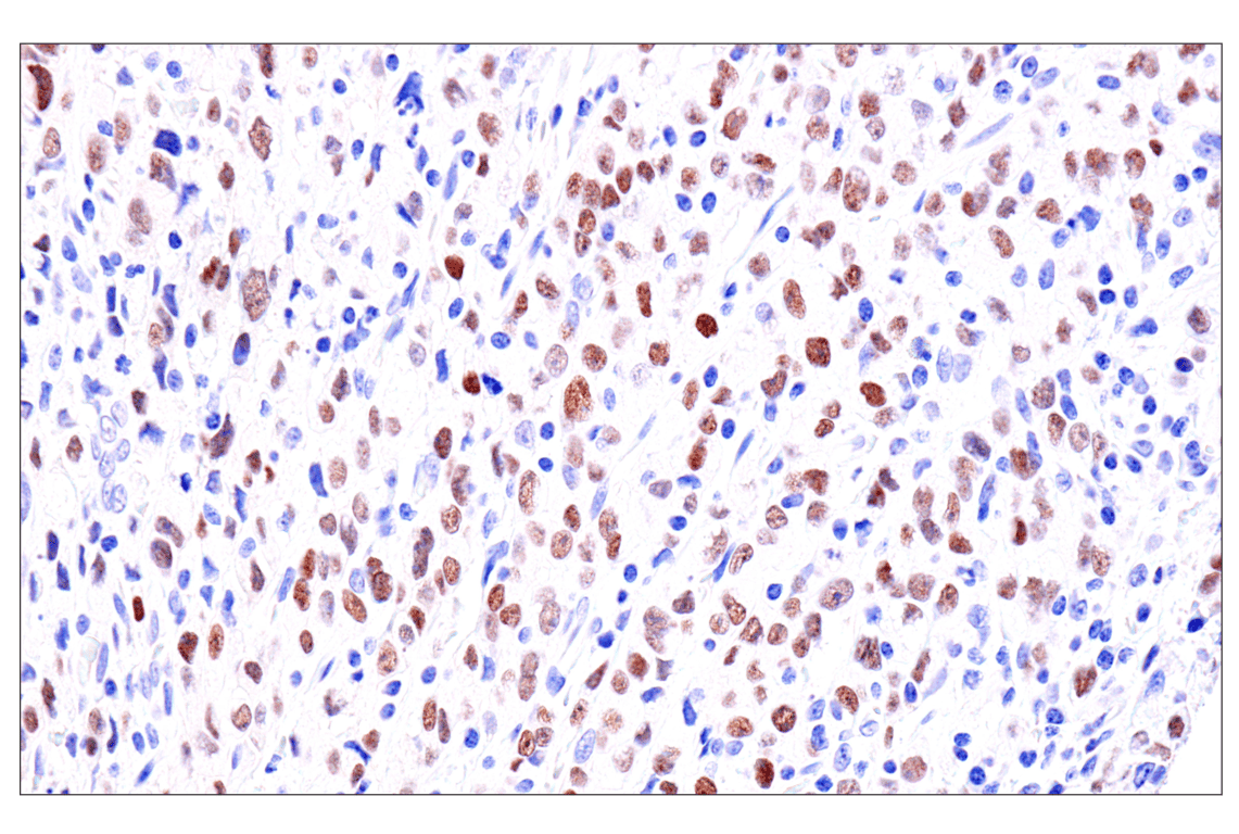
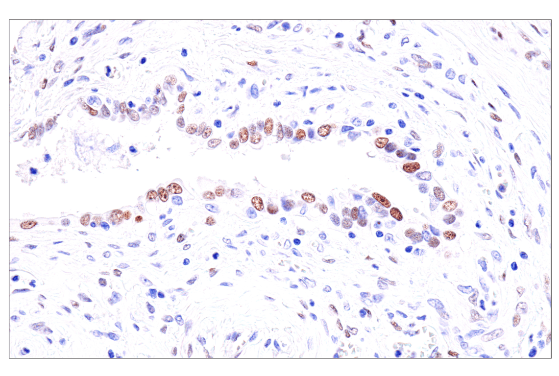
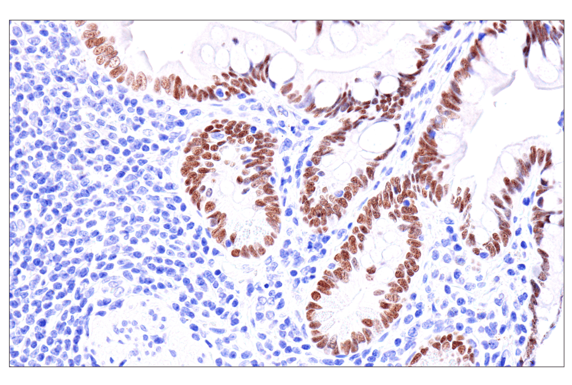
Immunohistochemical analysis of paraffin-embedded human gastric carcinoma using GATA-6 (D61E4) XP® Rabbit mAb. Data were generated using the standard formulation of this product.
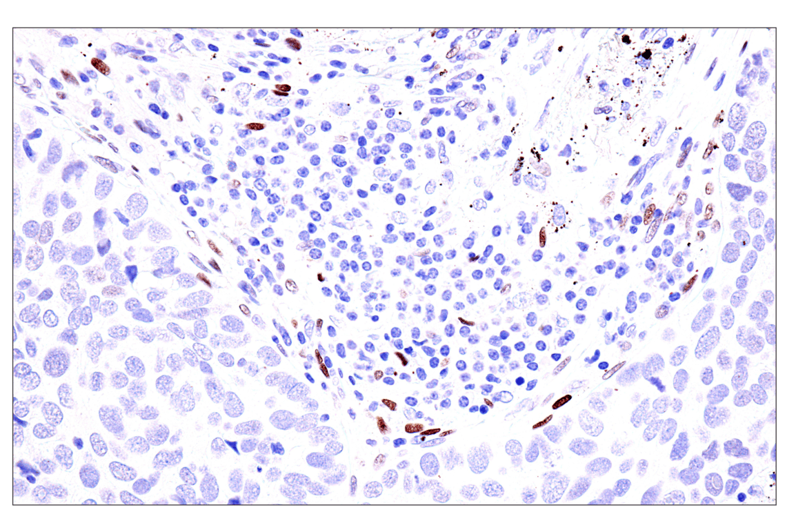
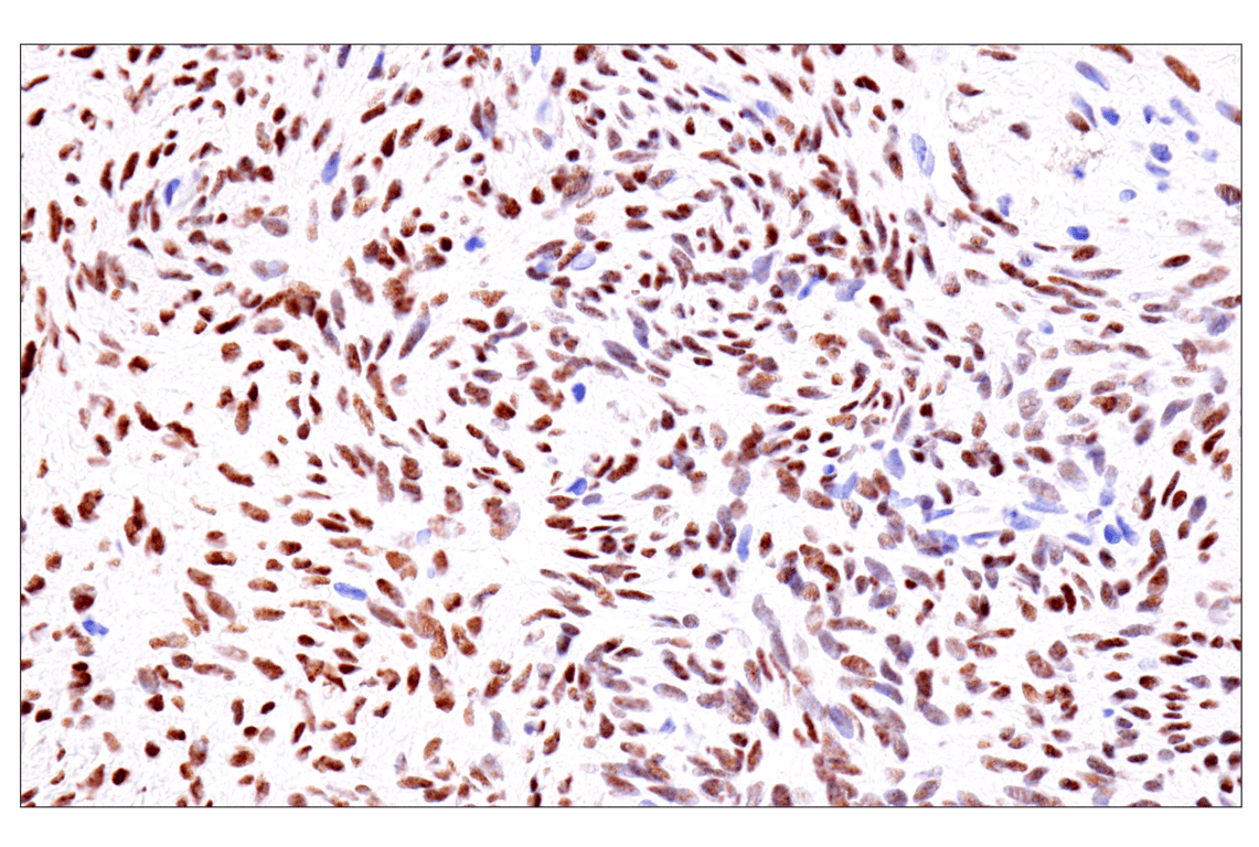
Immunohistochemical analysis of paraffin-embedded human serous papillary carcinoma of the ovary using GATA-6 (D61E4) XP® Rabbit mAb. Data were generated using the standard formulation of this product.
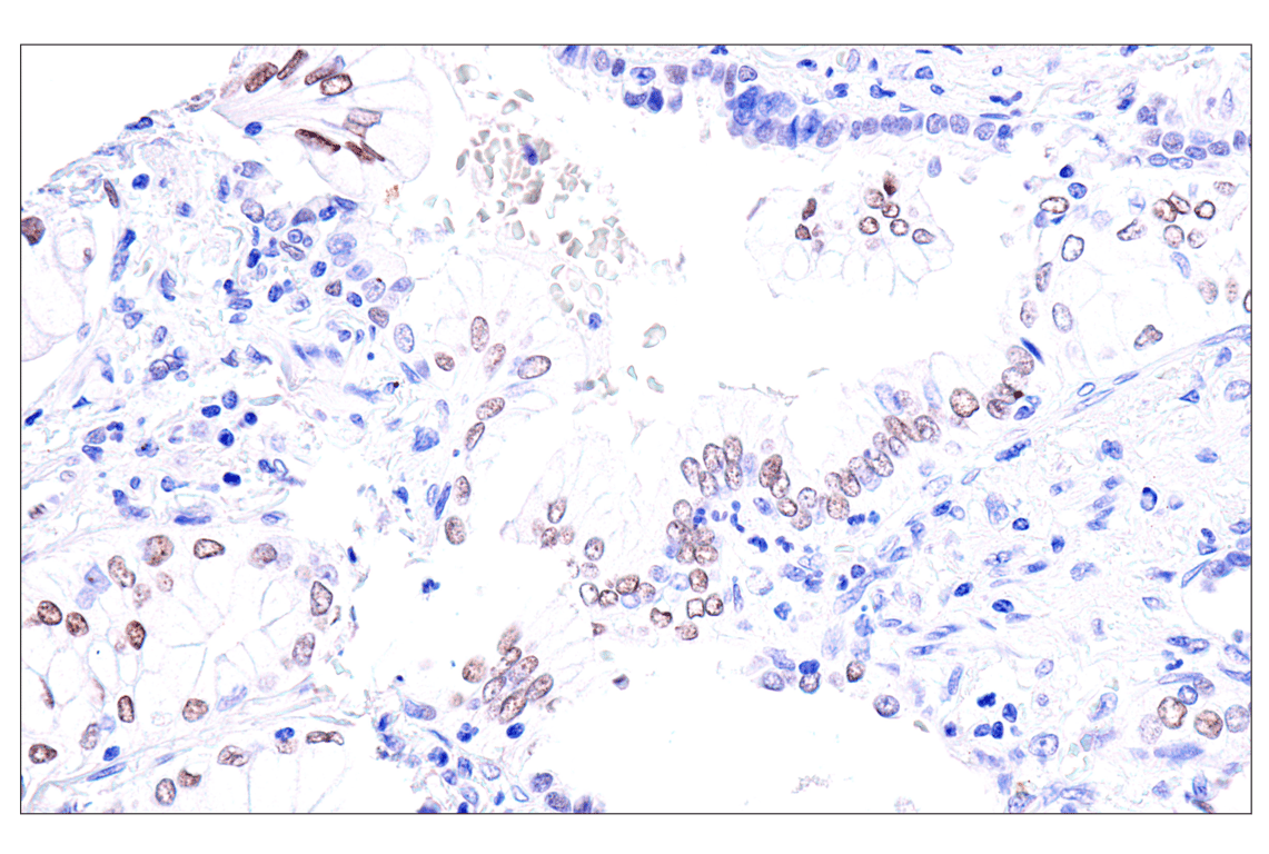
Immunohistochemical analysis of paraffin-embedded human squamous cell lung carcinoma using GATA-6 (D61E4) XP® Rabbit mAb. Data were generated using the standard formulation of this product.
Revision 2
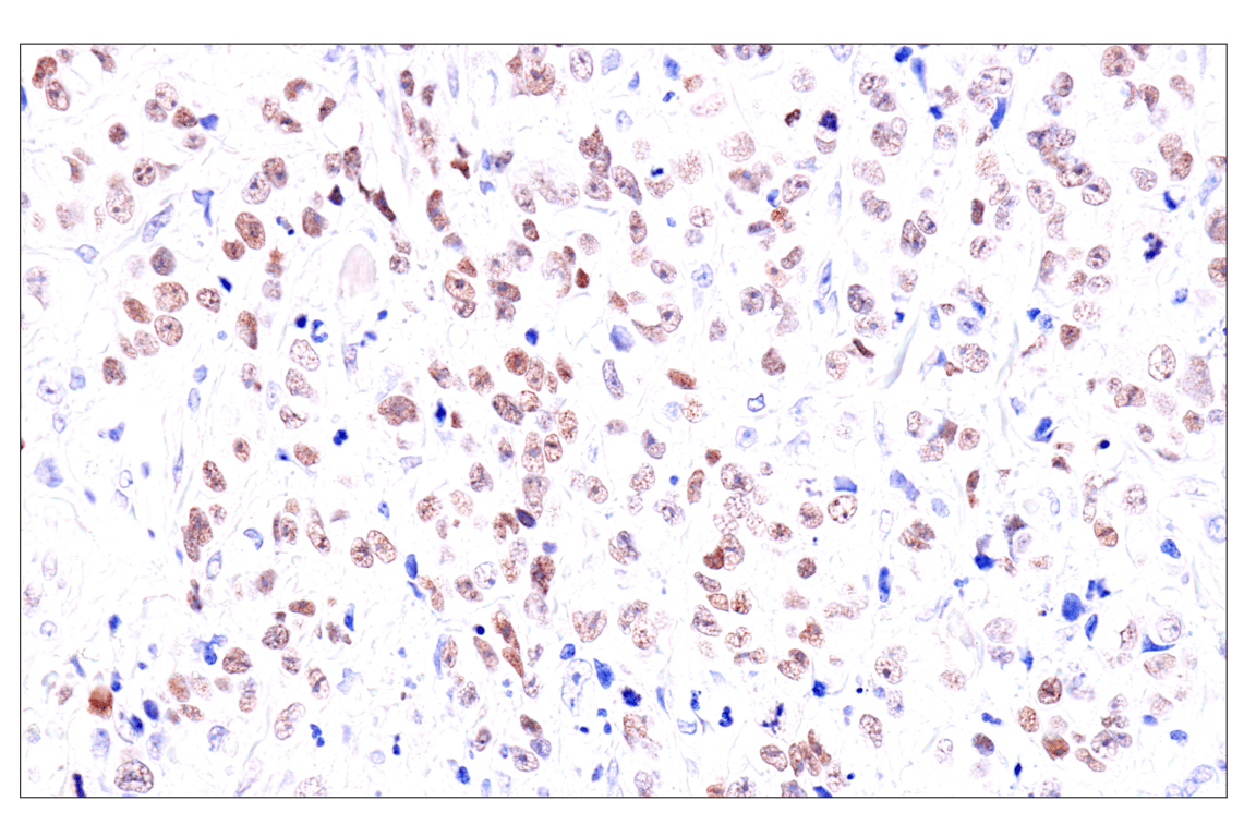
Immunohistochemical analysis of paraffin-embedded human lung adenocarcinoma using GATA-6 (D61E4) XP® Rabbit mAb. Data were generated using the standard formulation of this product.
Immunohistochemical analysis of paraffin-embedded human colon carcinoma using GATA-6 (D61E4) XP® Rabbit mAb. Data were generated using the standard formulation of this product.
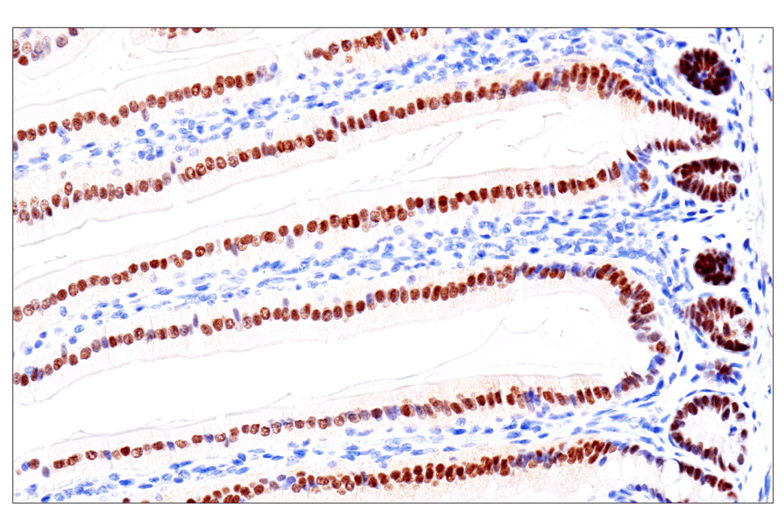
Immunohistochemical analysis of paraffin-embedded normal human stomach using GATA-6 (D61E4) XP® Rabbit mAb. Data were generated using the standard formulation of this product.
Revision 2
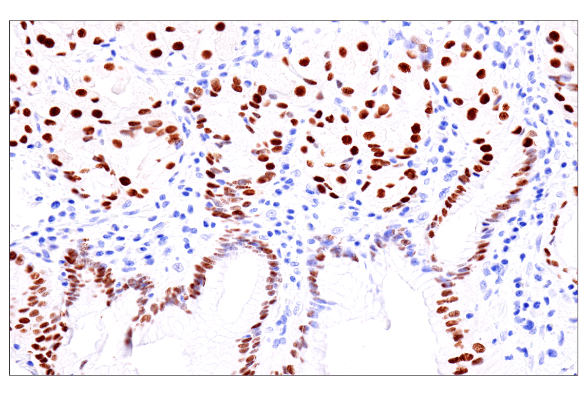
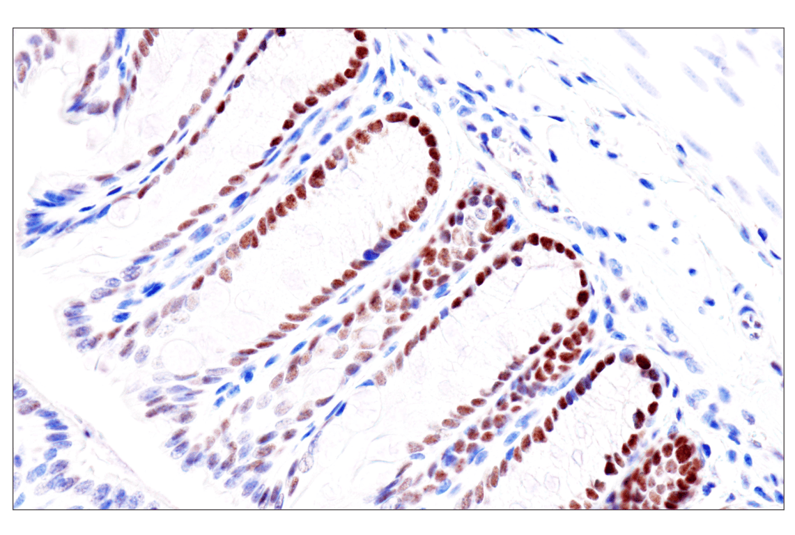
Immunohistochemical analysis of paraffin-embedded normal human small intestine using GATA-6 (D61E4) XP® Rabbit mAb. Data were generated using the standard formulation of this product.
Immunohistochemical analysis of paraffin-embedded normal human pancreas using GATA-6 (D61E4) XP® Rabbit mAb. Data were generated using the standard formulation of this product.
Immunohistochemical analysis of paraffin-embedded normal human ovary using GATA-6 (D61E4) XP® Rabbit mAb. Data were generated using the standard formulation of this product.
Revision 2
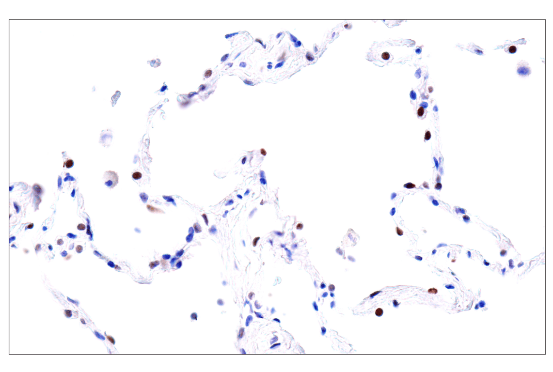
>Immunohistochemical analysis of paraffin-embedded normal human lung using GATA-6 (D61E4) XP® Rabbit mAb. Data were generated using the standard formulation of this product.
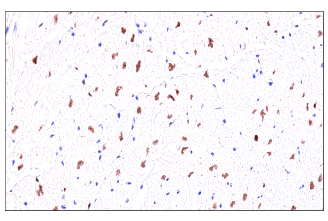
Immunohistochemical analysis of paraffin-embedded normal human heart using GATA-6 (D61E4) XP® Rabbit mAb. Data were generated using the standard formulation of this product.
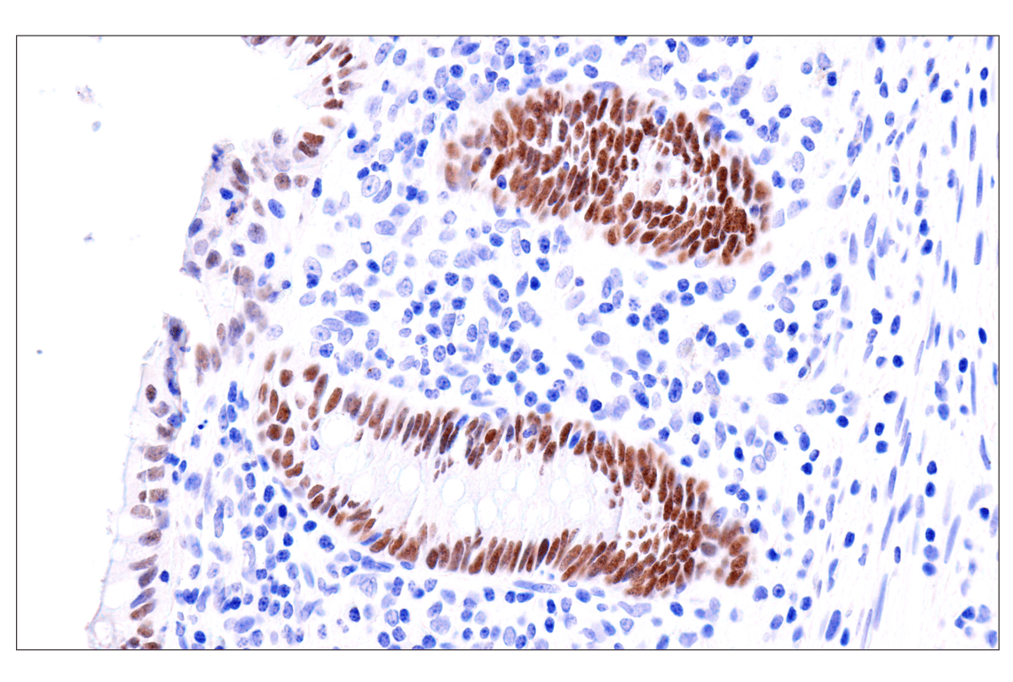
Immunohistochemical analysis of paraffin-embedded human appendix using GATA-6 (D61E4) XP® Rabbit mAb. Data were generated using the standard formulation of this product.
Revision 2
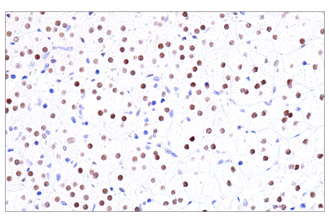
Immunohistochemical analysis of paraffin-embedded normal human adrenal gland using GATA-6 (D61E4) XP ® Rabbit mAb. Data were generated using the standard formulation of this product.
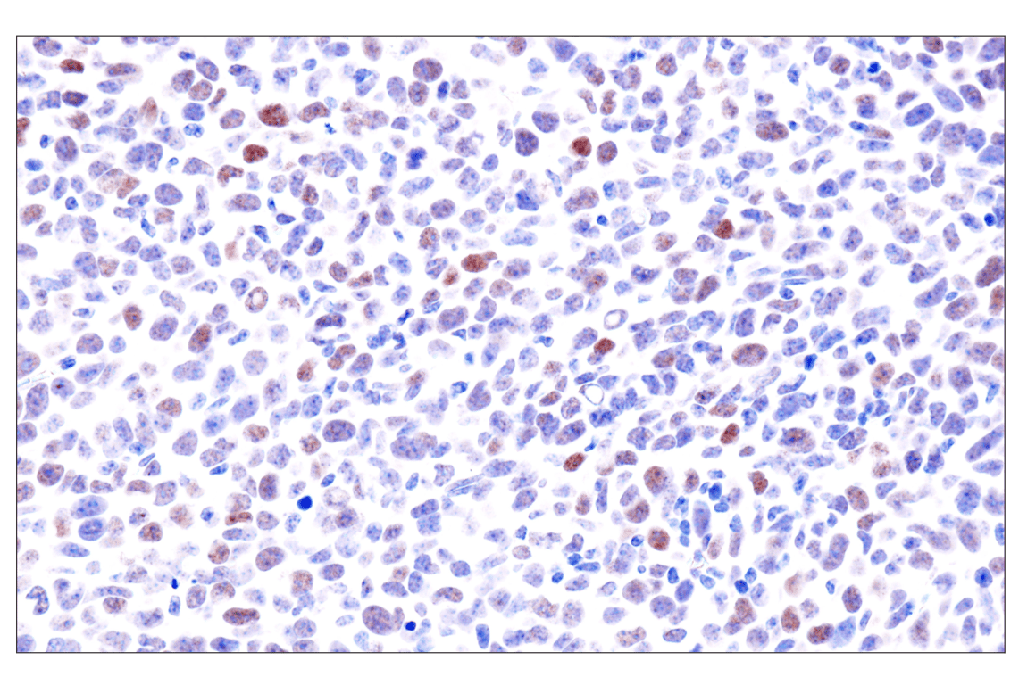
Immunohistochemical analysis of paraffin-embedded LL/2 syngeneic tumor using GATA-6 (D61E4) XP® Rabbit mAb. Data were generated using the standard formulation of this product.
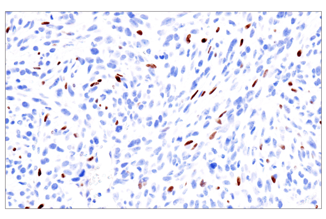
Immunohistochemical analysis of paraffin-embedded 4T1 syngeneic tumor using GATA-6 (D61E4) XP® Rabbit mAb. Data were generated using the standard formulation of this product.
Revision 2
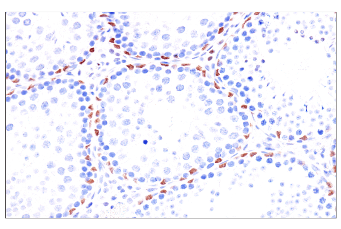
Immunohistochemical analysis of paraffin-embedded mouse testis using GATA-6 (D61E4) XP® Rabbit mAb. Data were generated using the standard formulation of this product.
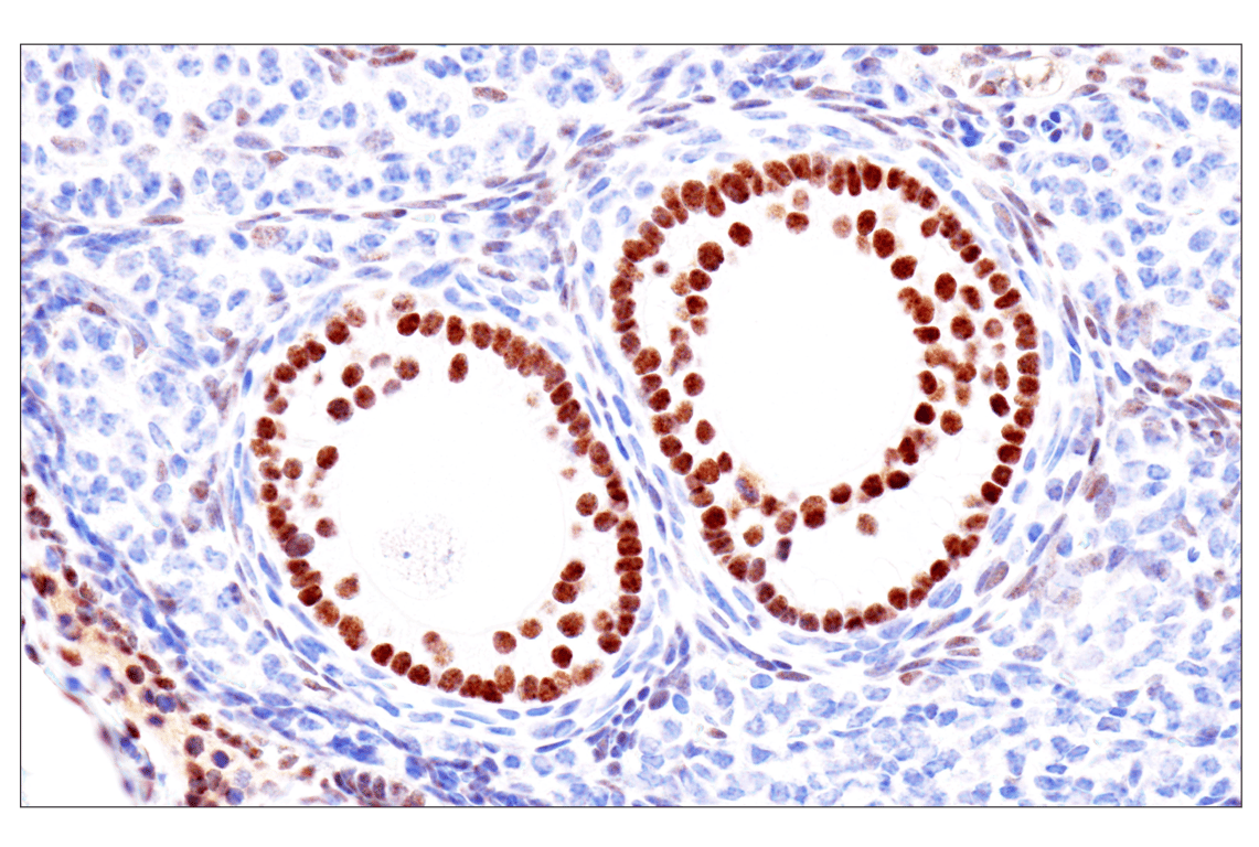
Immunohistochemical analysis of paraffin-embedded mouse ovary using GATA-6 (D61E4) XP® Rabbit mAb. Data were generated using the standard formulation of this product.
Immunohistochemical analysis of paraffin-embedded mouse colon using GATA-6 (D61E4) XP® Rabbit mAb Data were generated using the standard formulation of this product.
Revision 2
Immunohistochemical analysis of paraffin-embedded mouse small intestine using GATA-6 (D61E4) XP® Rabbit mAb. Data were generated using the standard formulation of this product.
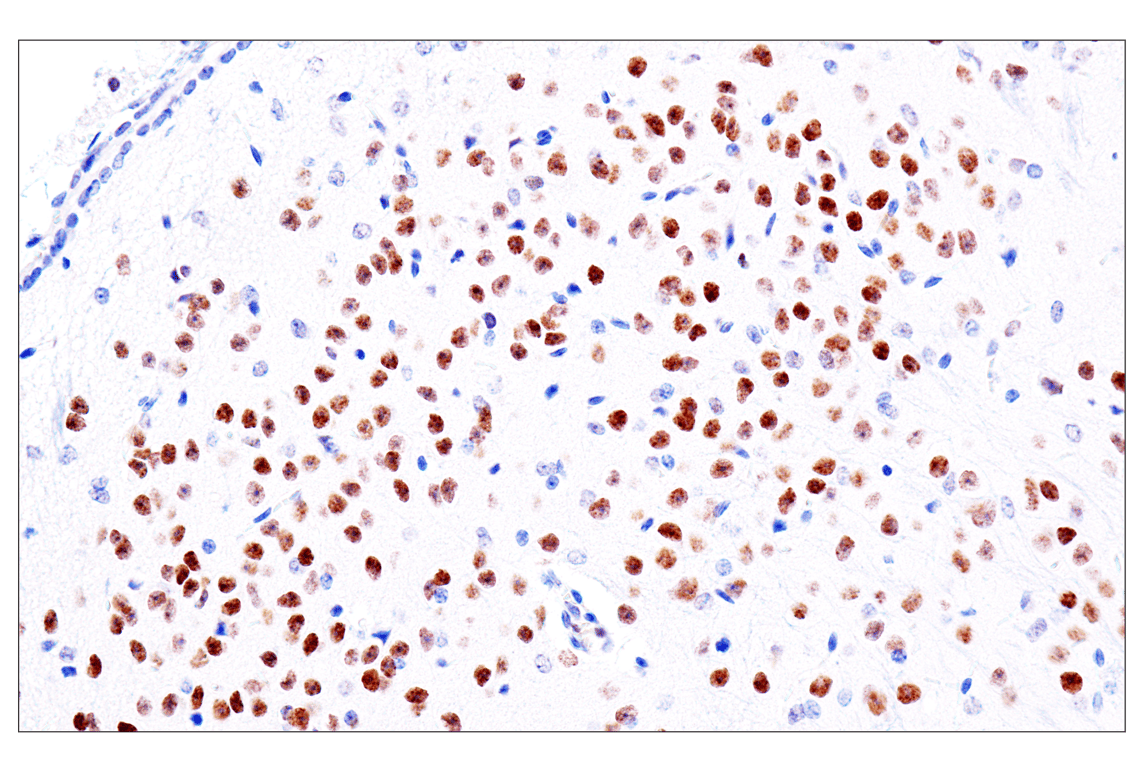
Immunohistochemical analysis of paraffin-embedded mouse brain using GATA-6 (D61E4) XP® Rabbit mAb. Data were generated using the standard formulation of this product.
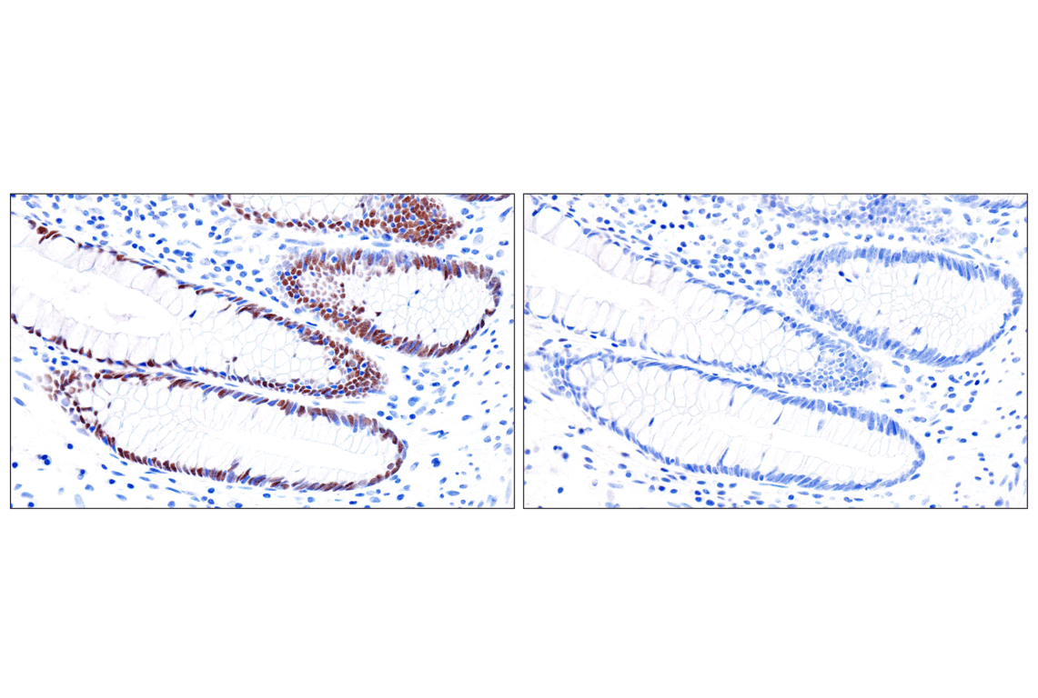
Immunohistochemical analysis of paraffin-embedded normal human colon using GATA-6 (D61E4) XP® Rabbit mAb (left) compared to concentration-matched Rabbit (DA1E) mAb IgG XP® Isotype Control #3900 (right). Data were generated using the standard formulation of this product.
Revision 2
Immunohistochemical analysis of paraffin-embedded HuH-7 cell pellet (left, positive) and SUP-B15 cell pellet (right, negative) using GATA-6 (D61E4) XP® Rabbit mAb. Data were generated using the standard formulation of this product.

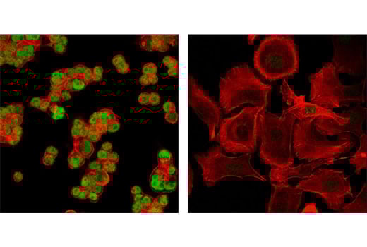
Confocal immunofluorescent analysis of KM12 (left) and SK-OV-3 cells (right) using GATA-6 (D61E4) XP® Rabbit mAb (green). Actin filaments were labeled with DY-554 phalloidin (red). Data were generated using the standard formulation of this product.
Flow cytometric analysis of Jurkat cells (blue, negative) and BT-549 cells (green, positive) using GATA-6 (D61E4) XP® Rabbit mAb (solid lines) or concentration-matched Rabbit (DA1E) mAb IgG XP® Isotype Control #3900 (dashed lines). Anti-rabbit IgG (H+L), F(ab')2 Fragment (Alexa Fluor® 488 Conjugate) #4412 was used as a secondary antibody.
