Revision 4
#37472
Store at -20C
877-616-CELL (2355)
877-678-TECH (8324)
3 Trask Lane | Danvers | Massachusetts | 01923 | USA
For Research Use Only. Not for Use in Diagnostic Procedures.
Applications:
W, IHC-Bond, IHC-P, IF-F, FC-FP, FC-L
Reactivity:
M
Sensitivity:
Endogenous
MW (kDa):
40-50
Source/Isotype:
Rabbit IgG
UniProt ID:
#O35714
Entrez-Gene Id:
21936
Product Usage Information
| Application | Dilution |
|---|---|
| Western Blotting | 1:1000 |
| IHC Leica Bond | 1:200 - 1:800 |
| Immunohistochemistry (Paraffin) | 1:100 - 1:400 |
| Immunofluorescence (Frozen) | 1:50 - 1:200 |
| Flow Cytometry (Fixed/Permeabilized) | 1:50 - 1:100 |
| Flow Cytometry (Live) | 1:50 - 1:100 |
Storage
For a carrier free (BSA and azide free) version of this product see product #34457.
Specificity/Sensitivity
GITR (E9O9H) Rabbit mAb recognizes endogenous levels of total mouse GITR protein. Non-specific staining was observed in mouse testis by immunohistochemistry.
Source / Purification
Monoclonal antibody is produced by immunizing animals with a synthetic peptide corresponding to residues surrounding Val95 of mouse GITR protein.
Background
TNFRSF18, also known as glucocorticoid-induced tumor necrosis factor-receptor (TNFR)-related protein (GITR) and activation-inducible TNFR family receptor, encodes a type 1 membrane protein of the TNF-receptor superfamily (1). Three alternatively spliced transcript variants encoding distinct isoforms have been reported (2). GITR is an immune cell co-stimulatory receptor expressed constitutively at high levels on CD4+CD25+ T regulatory cells (Tregs), at low levels on naïve and memory T cells, and is induced upon T cell activation (3-5). Studies show GITR can also be induced on NK cells, macrophages, and DCs (3,4,6). Although GITR does not have intrinsic enzymatic activity, TNFSF18 (also known as GITRL) expressed on antigen presenting cells binds to GITR, resulting in recruitment of TNFR-associated factor family members and activation of the NF-κB pathway in T cells (7). GITR ligation has been shown to play a role in CD8+ T cell activation, cytotoxicity, and memory T cell survival (8-10). In the thymus, GITR is thought to play a key role in dominant immunological self-tolerance through thymic Treg differentiation and expansion (11). Of note, GITR ligation inhibits Treg suppressive function (12-13) and promotes effector T cell resistance to Treg suppression (14-15). Due to the combined effects on both Treg suppression and effector cell activation, GITR represents a unique opportunity for immunotherapeutic intervention in cancer (16).
Background References
- Nocentini, G. et al. (1997) Proc Natl Acad Sci U S A 94, 6216-21.
- Nocentini, G. et al. (2000) Cell Death Differ 7, 408-10.
- Shimizu, J. et al. (2002) Nat Immunol 3, 135-42.
- Nocentini, G. and Riccardi, C. (2009) Adv Exp Med Biol 647, 156-73.
- McHugh, R.S. et al. (2002) Immunity 16, 311-23.
- Hanabuchi, S. et al. (2006) Blood 107, 3617-23.
- Snell, L.M. et al. (2011) Immunol Rev 244, 197-217.
- Ronchetti, S. et al. (2007) J Immunol 179, 5916-26.
- Kim, I.K. et al. (2015) Nat Med 21, 1010-7.
- Snell, L.M. et al. (2012) J Immunol 188, 5915-23.
- Petrillo, M.G. et al. (2015) Autoimmun Rev 14, 117-26.
- Kanamaru, F. et al. (2004) J Immunol 172, 7306-14.
- Valzasina, B. et al. (2005) Blood 105, 2845-51.
- Stephens, G.L. et al. (2004) J Immunol 173, 5008-20.
- Nishikawa, H. et al. (2008) Cancer Res 68, 5948-54.
- Knee, D.A. et al. (2016) Eur J Cancer 67, 1-10.
Species Reactivity
Species reactivity is determined by testing in at least one approved application (e.g., western blot).
Western Blot Buffer
IMPORTANT: For western blots, incubate membrane with diluted primary antibody in 5% w/v BSA, 1X TBS, 0.1% Tween® 20 at 4°C with gentle shaking, overnight.
Applications Key
W: Western Blotting IHC-Bond: IHC Leica Bond IF-F: Immunofluorescence (Frozen) FC-FP: Flow Cytometry (Fixed/Permeabilized)
Cross-Reactivity Key
H: Human M: Mouse R: Rat Hm: Hamster Mk: Monkey Vir: Virus Mi: Mink C: Chicken Dm: D. melanogaster X: Xenopus Z: Zebrafish B: Bovine Dg: Dog Pg: Pig Sc: S. cerevisiae Ce: C. elegans Hr: Horse GP: Guinea Pig Rab: Rabbit G: Goat All: All Species Expected
Trademarks and Patents
Cell Signaling Technology is a trademark of Cell Signaling Technology, Inc.
Alexa Fluor is a registered trademark of Life Technologies Corporation.
All other trademarks are the property of their respective owners. Visit cellsignal.com/trademarks for more information.
Limited Uses
Except as otherwise expressly agreed in a writing signed by a legally authorized representative of CST, the following terms apply to Products provided by CST, its affiliates or its distributors. Any Customer's terms and conditions that are in addition to, or different from, those contained herein, unless separately accepted in writing by a legally authorized representative of CST, are rejected and are of no force or effect.
Products are labeled with For Research Use Only or a similar labeling statement and have not been approved, cleared, or licensed by the FDA or other regulatory foreign or domestic entity, for any purpose. Customer shall not use any Product for any diagnostic or therapeutic purpose, or otherwise in any manner that conflicts with its labeling statement. Products sold or licensed by CST are provided for Customer as the end-user and solely for research and development uses. Any use of Product for diagnostic, prophylactic or therapeutic purposes, or any purchase of Product for resale (alone or as a component) or other commercial purpose, requires a separate license from CST. Customer shall (a) not sell, license, loan, donate or otherwise transfer or make available any Product to any third party, whether alone or in combination with other materials, or use the Products to manufacture any commercial products, (b) not copy, modify, reverse engineer, decompile, disassemble or otherwise attempt to discover the underlying structure or technology of the Products, or use the Products for the purpose of developing any products or services that would compete with CST products or services, (c) not alter or remove from the Products any trademarks, trade names, logos, patent or copyright notices or markings, (d) use the Products solely in accordance with CST Product Terms of Sale and any applicable documentation, and (e) comply with any license, terms of service or similar agreement with respect to any third party products or services used by Customer in connection with the Products.
Revision 4
#37472
GITR (E9O9H) Rabbit mAb
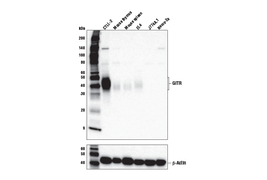
Western blot analysis of extracts from various tissues and cell lines using GITR (E9O9H) Rabbit mAb (upper) and β-Actin (D6A8) Rabbit mAb #8457 (lower).
Immunohistochemical analysis of paraffin-embedded mouse spleen using GITR (E9O9H) Rabbit mAb performed on the Leica® BOND™ Rx.

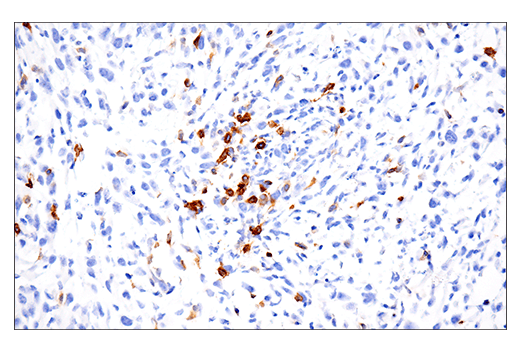
Immunohistochemical analysis of paraffin-embedded 4T1 syngeneic mammary tumor using GITR (E9O9H) Rabbit mAb.

Revision 4
#37472
GITR (E9O9H) Rabbit mAb
Immunohistochemical analysis of paraffin-embedded GL-261 syngeneic tumor using GITR (E9O9H) Rabbit mAb.

Immunohistochemical analysis of paraffin-embedded MC38 syngeneic tumor using GITR (E9O9H) Rabbit mAb.
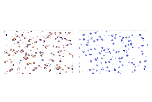
Immunohistochemical analysis of paraffin-embedded CTLL-2 cell pellet (left, positive) or J774A.1 cell pellet (right, negative) using GITR (E9O9H) Rabbit mAb.
Revision 4
#37472
GITR (E9O9H) Rabbit mAb
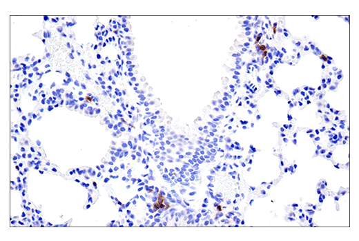
Immunohistochemical analysis of paraffin-embedded mouse lung using GITR (E9O9H) Rabbit mAb.
Confocal immunofluorescent analysis of mouse spleen (left, positive) and pancreas (right, negative) using GITR (E9O9H) Rabbit mAb (green) and S6 Ribosomal Protein (54D2) Mouse mAb (Alexa Fluor® 647 Conjugate) #5548 (red). Samples were mounted in ProLong® Gold Antifade Reagent with DAPI #8961 (blue).

Flow cytometric analysis of live J774A.1 cells (blue) and CTLL-2 cells (green) using GITR (E9O9H) Rabbit mAb (solid lines) or a concentration-matched Rabbit (DA1E) mAb IgG XP® Isotype Control #3900 (dashed lines). Anti-rabbit IgG (H+L), F(ab')2 Fragment (Alexa Fluor® 488 Conjugate) #4412 was used as a secondary antibody.
