Revision 3
#39627
Store at -20C
877-616-CELL (2355)
877-678-TECH (8324)
3 Trask Lane | Danvers | Massachusetts | 01923 | USA
For Research Use Only. Not for Use in Diagnostic Procedures.
Applications:
W, IHC-P, IF-IC
Reactivity:
H M R
Sensitivity:
Endogenous
MW (kDa):
41
Source/Isotype:
Rabbit IgG
UniProt ID:
#P00505
Entrez-Gene Id:
2806
Product Usage Information
| Application | Dilution |
|---|---|
| Western Blotting | 1:1000 |
| Immunohistochemistry (Paraffin) | 1:50 - 1:200 |
| Immunofluorescence (Immunocytochemistry) | 1:100 - 1:400 |
Storage
For a carrier free (BSA and azide free) version of this product see product #66911.
Specificity/Sensitivity
GOT2 (F4P3R) Rabbit mAb recognizes endogenous levels of total GOT2 protein. This antibody does not cross-react with GOT1 protein.
Source / Purification
Monoclonal antibody is produced by immunizing animals with a synthetic peptide corresponding to residues surrounding Gly39 of human GOT2 protein.
Background
Glutamate oxaloacetate transaminase 2 (GOT2), a mitochondrial enzyme, catalyzes the conversion of oxaloacetate to aspartate. Studies show that the metabolite pyridoxal phosphate (PLP) is selectively required by acute myeloid leukemia. PLP-dependent GOT2 selectively sustains leukemic cell proliferation. Depletion of GOT2 suppresses the proliferation of acute myeloid leukemia cells (1). In addition, mouse hematopoietic stem cells are dependent on cell-autonomous aspartate synthesis. Got2 deletion leads to decreased aspartate levels and reduced hematopoietic stem cell function (2). Furthermore, GOT2-dependent aspartate synthesis is also necessary for protein and collagen biosynthesis in chondrocytes and is critical for chondrocyte proliferation and growth (3).
Species Reactivity
Species reactivity is determined by testing in at least one approved application (e.g., western blot).
Western Blot Buffer
IMPORTANT: For western blots, incubate membrane with diluted primary antibody in 5% w/v nonfat dry milk, 1X TBS, 0.1% Tween® 20 at 4°C with gentle shaking, overnight.
Applications Key
W: Western Blotting IHC-P: Immunohistochemistry (Paraffin) IF-IC: Immunofluorescence (Immunocytochemistry)
Cross-Reactivity Key
H: Human M: Mouse R: Rat Hm: Hamster Mk: Monkey Vir: Virus Mi: Mink C: Chicken Dm: D. melanogaster X: Xenopus Z: Zebrafish B: Bovine Dg: Dog Pg: Pig Sc: S. cerevisiae Ce: C. elegans Hr: Horse GP: Guinea Pig Rab: Rabbit G: Goat All: All Species Expected
Trademarks and Patents
Cell Signaling Technology is a trademark of Cell Signaling Technology, Inc.
All other trademarks are the property of their respective owners. Visit cellsignal.com/trademarks for more information.
Limited Uses
Except as otherwise expressly agreed in a writing signed by a legally authorized representative of CST, the following terms apply to Products provided by CST, its affiliates or its distributors. Any Customer's terms and conditions that are in addition to, or different from, those contained herein, unless separately accepted in writing by a legally authorized representative of CST, are rejected and are of no force or effect.
Products are labeled with For Research Use Only or a similar labeling statement and have not been approved, cleared, or licensed by the FDA or other regulatory foreign or domestic entity, for any purpose. Customer shall not use any Product for any diagnostic or therapeutic purpose, or otherwise in any manner that conflicts with its labeling statement. Products sold or licensed by CST are provided for Customer as the end-user and solely for research and development uses. Any use of Product for diagnostic, prophylactic or therapeutic purposes, or any purchase of Product for resale (alone or as a component) or other commercial purpose, requires a separate license from CST. Customer shall (a) not sell, license, loan, donate or otherwise transfer or make available any Product to any third party, whether alone or in combination with other materials, or use the Products to manufacture any commercial products, (b) not copy, modify, reverse engineer, decompile, disassemble or otherwise attempt to discover the underlying structure or technology of the Products, or use the Products for the purpose of developing any products or services that would compete with CST products or services, (c) not alter or remove from the Products any trademarks, trade names, logos, patent or copyright notices or markings, (d) use the Products solely in accordance with CST Product Terms of Sale and any applicable documentation, and (e) comply with any license, terms of service or similar agreement with respect to any third party products or services used by Customer in connection with the Products.
Revision 3
#39627
GOT2 (F4P3R) Rabbit mAb
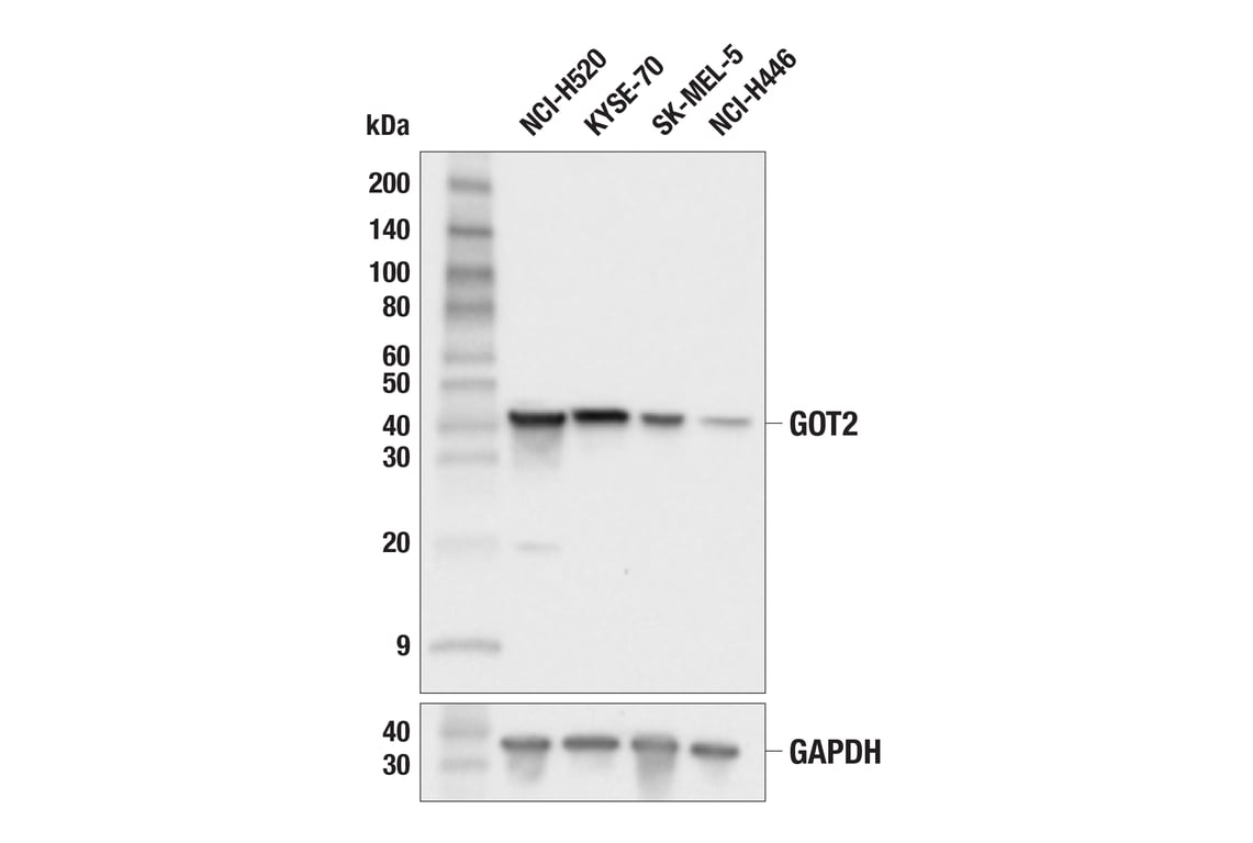
Western blot analysis of extracts from various cell lines using GOT2 (F4P3R) Rabbit mAb (upper) or GAPDH (D16H11) XP® Rabbit mAb #5174 (lower). Low expression of GOT2 protein in NCI-H446 cells is consistent with the predicted expression pattern.
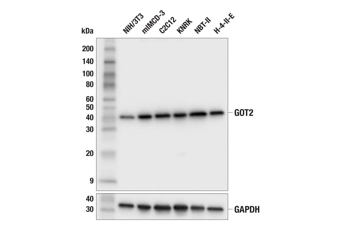
Western blot analysis of extracts from various cell lines using GOT2 (F4P3R) Rabbit mAb (upper) or GAPDH (D16H11) XP® Rabbit mAb #5174 (lower).
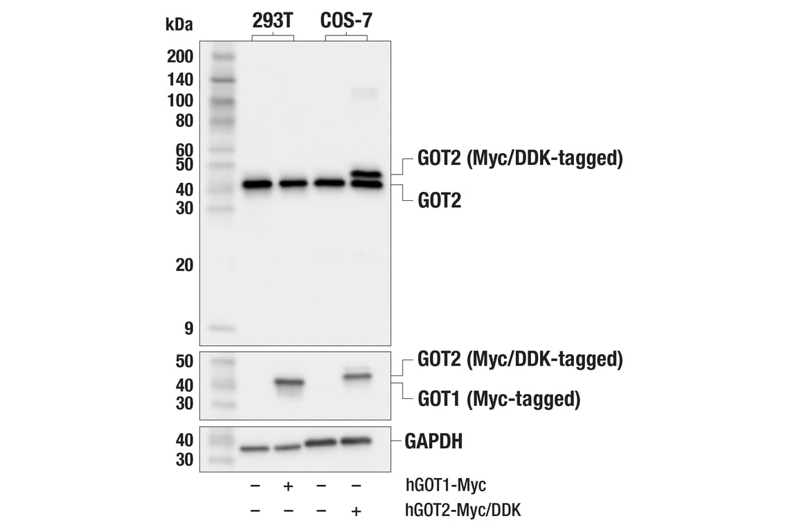
Western blot analysis of extracts from 293T cells, mock transfected (-) or transfected with a construct expressing Myc-tagged human GOT1 protein (hGOT1-Myc; +), and COS-7 cells, mock transfected (-) or transfected with a construct expressing Myc/DDK-tagged human GOT2 protein (hGOT2-Myc/DDK; +), using GOT2 (F4P3R) Rabbit mAb (upper), Myc-Tag (71D10) Rabbit mAb #2278 (middle), or GAPDH (D16H11) XP® Rabbit mAb #5174 (lower).
Revision 3
#39627
GOT2 (F4P3R) Rabbit mAb
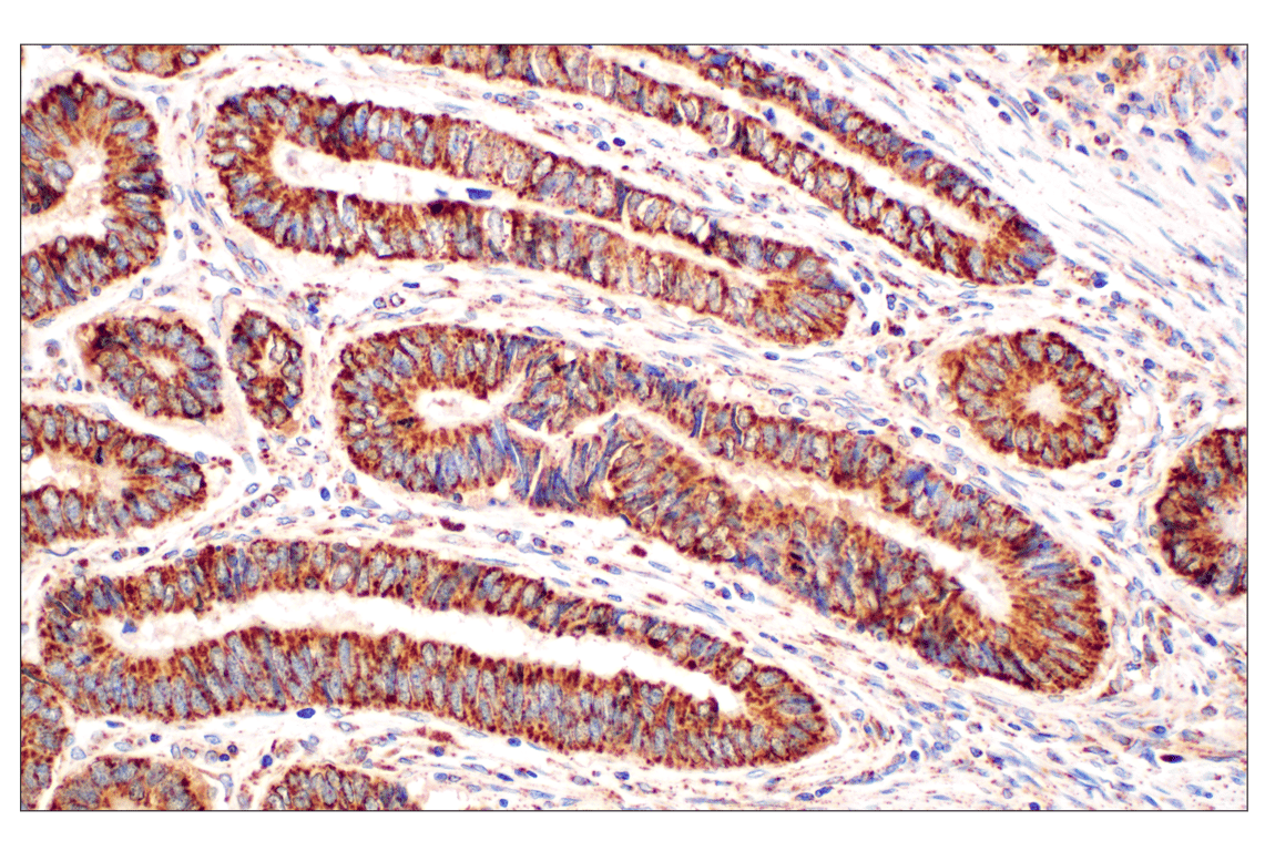
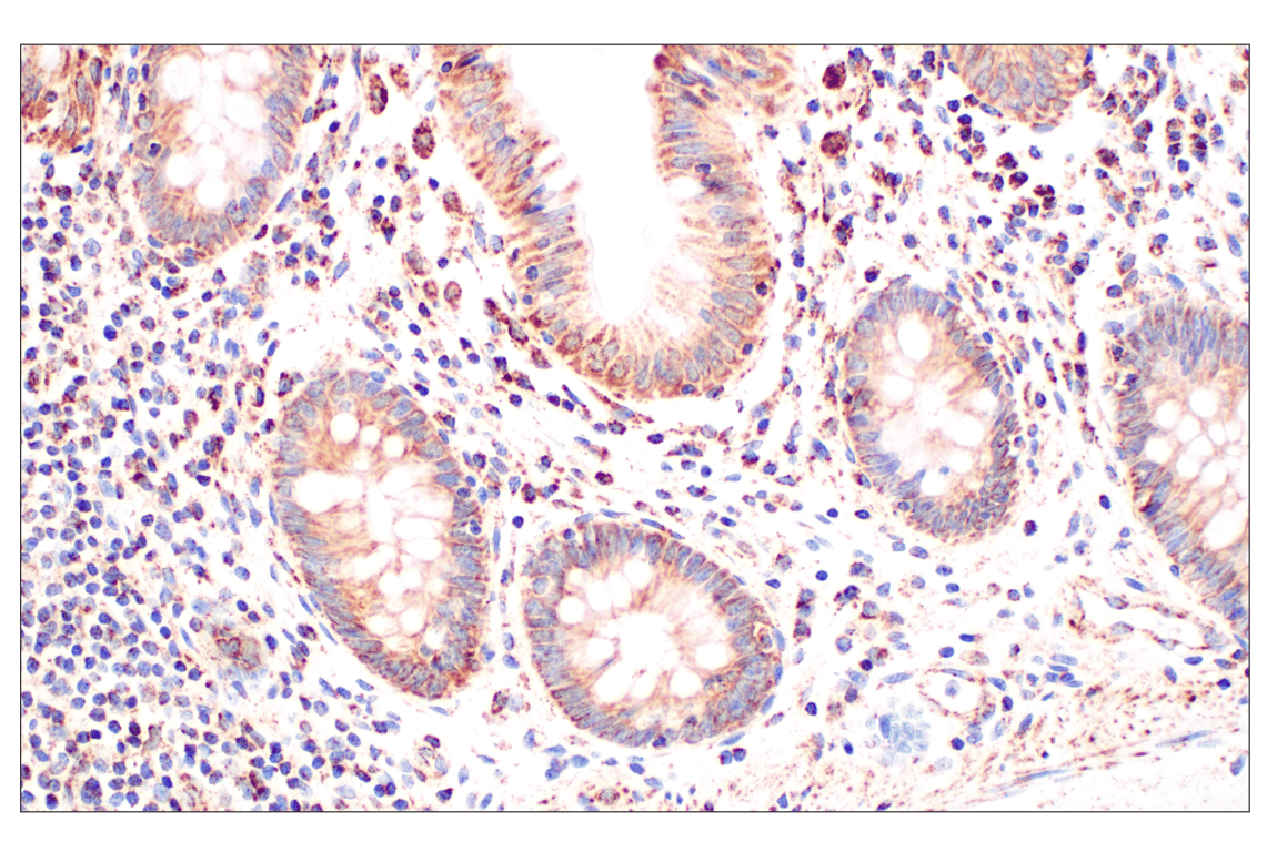
Immunohistochemical analysis of paraffin-embedded human colon adenocarcinoma using GOT2 (F4P3R) Rabbit mAb.
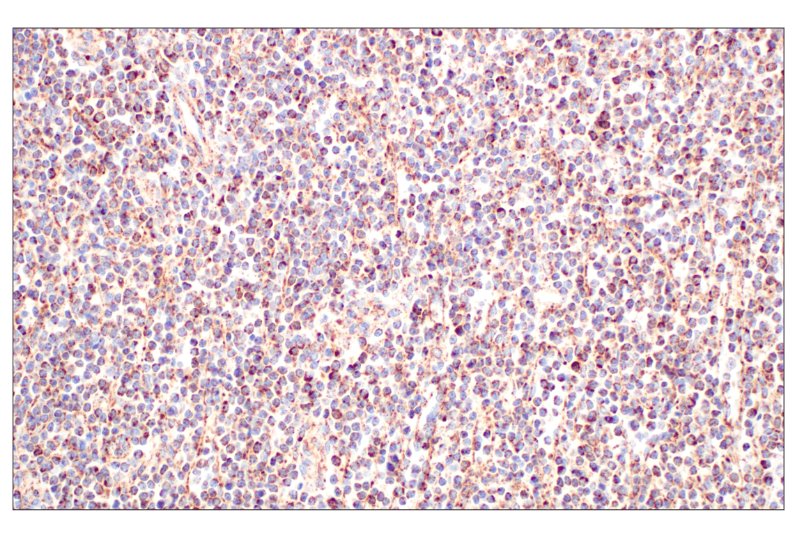
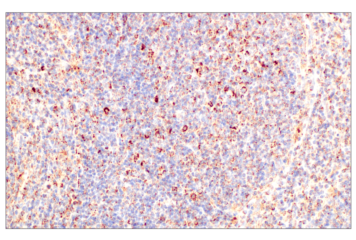
Immunohistochemical analysis of paraffin-embedded human B-cell non-Hodgkin lymphoma using GOT2 (F4P3R) Rabbit mAb.
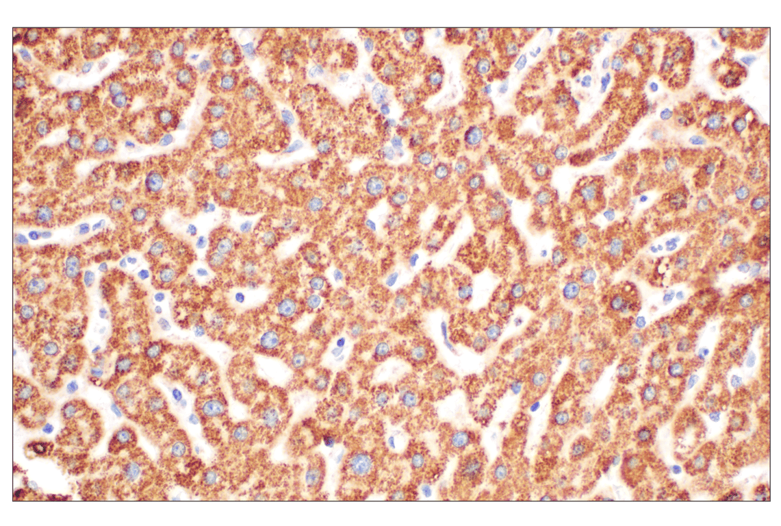
Immunohistochemical analysis of paraffin-embedded normal human liver using GOT2 (F4P3R) Rabbit mAb.
Revision 3
#39627
GOT2 (F4P3R) Rabbit mAb
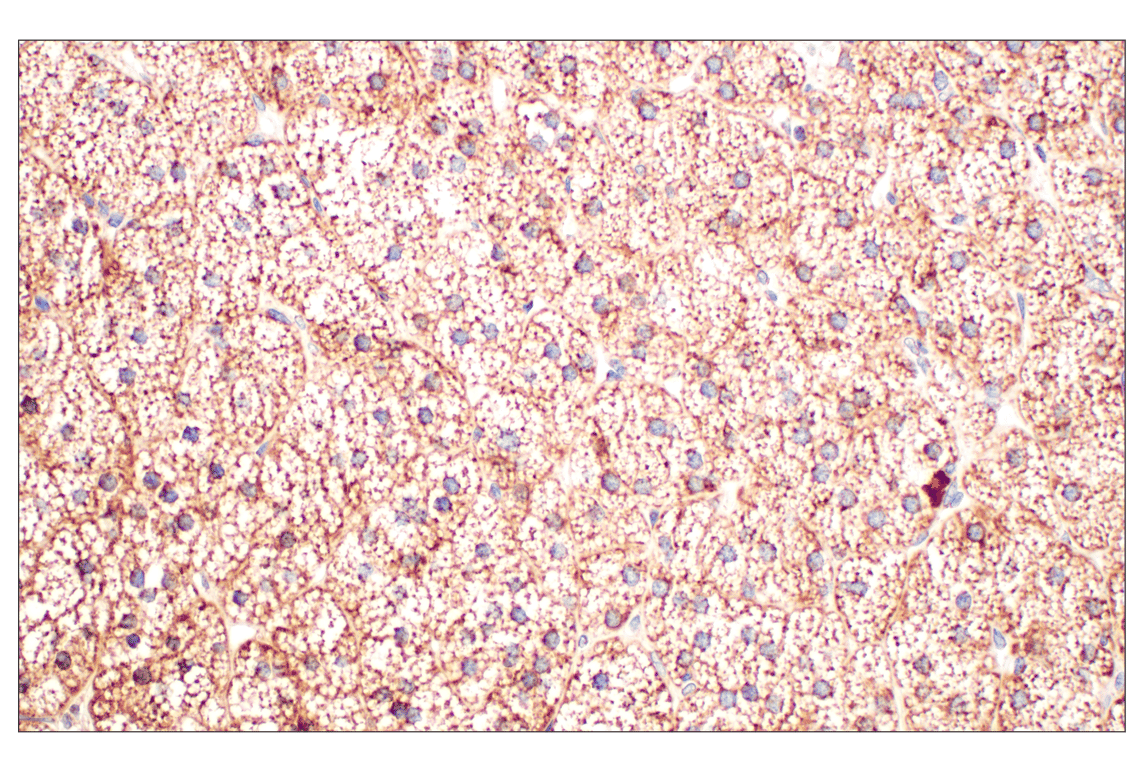
Immunohistochemical analysis of paraffin-embedded normal human adrenal gland using GOT2 (F4P3R) Rabbit mAb.
Immunohistochemical analysis of paraffin-embedded Peyer's patch within normal human small intestine using GOT2 (F4P3R) Rabbit mAb.
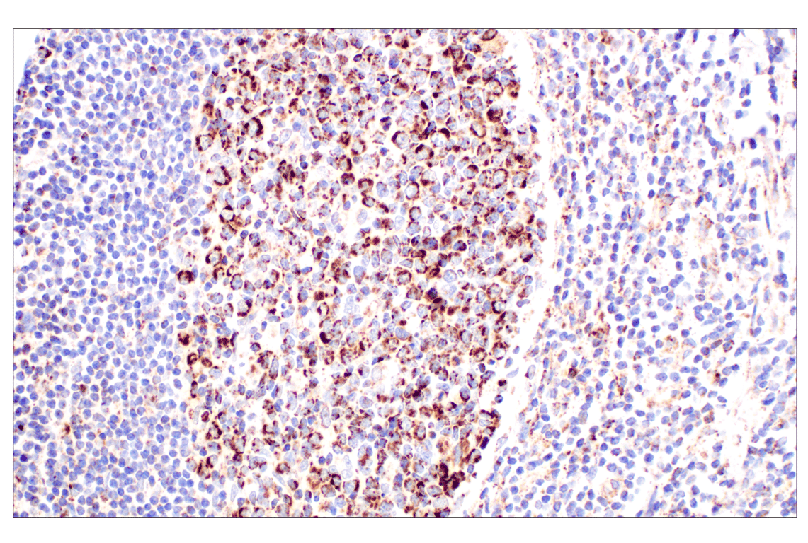
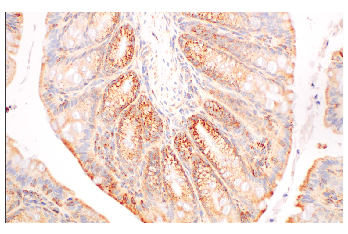
Immunohistochemical analysis of paraffin-embedded normal human colon using GOT2 (F4P3R) Rabbit mAb.
Revision 3
#39627
GOT2 (F4P3R) Rabbit mAb
Immunohistochemical analysis of paraffin-embedded Renca syngeneic tumor using GOT2 (F4P3R) Rabbit mAb.
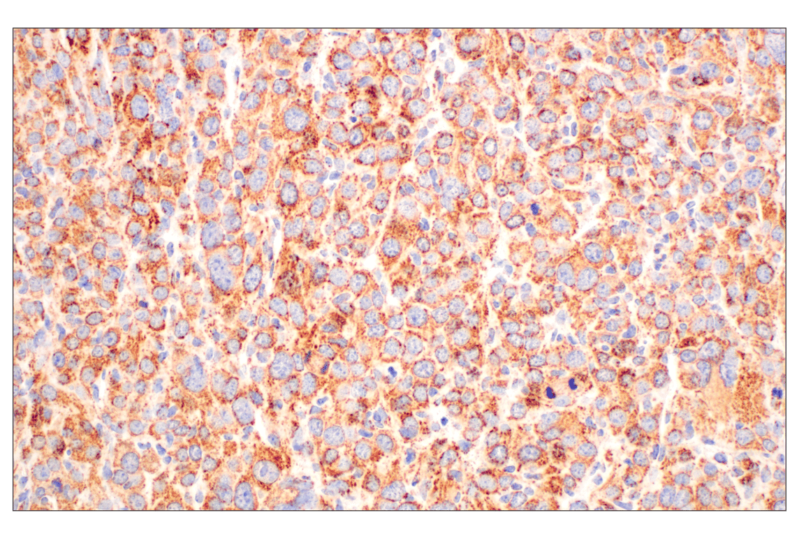
Immunohistochemical analysis of paraffin-embedded LL/2 syngeneic tumor using GOT2 (F4P3R) Rabbit mAb.
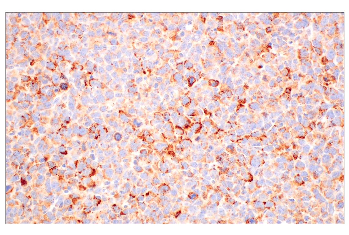
Immunohistochemical analysis of paraffin-embedded 4T1 syngeneic mammary tumor using GOT2 (F4P3R) Rabbit mAb.
Revision 3
#39627
GOT2 (F4P3R) Rabbit mAb
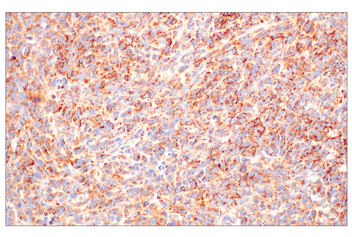
Immunohistochemical analysis of paraffin-embedded mouse colon using GOT2 (F4P3R) Rabbit mAb.
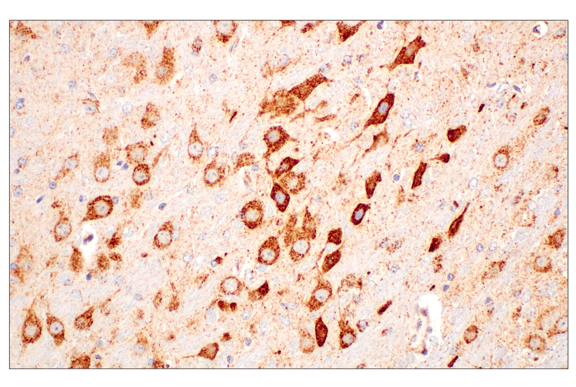
Immunohistochemical analysis of paraffin-embedded mouse brain using GOT2 (F4P3R) Rabbit mAb.
Immunohistochemical analysis of paraffin-embedded mouse spleen using GOT2 (F4P3R) Rabbit mAb.
Revision 3
#39627
GOT2 (F4P3R) Rabbit mAb
Immunohistochemical analysis of paraffin-embedded human tonsil using GOT2 (F4P3R) Rabbit mAb (left) compared to concentration-matched Rabbit (DA1E) mAb IgG XP® Isotype Control #3900 (right).

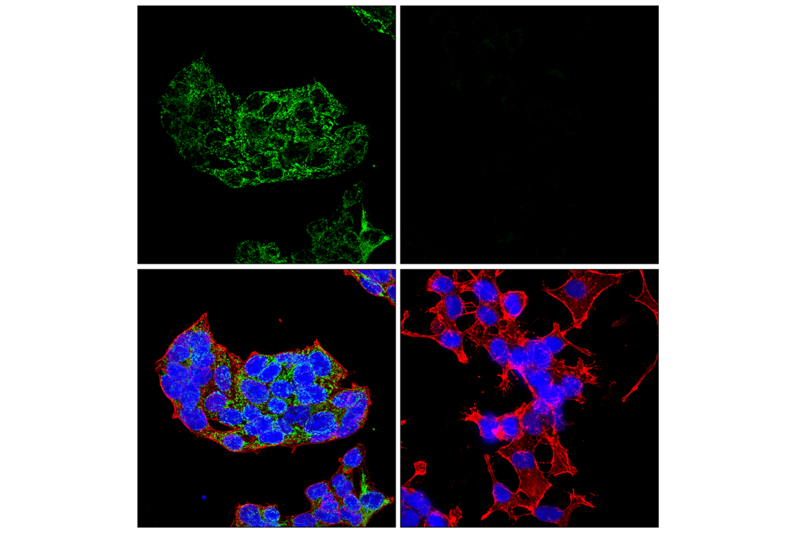
Confocal immunofluorescent analysis of NCI-H520 cells (left, high-expressing) and NCI-H446 cells (right, low-expressing) using GOT2 (F4P3R) Rabbit mAb (green), β-Actin (8H10D10) Mouse mAb #3700 (red), and DAPI #4083 (blue).