Revision 1
#90488
Store at -20C
877-616-CELL (2355)
877-678-TECH (8324)
3 Trask Lane | Danvers | Massachusetts | 01923 | USA
For Research Use Only. Not for Use in Diagnostic Procedures.
Applications:
W, IHC-P, IF-IC, FC-L
Reactivity:
H
Sensitivity:
Endogenous
MW (kDa):
55, 80-300
Source/Isotype:
Mouse IgG1 kappa
UniProt ID:
#Q8N158
Entrez-Gene Id:
221914
Product Usage Information
| Application | Dilution |
|---|---|
| Western Blotting | 1:1000 |
| Immunohistochemistry (Paraffin) | 1:200 - 1:800 |
| Immunofluorescence (Immunocytochemistry) | 1:50 - 1:200 |
| Flow Cytometry (Live) | 1:50 - 1:200 |
Storage
Specificity/Sensitivity
GPC2 (CT3) Mouse mAb recognizes endogenous levels of total GPC2 protein. This antibody reacts with the core GPC2 protein and glycanated forms of GPC2 protein. Based upon sequence alignment, this antibody is not predicted to cross-react with other GPC family members.
Source / Purification
Monoclonal antibody is produced by immunizing animals with a synthetic peptide corresponding to residues near the carboxy terminus of human GPC2 protein.
Background
Glypican 2 (GPC2) is a heparan sulfate proteoglycan that is linked to the outer leaflet of the plasma membrane via a glycosylphosphatidylinositol (GPI) linkage (1). Unlike other members of the glypican family, GPC2 is expressed in the developing nervous system, where it is thought to function as a signaling co-receptor to promote neuronal growth and axon guidance (2,3). GPC2 is considered to be an oncofetal antigen as it is highly expressed in the developing nervous system and then largely silenced in adult, normal tissues. Research studies have shown, however, that GPC2 is highly overexpressed in a high percentage of human neuroblastomas, medulloblastomas, and retinoblastomas. Given the restricted expression pattern of GPC2 in healthy, postnatal tissues, there is significant interest in targeting this antigen using a variety of immunotherapeutic approaches (4-6).
Background References
Species Reactivity
Species reactivity is determined by testing in at least one approved application (e.g., western blot).
Western Blot Buffer
IMPORTANT: For western blots, incubate membrane with diluted primary antibody in 5% w/v BSA, 1X TBS, 0.1% Tween® 20 at 4°C with gentle shaking, overnight.
Applications Key
W: Western Blotting IHC-P: Immunohistochemistry (Paraffin) IF-IC: Immunofluorescence (Immunocytochemistry) FC-L: Flow Cytometry (Live)
Cross-Reactivity Key
H: Human M: Mouse R: Rat Hm: Hamster Mk: Monkey Vir: Virus Mi: Mink C: Chicken Dm: D. melanogaster X: Xenopus Z: Zebrafish B: Bovine Dg: Dog Pg: Pig Sc: S. cerevisiae Ce: C. elegans Hr: Horse GP: Guinea Pig Rab: Rabbit G: Goat All: All Species Expected
Trademarks and Patents
Cell Signaling Technology is a trademark of Cell Signaling Technology, Inc.
All other trademarks are the property of their respective owners. Visit cellsignal.com/trademarks for more information.
Limited Uses
Except as otherwise expressly agreed in a writing signed by a legally authorized representative of CST, the following terms apply to Products provided by CST, its affiliates or its distributors. Any Customer's terms and conditions that are in addition to, or different from, those contained herein, unless separately accepted in writing by a legally authorized representative of CST, are rejected and are of no force or effect.
Products are labeled with For Research Use Only or a similar labeling statement and have not been approved, cleared, or licensed by the FDA or other regulatory foreign or domestic entity, for any purpose. Customer shall not use any Product for any diagnostic or therapeutic purpose, or otherwise in any manner that conflicts with its labeling statement. Products sold or licensed by CST are provided for Customer as the end-user and solely for research and development uses. Any use of Product for diagnostic, prophylactic or therapeutic purposes, or any purchase of Product for resale (alone or as a component) or other commercial purpose, requires a separate license from CST. Customer shall (a) not sell, license, loan, donate or otherwise transfer or make available any Product to any third party, whether alone or in combination with other materials, or use the Products to manufacture any commercial products, (b) not copy, modify, reverse engineer, decompile, disassemble or otherwise attempt to discover the underlying structure or technology of the Products, or use the Products for the purpose of developing any products or services that would compete with CST products or services, (c) not alter or remove from the Products any trademarks, trade names, logos, patent or copyright notices or markings, (d) use the Products solely in accordance with CST Product Terms of Sale and any applicable documentation, and (e) comply with any license, terms of service or similar agreement with respect to any third party products or services used by Customer in connection with the Products.
Revision 1
#90488
GPC2 (CT3) Mouse mAb
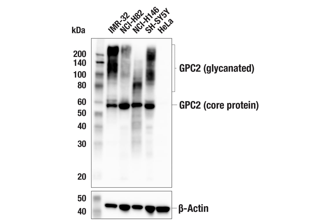
Western blot analysis of extracts from various cell lines using GPC2 (CT3) Mouse mAb (upper) or β-Actin (D6A8) Rabbit mAb #8457 (lower). Negative expression of GPC2 protein in HeLa cells is consistent with published observations.
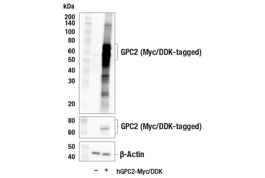
Western blot analysis of extracts from 293T cells, mock transfected (-) or transfected with a construct expressing Myc/DDK-tagged full-length human GPC2 (hGPC2-Myc/DDK; +), using GPC2 (CT3) Mouse mAb (upper), Myc-Tag (71D10) Rabbit mAb #2278 (middle), or β-Actin (D6A8) Rabbit mAb #8457 (lower).
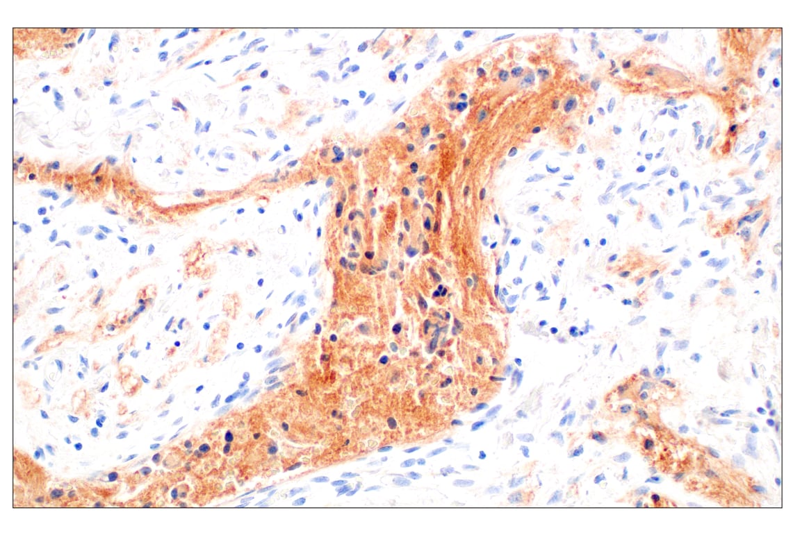
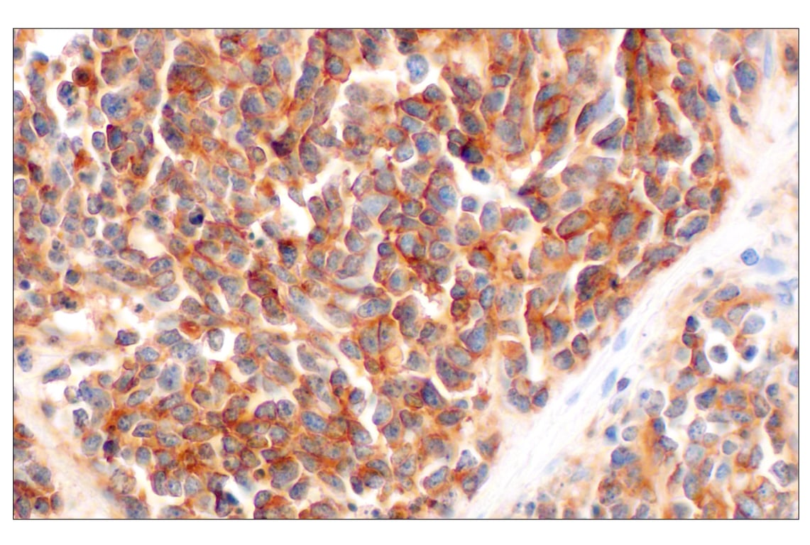
Immunohistochemical analysis of paraffin-embedded human neuroblastoma using GPC2 (CT3) Mouse mAb.
Revision 1
#90488
GPC2 (CT3) Mouse mAb
Immunohistochemical analysis of paraffin-embedded human medulloblastoma using GPC2 (CT3) Mouse mAb.
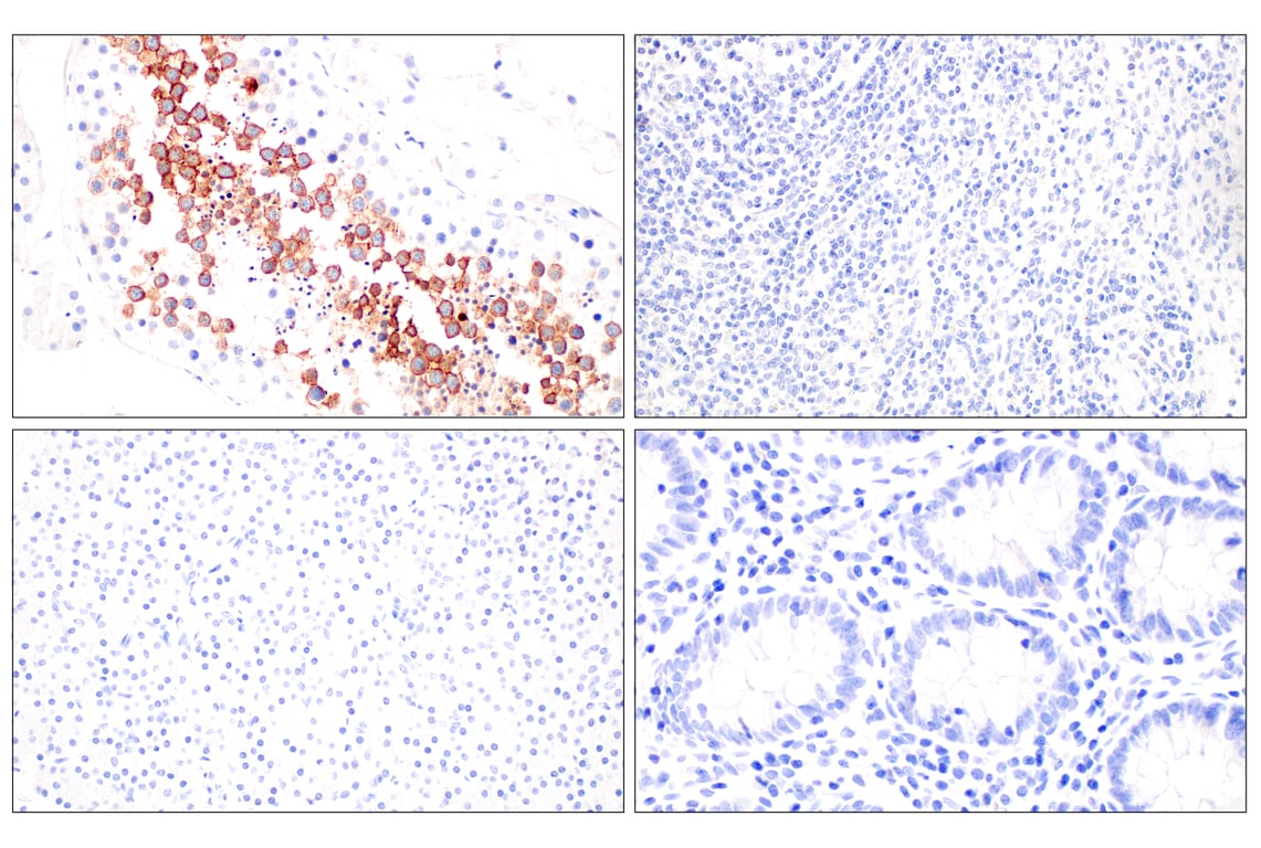
Immunohistochemical analysis of various paraffin-embedded normal human tissues: testes (left-top), spleen (right-top), pancreas (left-bottom), and colon (right-bottom) using GPC2 (CT3) Mouse mAb.
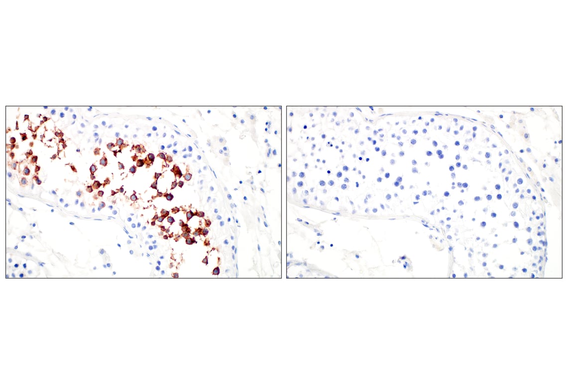
Immunohistochemical analysis of paraffin-embedded normal human testes using GPC2 (CT3) Mouse mAb (left) compared to concentration-matched Mouse (G3A1) mAb IgG1 Isotype Control #5415 (right).
Revision 1
#90488
GPC2 (CT3) Mouse mAb
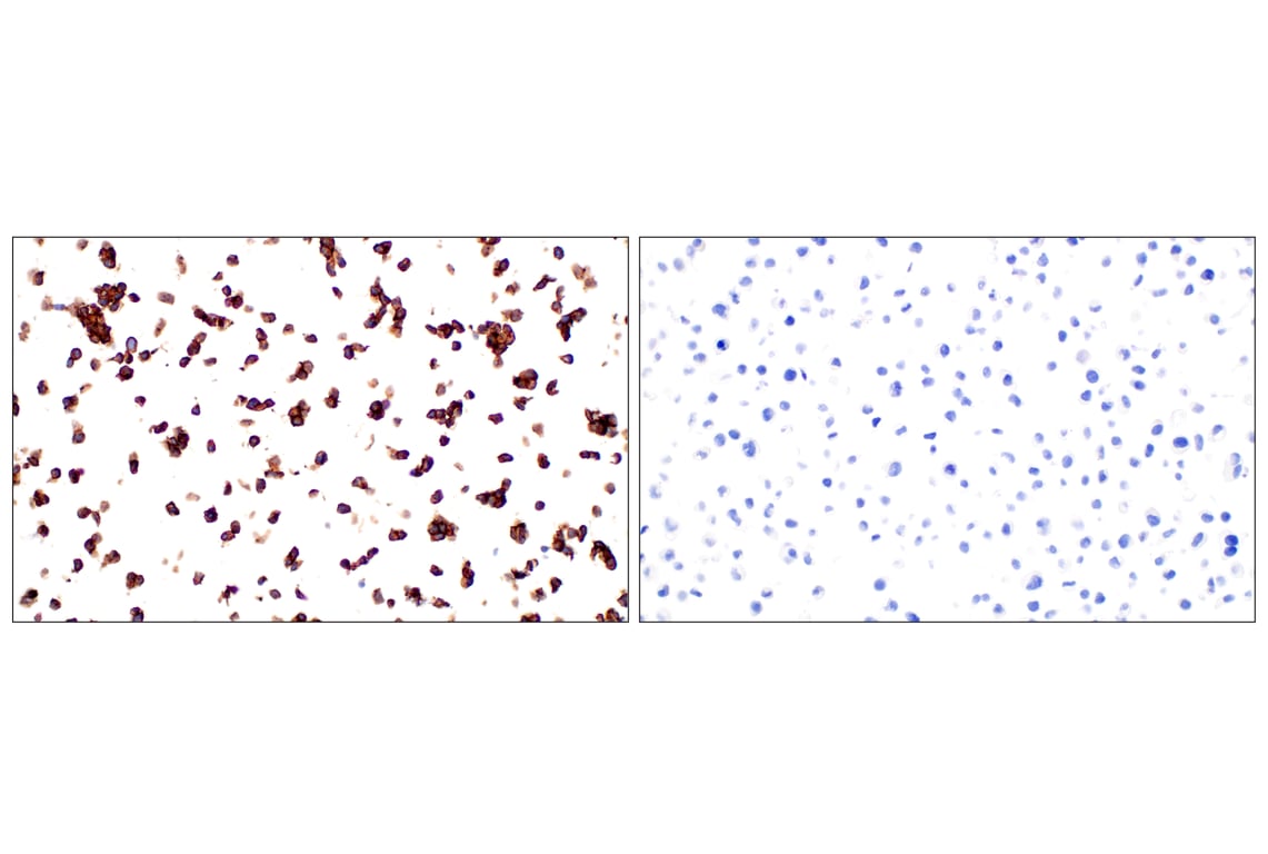
Immunohistochemical analysis of paraffin-embedded IMR-32 cell pellet (left, positive) or HeLa cell pellet (right, negative) using GPC2 (CT3) Mouse mAb.
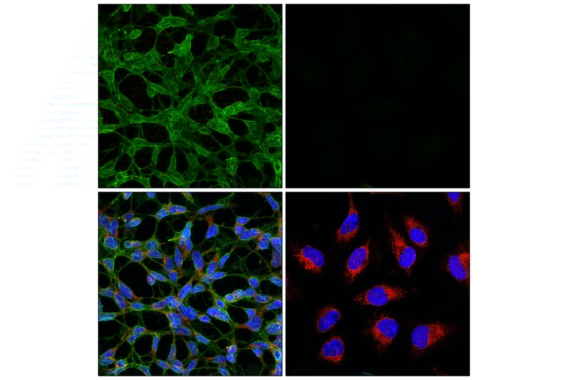
Confocal immunofluorescent analysis of IMR-32 cells (left, positive) or HeLa cells (right, negative) using GPC2 (CT3) Mouse mAb (green), COX IV (3E11) Rabbit mAb (Alexa Fluor® 647 Conjugate) #7561 (red), and DAPI #4083 (blue).
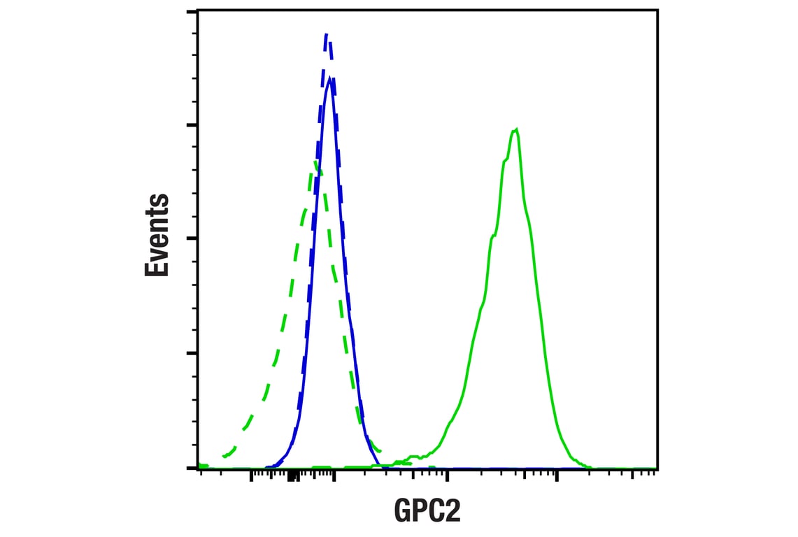
Flow cytometric analysis of live HeLa cells (blue, negative) and IMR-32 cells (green, positive) using GPC2 (CT3) Mouse mAb (solid lines) or concentration-matched Mouse (G3A1) mAb IgG1 Isotype Control #5415 (dashed lines). Anti-mouse IgG (H+L), F(ab')2 Fragment (Alexa Fluor® 488 Conjugate) #4408 was used as a secondary antibody.