Revision 1
#87697
Store at -20C
877-616-CELL (2355)
877-678-TECH (8324)
3 Trask Lane | Danvers | Massachusetts | 01923 | USA
For Research Use Only. Not for Use in Diagnostic Procedures.
Applications:
W, IHC-Bond, IHC-P, IF-IC
Reactivity:
H M
Sensitivity:
Endogenous
MW (kDa):
120
Source/Isotype:
Rabbit IgG
UniProt ID:
#Q16665
Entrez-Gene Id:
3091
Product Usage Information
This formulation is ideal for use with technologies requiring specialized or custom antibody labeling, including fluorophores, metals, lanthanides, and oligonucleotides. It is not recommended for ChIP, ChIP-seq, CUT&RUN or CUT&Tag assays. If you require a carrier free formulation for chromatin profiling, please contact us. Optimal dilutions/concentrations should be determined by the end user.
Formulation
Supplied in 1X PBS, BSA and Azide Free.
For standard formulation of this product see product #48085.
Storage
Specificity/Sensitivity
HIF-1α (E1V6A) Rabbit mAb (BSA and Azide Free) recognizes endogenous levels of total HIF-1α protein. This antibody does not cross-react with HIF-2α protein.
Source / Purification
Monoclonal antibody is produced by immunizing animals with a synthetic peptide corresponding to residues surrounding Ala475 of human HIF-1α protein.
Background
Hypoxia-inducible factor 1 (HIF1) is a heterodimeric transcription factor that plays a critical role in the cellular response to hypoxia (1). The HIF1 complex consists of two subunits, HIF-1α and HIF-1β, which are basic helix-loop-helix proteins of the PAS (Per, ARNT, Sim) family (2). HIF1 regulates the transcription of a broad range of genes that facilitate responses to the hypoxic environment, including genes regulating angiogenesis, erythropoiesis, cell cycle, metabolism, and apoptosis. The widely expressed HIF-1α is typically degraded rapidly in normoxic cells by the ubiquitin/proteasomal pathway. Under normoxic conditions, HIF-1α is proline hydroxylated leading to a conformational change that promotes binding to the von Hippel-Lindau protein (VHL) E3 ligase complex; ubiquitination and proteasomal degradation follows (3,4). Both hypoxic conditions and chemical hydroxylase inhibitors (such as desferrioxamine and cobalt) inhibit HIF-1α degradation and lead to its stabilization. In addition, HIF-1α can be induced in an oxygen-independent manner by various cytokines through the PI3K-AKT-mTOR pathway (5-7).
HIF-1β is also known as AhR nuclear translocator (ARNT) due to its ability to partner with the aryl hydrocarbon receptor (AhR) to form a heterodimeric transcription factor complex (8). Together with AhR, HIF-1β plays an important role in xenobiotics metabolism (8). In addition, a chromosomal translocation leading to a TEL-ARNT fusion protein is associated with acute myeloblastic leukemia (9). Studies also found that ARNT/HIF-1β expression levels decrease significantly in pancreatic islets from patients with type 2 diabetes, suggesting that HIF-1β plays an important role in pancreatic β-cell function (10).
Background References
- Sharp, F.R. and Bernaudin, M. (2004) Nat Rev Neurosci 5, 437-48.
- Wang, G.L. et al. (1995) Proc Natl Acad Sci U S A 92, 5510-4.
- Jaakkola, P. et al. (2001) Science 292, 468-72.
- Maxwell, P.H. et al. (1999) Nature 399, 271-5.
- Fukuda, R. et al. (2002) J Biol Chem 277, 38205-11.
- Jiang, B.H. et al. (2001) Cell Growth Differ 12, 363-9.
- Laughner, E. et al. (2001) Mol Cell Biol 21, 3995-4004.
- Walisser, J.A. et al. (2004) Proc Natl Acad Sci U S A 101, 16677-82.
- Salomon-Nguyen, F. et al. (2000) Proc Natl Acad Sci U S A 97, 6757-62.
- Gunton, J.E. et al. (2005) Cell 122, 337-49.
Species Reactivity
Species reactivity is determined by testing in at least one approved application (e.g., western blot).
Applications Key
W: Western Blotting IHC-Bond: IHC Leica Bond IF-IC: Immunofluorescence (Immunocytochemistry)
Cross-Reactivity Key
H: Human M: Mouse R: Rat Hm: Hamster Mk: Monkey Vir: Virus Mi: Mink C: Chicken Dm: D. melanogaster X: Xenopus Z: Zebrafish B: Bovine Dg: Dog Pg: Pig Sc: S. cerevisiae Ce: C. elegans Hr: Horse GP: Guinea Pig Rab: Rabbit G: Goat All: All Species Expected
Trademarks and Patents
Cell Signaling Technology is a trademark of Cell Signaling Technology, Inc.
SignalStain is a registered trademark of Cell Signaling Technology, Inc.
XP is a registered trademark of Cell Signaling Technology, Inc.
All other trademarks are the property of their respective owners. Visit cellsignal.com/trademarks for more information.
Limited Uses
Except as otherwise expressly agreed in a writing signed by a legally authorized representative of CST, the following terms apply to Products provided by CST, its affiliates or its distributors. Any Customer's terms and conditions that are in addition to, or different from, those contained herein, unless separately accepted in writing by a legally authorized representative of CST, are rejected and are of no force or effect.
Products are labeled with For Research Use Only or a similar labeling statement and have not been approved, cleared, or licensed by the FDA or other regulatory foreign or domestic entity, for any purpose. Customer shall not use any Product for any diagnostic or therapeutic purpose, or otherwise in any manner that conflicts with its labeling statement. Products sold or licensed by CST are provided for Customer as the end-user and solely for research and development uses. Any use of Product for diagnostic, prophylactic or therapeutic purposes, or any purchase of Product for resale (alone or as a component) or other commercial purpose, requires a separate license from CST. Customer shall (a) not sell, license, loan, donate or otherwise transfer or make available any Product to any third party, whether alone or in combination with other materials, or use the Products to manufacture any commercial products, (b) not copy, modify, reverse engineer, decompile, disassemble or otherwise attempt to discover the underlying structure or technology of the Products, or use the Products for the purpose of developing any products or services that would compete with CST products or services, (c) not alter or remove from the Products any trademarks, trade names, logos, patent or copyright notices or markings, (d) use the Products solely in accordance with CST Product Terms of Sale and any applicable documentation, and (e) comply with any license, terms of service or similar agreement with respect to any third party products or services used by Customer in connection with the Products.
Revision 1
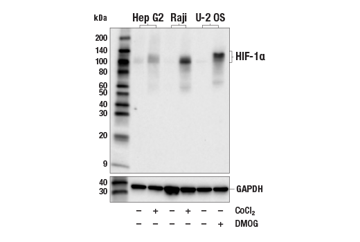
Western blot analysis of extracts from Hep G2 cells untreated (-) or treated with cobalt chloride (CoCl2) (0.1 mM, 4 hr; +), Raji cells untreated (-) or treated with CoCl2 (0.1 mM, 4 hr; +), and U-2 OS cells untreated (-) or treated with DMOG (1 mM, 6 hr; +), using HIF-1α (E1V6A) Rabbit mAb (upper) or GAPDH (D16H11) XP® Rabbit mAb #5174 (lower). Data were generated using the standard formulation of this product.
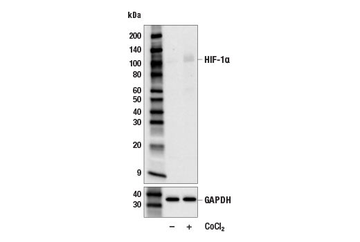
Western blot analysis of extracts from C2C12 cells, untreated (-) or treated with cobalt chloride (CoCl2) (0.1 mM, 4 hr; +), using HIF-1α (E1V6A) Rabbit mAb (upper) or GAPDH (D16H11) XP® Rabbit mAb #5174 (lower). Data were generated using the standard formulation of this product.
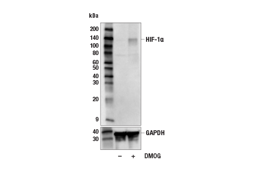
Western blot analysis of extracts from NCI-H3122 cells, untreated (-) or treated with DMOG (1 mM, 6 hr; +), using HIF-1α (E1V6A) Rabbit mAb (upper) or GAPDH (D16H11) XP® Rabbit mAb #5174 (lower). Data were generated using the standard formulation of this product.
Revision 1
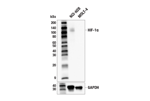
Western blot analysis of extracts from NCI-H28 and MOLT-4 cells using HIF-1α (E1V6A) Rabbit mAb (upper) or GAPDH (D16H11) XP® Rabbit mAb #5174 (lower). Data were generated using the standard formulation of this product.
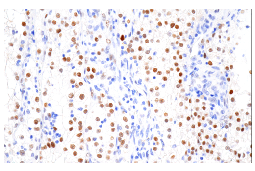
Immunohistochemical analysis of paraffin-embedded human renal cell carcinoma using HIF-1α (E1V6A) Rabbit mAb performed on the Leica® BOND™ Rx. Data were generated using the standard formulation of this product.
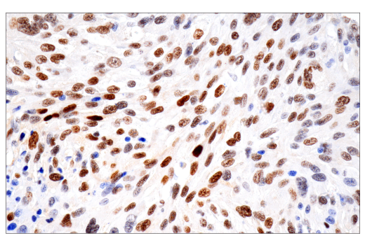
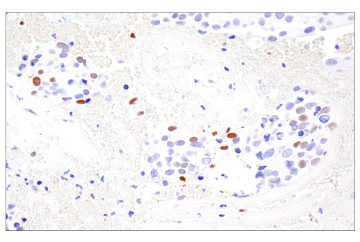
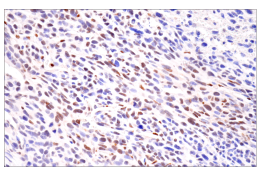
Immunohistochemical analysis of paraffin-embedded human gastrointestinal stromal tumor using HIF-1α (E1V6A) Rabbit mAb performed on the Leica® BOND™ Rx. Data were generated using the standard formulation of this product.
Revision 1
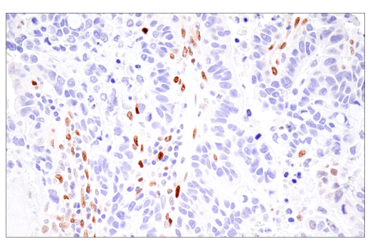
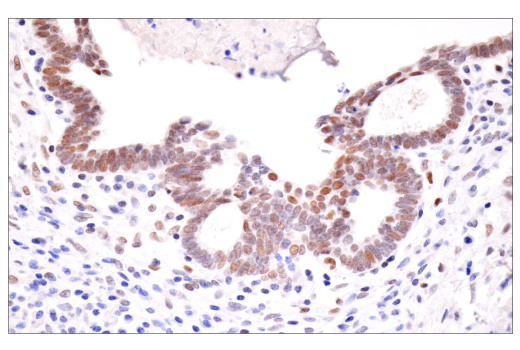
Immunohistochemical analysis of paraffin-embedded human esophageal adenocarcinoma using HIF-1α (E1V6A) Rabbit mAb. Data were generated using the standard formulation of this product.
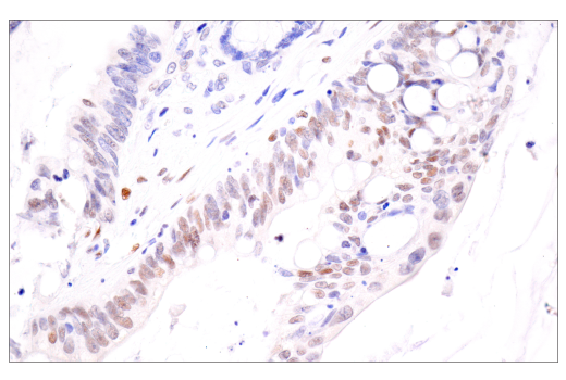
Immunohistochemical analysis of paraffin-embedded human endometrioid adenocarcinoma using HIF-1α (E1V6A) Rabbit mAb. Data were generated using the standard formulation of this product.
Immunohistochemical analysis of paraffin-embedded human ductal breast carcinoma using HIF-1α (E1V6A) Rabbit mAb. Data were generated using the standard formulation of this product.
Revision 1
Immunohistochemical analysis of paraffin-embedded human colon carcinoma using HIF-1α (E1V6A) Rabbit mAb. Data were generated using the standard formulation of this product.
Immunohistochemical analysis of paraffin-embedded LL/2 syngeneic tumor using HIF-1α (E1V6A) Rabbit mAb. Data were generated using the standard formulation of this product.
Immunohistochemical analysis of paraffin-embedded 4T1 syngeneic mammary tumor using HIF-1α (E1V6A) Rabbit mAb. Data were generated using the standard formulation of this product.
Revision 1
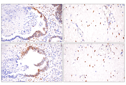
Immunohistochemical analysis of paraffin-embedded human endometrioid adenocarcinoma (left) and gastric carcinoma (right) using HIF-1α (E1V6A) Rabbit mAb (top) or a HIF-1α Rabbit mAb (bottom). These two antibodies detect unique, non-overlapping epitopes on human HIF-1α. The similar staining patterns obtained with both antibodies help to confirm the specificity of the staining. Data were generated using the standard formulation of this product.
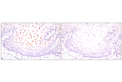
Immunohistochemical analysis of paraffin-embedded normal human esophagus using HIF-1α (E1V6A) Rabbit mAb (left) compared to concentration-matched Rabbit (DA1E) mAb IgG XP® Isotype Control #3900 (right). Data were generated using the standard formulation of this product.
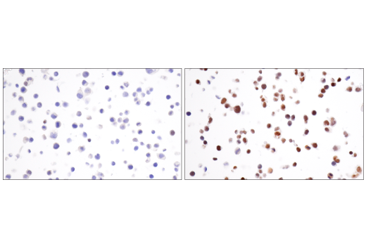
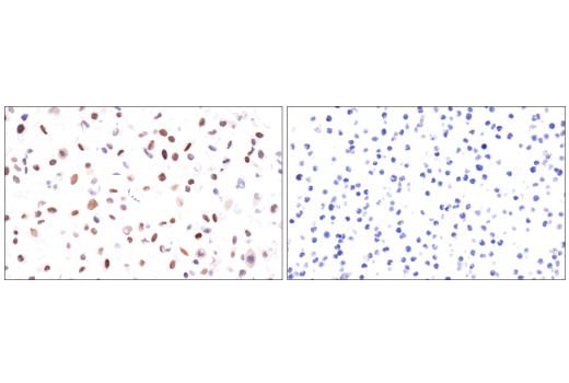
Immunohistochemical analysis of paraffin-embedded NCI-H3122 cell pellets, untreated (left) or treated with DMOG (1 mM, 6 hr; right), using HIF-1α (E1V6A) Rabbit mAb. Data were generated using the standard formulation of this product.
Revision 1
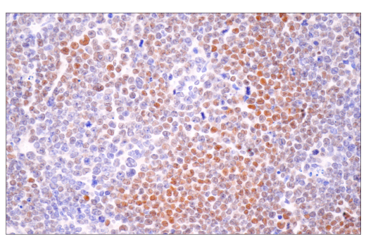
Immunohistochemical analysis of paraffin-embedded NCI-H28 cell pellet (left, positive) or MOLT-4 cell pellet (right, negative) using HIF-1α (E1V6A) Rabbit mAb. Data were generated using the standard formulation of this product.
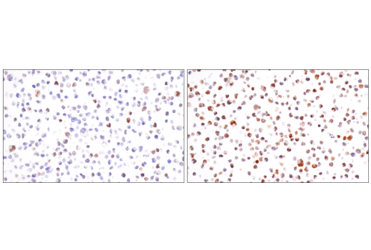
Immunohistochemical analysis of paraffin-embedded Raji cell pellets, untreated (left) or treated with cobalt chloride (CoCl2) (100 μM, 4 hr; right), using HIF-1α (E1V6A) Rabbit mAb. Data were generated using the standard formulation of this product.
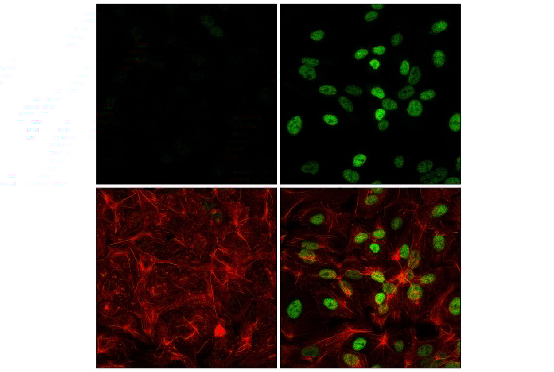
Confocal immunofluorescent analysis of Hep G2 cells, untreated (left) or treated with cobalt chloride (500 μM, 24 h; right), using HIF-1α (E1V6A) Rabbit mAb (green) and DyLight™ 650 Phalloidin #12956 (red). Data were generated using the standard formulation of this product.