Revision 1
#8579
Store at -20C
Hippo Signaling Antibody Sampler Kit
1 Kit
(9 x 20 microliters)
877-616-CELL (2355)
877-678-TECH (8324)
3 Trask Lane | Danvers | Massachusetts | 01923 | USA
For Research Use Only. Not for Use in Diagnostic Procedures.
| Product Includes | Product # | Quantity | Mol. Wt | Isotype/Source |
|---|---|---|---|---|
| Phospho-YAP (Ser397) (D1E7Y) Rabbit mAb | 13619 | 20 µl | 65-78 kDa | Rabbit IgG |
| LATS1 (C66B5) Rabbit mAb | 3477 | 20 µl | 140 kDa | Rabbit IgG |
| Phospho-MOB1 (Thr35) (D2F10) Rabbit mAb | 8699 | 20 µl | 24 kDa | Rabbit IgG |
| MOB1 (E1N9D) Rabbit mAb | 13730 | 20 µl | 24 kDa | Rabbit IgG |
| MST1 Antibody | 3682 | 20 µl | 59 kDa | Rabbit |
| MST2 Antibody | 3952 | 20 µl | 60 kDa | Rabbit |
| SAV1 (D6M6X) Rabbit mAb | 13301 | 20 µl | 45 kDa | Rabbit IgG |
| Phospho-YAP (Ser127) (D9W2I) Rabbit mAb | 13008 | 20 µl | 65-78 kDa | Rabbit IgG |
| YAP/TAZ (D24E4) Rabbit mAb | 8418 | 20 µl | 55, 78 kDa | Rabbit IgG |
| Anti-rabbit IgG, HRP-linked Antibody | 7074 | 100 µl | Goat |
Please visit cellsignal.com for individual component applications, species cross-reactivity, dilutions, protocols, and additional product information.
Description
The Hippo Signaling Antibody Sampler Kit provides an economical means of detecting target proteins of the Hippo signaling pathway. The kit contains enough primary antibody to perform two western blots per primary.
Storage
Background
Hippo signaling is an evolutionarily conserved pathway that controls cell proliferation, apoptosis, and organ size in response to changing cell density levels (1,2). At relative low cell density, transcription co-activators YAP and TAZ bind transcription factors to induce expression of genes that favor cell growth and proliferation. As cell density increases, interaction between membrane-bound upstream hippo pathway regulators trigger activation of cytoplasmic kinases Mst1/2 and LATS1/2. Activated Mst kinase (the eponymous Hippo in Drosophila) associates with the adaptor Sav1 and phosphorylates MOB1 to activate LATS kinase, which phosphorylates YAP and TAZ to suppress cell proliferation (3).
Trademarks and Patents
Cell Signaling Technology is a trademark of Cell Signaling Technology, Inc.
U.S. Patent No. 7,429,487, foreign equivalents, and child patents deriving therefrom.
All other trademarks are the property of their respective owners. Visit cellsignal.com/trademarks for more information.
Limited Uses
Except as otherwise expressly agreed in a writing signed by a legally authorized representative of CST, the following terms apply to Products provided by CST, its affiliates or its distributors. Any Customer's terms and conditions that are in addition to, or different from, those contained herein, unless separately accepted in writing by a legally authorized representative of CST, are rejected and are of no force or effect.
Products are labeled with For Research Use Only or a similar labeling statement and have not been approved, cleared, or licensed by the FDA or other regulatory foreign or domestic entity, for any purpose. Customer shall not use any Product for any diagnostic or therapeutic purpose, or otherwise in any manner that conflicts with its labeling statement. Products sold or licensed by CST are provided for Customer as the end-user and solely for research and development uses. Any use of Product for diagnostic, prophylactic or therapeutic purposes, or any purchase of Product for resale (alone or as a component) or other commercial purpose, requires a separate license from CST. Customer shall (a) not sell, license, loan, donate or otherwise transfer or make available any Product to any third party, whether alone or in combination with other materials, or use the Products to manufacture any commercial products, (b) not copy, modify, reverse engineer, decompile, disassemble or otherwise attempt to discover the underlying structure or technology of the Products, or use the Products for the purpose of developing any products or services that would compete with CST products or services, (c) not alter or remove from the Products any trademarks, trade names, logos, patent or copyright notices or markings, (d) use the Products solely in accordance with CST Product Terms of Sale and any applicable documentation, and (e) comply with any license, terms of service or similar agreement with respect to any third party products or services used by Customer in connection with the Products.
Revision 1
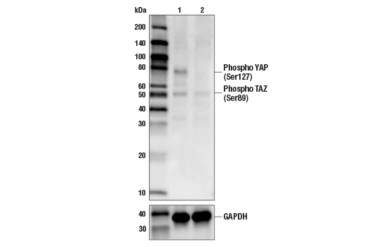
Western blot analysis of extracts from HeLa cells (lane 1) or YAP knock-out cells (lane 2) using Phospho-YAP (Ser127) (D9W2I) Rabbit mAb #13008 (upper), and GAPDH (D6H11) XP® Rabbit mAb #5174 (lower). The absence of signal in the YAP knock-out HeLa cells confirms specificity of the antibody for YAP.
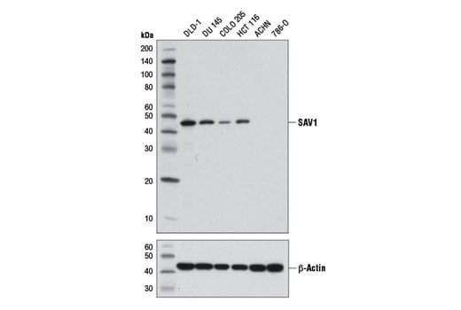
Western blot analysis of extracts from various cell lines using SAV1 (D6M6X) Rabbit mAb (upper) and β-Actin (D6A8) Rabbit mAb #8457 (lower).
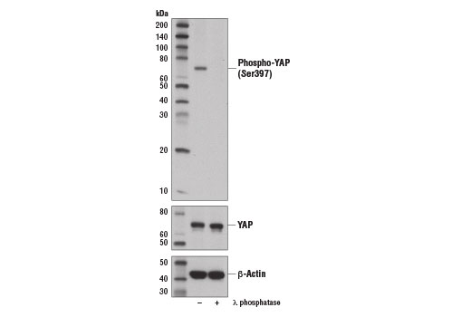
Western blot analysis of extracts from Hep G2 cells, untreated (-) or λ-phosphatase-treated (+), using Phospho-YAP (Ser397) (D1E7Y) Rabbit mAb (upper), YAP Antibody #4912 (middle), and β-Actin (D6A8) Rabbit mAb #8457 (lower). YAP protein isoform 1 Ser397 corresponds to Ser381 of YAP isoform 2, as reported by Zhao et al. (2010) Genes Dev 24, 72-85 (9).
Revision 1
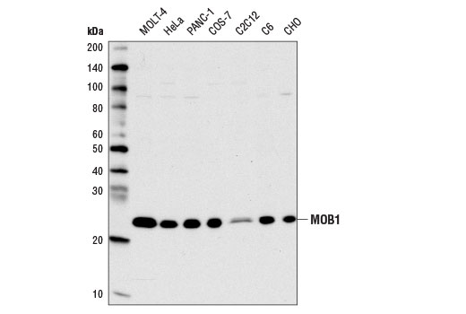
Western blot analysis of extracts from various cell lines using MOB1 (E1N9D) Rabbit mAb.
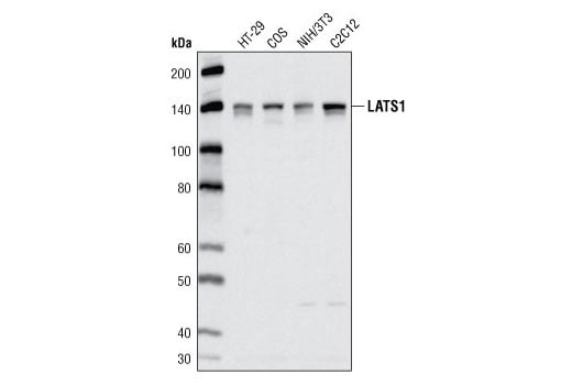
Western blot analysis of extracts from various cell lines using LATS1 (C66B5) Rabbit mAb.
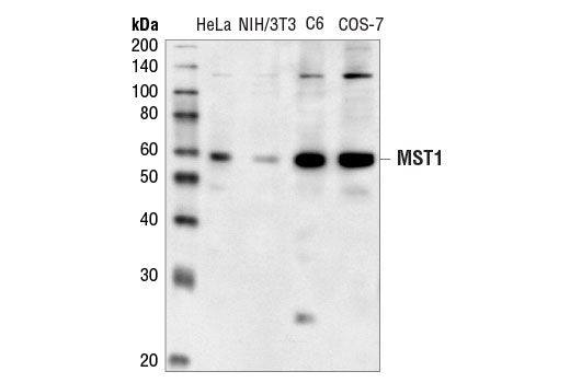
Western blot analysis of extracts from various cell lines using MST1 Antibody.
Revision 1
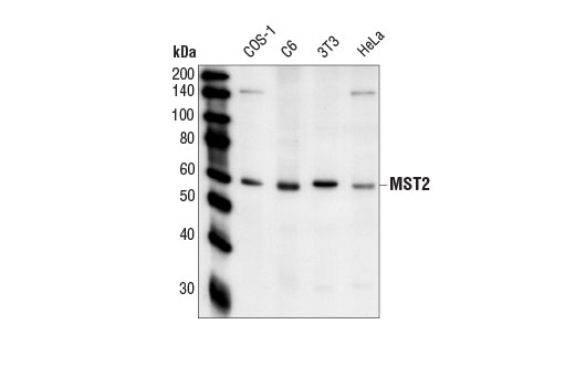
Western blot analysis of extracts from various cell lines using MST2 Antibody.
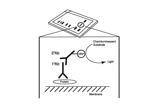
After the primary antibody is bound to the target protein, a complex with HRP-linked secondary antibody is formed. The LumiGLO® is added and emits light during enzyme catalyzed decomposition.
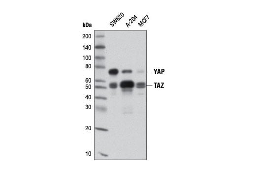
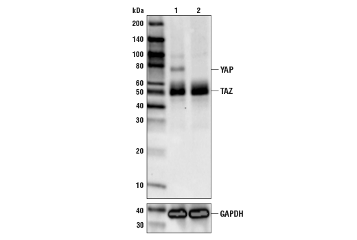
Western blot analysis of extracts from various cell lines using YAP/TAZ (D24E4) Rabbit mAb.
Revision 1
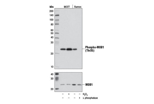
Western blot analysis of extracts from MCF7 cells, either untreated (-) or treated (+) with H2O2 (2.5 mM, 30 min) and Ramos cells, either untreated (-) or treated (+) with λ phosphatase, using Phospho-MOB1 (Thr35) (D2F10) Rabbit mAb (upper) and MOB1 Antibody #3863 (lower).
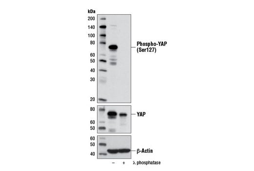
Western blot analysis of extracts from PANC-1 cells, untreated (-) or λ-phosphatase-treated (+), using Phospho-YAP (Ser127) (D9W2I) Rabbit mAb (upper), YAP Antibody #4912 (middle), and β-Actin (D6A8) Rabbit mAb #8457 (lower).
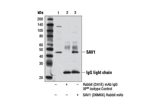
Immunoprecipitation of SAV1 protein from A-204 cell extracts using Rabbit (DA1E) mAb IgG XP® Isotype Control #3900 (lane 2) or SAV1 (D6M6X) Rabbit mAb (lane 3). Lane 1 is 10% input. Western blot analysis was performed using SAV1 (D6M6X) Rabbit mAb.
Revision 1
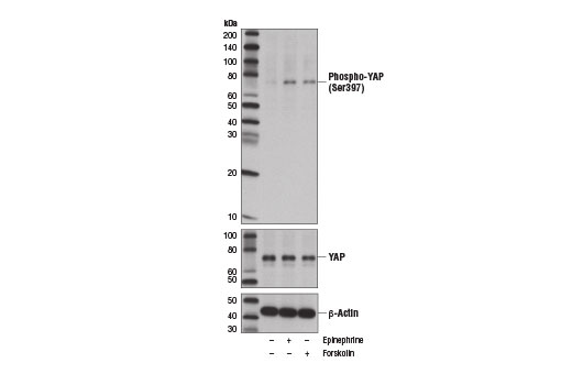
Western blot analysis of extracts from MDA-MB-231 cells, vehicle treated (-) or treated with epinephrine (10 μM, 60 min; +) or Forskolin #3828 (10 μm, 60 min; +), using Phospho-YAP (Ser397) (D1E7Y) Rabbit mAb (upper), YAP Antibody #4912 (middle), and β-Actin (D6A8) Rabbit mAb #8547 (lower). YAP protein isoform 1 Ser397 corresponds to Ser381 of YAP isoform 2, as reported by Zhao, B. et al. (2010) Genes Dev 24, 72-85 (9).
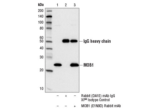
Immunoprecipitation of MOB1 protein from HeLa cell extracts using Rabbit (DA1E) mAb IgG XP® Isotype Control #3900 (lane 2) or MOB1 (E1N9D) Rabbit mAb (lane 3). Lane 1 is 10% input. Western blot analysis was performed using MOB1 (E1N9D) Rabbit mAb.
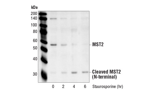
Western blot analysis of extracts from HeLa cells treated with Staurosporine #9953 for the indicated times using MST2 Antibody.
Revision 1
Western blot analysis of extracts from HeLa cells (lane 1) or YAP knock-out cells (lane 2) using YAP/TAZ (D24E4) Rabbit mAb #8418 (upper), and GAPDH (D6H11) XP® Rabbit mAb #5174 (lower). The absence of signal in the YAP knock-out HeLa cells confirms specificity of the antibody for YAP.
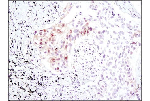
Immunohistochemical analysis of paraffin-embedded human lung carcinoma using Phospho-MOB1 (Thr35) (D2F10) Rabbit mAb.
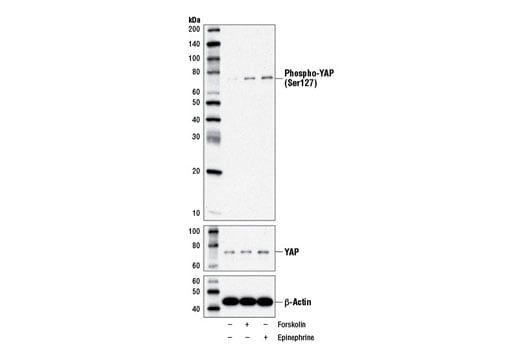
Western blot analysis of MDA-MB-231 cells, vehicle-treated (-) or treated with Forskolin #3828 (10 μM, 60 min; +) or epinephrine (10 μM, 60 min; +), using Phospho-YAP (Ser127) (D9W2I) Rabbit mAb (upper), YAP Antibody #4912 (middle), and β-Actin (D6A8) Rabbit mAb #8457 (lower). Note the induction of YAP (Ser127) phosphorylation after treatment with forskolin or epinephrine, consistent with the findings reported in Xu et al. (2012) [9].
Revision 1
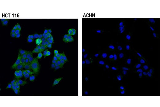
Confocal immunofluorescent analysis of HCT 116 (higher expressing, left) and ACHN (lower expressing, right) cells using SAV1 (D6M6X) Rabbit mAb (green). Blue pseudocolor = DRAQ5® #4084 (fluorescent DNA dye).
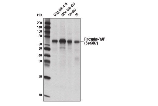
Western blot analysis of extracts from various cell lines using Phospho-YAP (Ser397) (D1E7Y) Rabbit mAb.
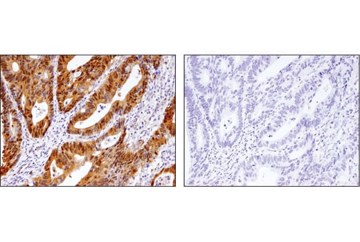
Immunohistochemical analysis of paraffin-embedded human ovarian carcinoma control (left) or lambda phosphatase-treated (right) using Phospho-MOB1 (Thr35) (D2F10) Rabbit mAb.
Revision 1
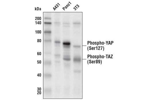
Western blot analysis of extracts from various cell lines using Phospho-YAP (Ser127) (D9W2I) Rabbit mAb.
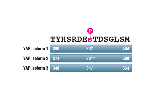
Schematic diagram showing annotation of the amino acid sequence surrounding Ser397 in different human YAP isoforms. Asterisk (*) indicates annotation of the phosphorylation site as described in Zhao et al. 2010 [9].
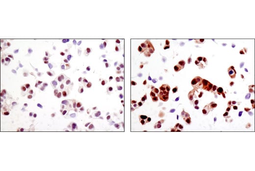
Immunohistochemical analysis of paraffin-embedded MCF7 cell pellets, control (left) or H2O2-treated (right), using Phospho-MOB1 (Thr35) (D2F10) Rabbit mAb.
Revision 1
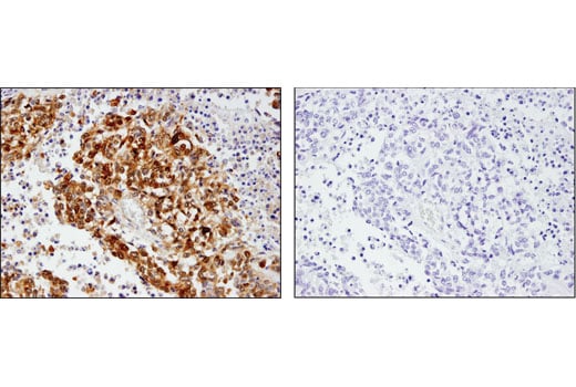
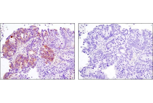
Immunohistochemical analysis of paraffin-embedded human colon adenocarcinoma, control (left) or λ-phosphatase treated (right), using Phospho-YAP (Ser127) (D9W2I) Rabbit mAb.
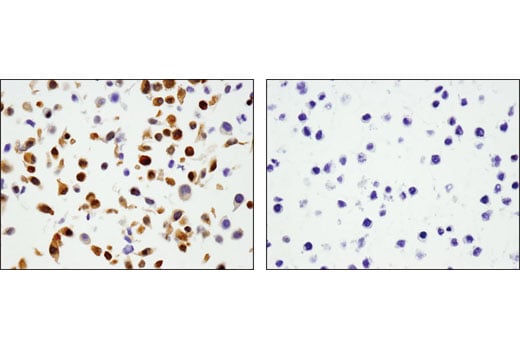
Immunohistochemical analysis of paraffin-embedded cell pellets, A-204 (left) and RL (right), using Phospho-YAP (Ser127) (D9W2I) Rabbit mAb.
Immunohistochemical analysis of paraffin-embedded human non-small cell lung carcinoma using Phospho-YAP (Ser127) (D9W2I) Rabbit mAb in the presence of control peptide (left) or antigen-specific peptide (right).