Revision 4
#26416
Store at -20C
877-616-CELL (2355)
877-678-TECH (8324)
3 Trask Lane | Danvers | Massachusetts | 01923 | USA
For Research Use Only. Not for Use in Diagnostic Procedures.
Applications:
W, IHC-P
Reactivity:
H
Sensitivity:
Endogenous
MW (kDa):
28
Source/Isotype:
Rabbit IgG
UniProt ID:
#P09601
Entrez-Gene Id:
3162
Product Usage Information
| Application | Dilution |
|---|---|
| Western Blotting | 1:1000 |
| Immunohistochemistry (Paraffin) | 1:200 - 1:800 |
Storage
For a carrier free (BSA and azide free) version of this product see product #56629.
Specificity/Sensitivity
HO-1 (E8B7A) XP® recognizes endogenous levels of total HO-1 protein.
Source / Purification
Monoclonal antibody is produced by immunizing animals with recombinant human HO-1 protein.
Background
Heme oxygenase (HO) is the rate-limiting enzyme in the catabolism of heme that results in the release of carbon monoxide, iron, and biliverdin (1). The products of this enzymatic reaction play important biological roles in antioxidant, anti-inflammatory, and cytoprotective functions (2). HO comprises two isozymes, including the constitutively expressed HO-2 isozyme and the inducible HO-1 isozyme (3). Inducible HO-1 is expressed as an adaptive response to several stimuli, including heme, metals, and hormones (4). The induction of HO-1 has been implicated in numerous disease states, such as transplant rejection, hypertension, atherosclerosis, Alzheimer's disease, endotoxic shock, diabetes, inflammation, and neurological disorders (1,5).
Species Reactivity
Species reactivity is determined by testing in at least one approved application (e.g., western blot).
Western Blot Buffer
IMPORTANT: For western blots, incubate membrane with diluted primary antibody in 5% w/v BSA, 1X TBS, 0.1% Tween® 20 at 4°C with gentle shaking, overnight.
Applications Key
W: Western Blotting IHC-P: Immunohistochemistry (Paraffin)
Cross-Reactivity Key
H: Human M: Mouse R: Rat Hm: Hamster Mk: Monkey Vir: Virus Mi: Mink C: Chicken Dm: D. melanogaster X: Xenopus Z: Zebrafish B: Bovine Dg: Dog Pg: Pig Sc: S. cerevisiae Ce: C. elegans Hr: Horse GP: Guinea Pig Rab: Rabbit G: Goat All: All Species Expected
Trademarks and Patents
Cell Signaling Technology is a trademark of Cell Signaling Technology, Inc.
SignalStain is a registered trademark of Cell Signaling Technology, Inc.
XP is a registered trademark of Cell Signaling Technology, Inc.
All other trademarks are the property of their respective owners. Visit cellsignal.com/trademarks for more information.
Limited Uses
Except as otherwise expressly agreed in a writing signed by a legally authorized representative of CST, the following terms apply to Products provided by CST, its affiliates or its distributors. Any Customer's terms and conditions that are in addition to, or different from, those contained herein, unless separately accepted in writing by a legally authorized representative of CST, are rejected and are of no force or effect.
Products are labeled with For Research Use Only or a similar labeling statement and have not been approved, cleared, or licensed by the FDA or other regulatory foreign or domestic entity, for any purpose. Customer shall not use any Product for any diagnostic or therapeutic purpose, or otherwise in any manner that conflicts with its labeling statement. Products sold or licensed by CST are provided for Customer as the end-user and solely for research and development uses. Any use of Product for diagnostic, prophylactic or therapeutic purposes, or any purchase of Product for resale (alone or as a component) or other commercial purpose, requires a separate license from CST. Customer shall (a) not sell, license, loan, donate or otherwise transfer or make available any Product to any third party, whether alone or in combination with other materials, or use the Products to manufacture any commercial products, (b) not copy, modify, reverse engineer, decompile, disassemble or otherwise attempt to discover the underlying structure or technology of the Products, or use the Products for the purpose of developing any products or services that would compete with CST products or services, (c) not alter or remove from the Products any trademarks, trade names, logos, patent or copyright notices or markings, (d) use the Products solely in accordance with CST Product Terms of Sale and any applicable documentation, and (e) comply with any license, terms of service or similar agreement with respect to any third party products or services used by Customer in connection with the Products.
Revision 4
#26416
HO-1 (E8B7A) XP® Rabbit mAb
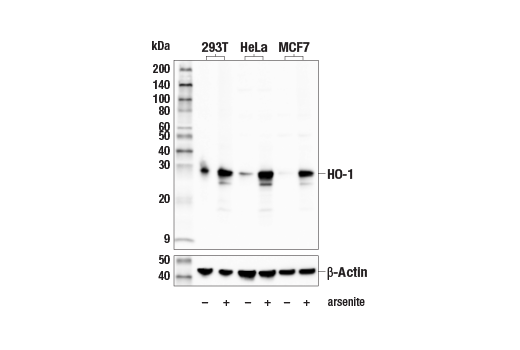
Western blot analysis of extracts from 293T, HeLa, and MCF7 cells, untreated (-) or treated with arsenite (50 μM, 8 hr; +), using HO-1 (E8B7A) XP® Rabbit mAb (upper) or β-Actin (D6A8) Rabbit mAb #8457 (lower).
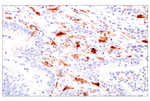
Immunohistochemical analysis of paraffin-embedded human small cell carcinoma of the salivary gland using HO-1 (E8B7A) XP® Rabbit mAb.
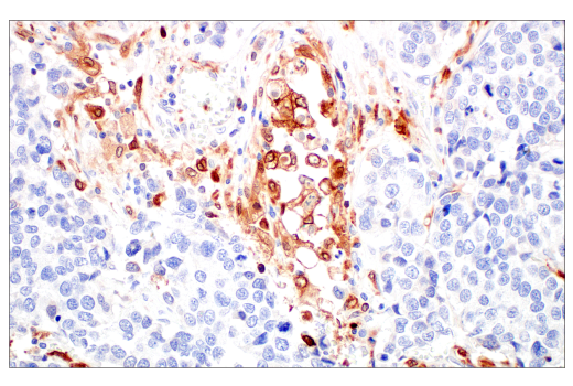
Immunohistochemical analysis of paraffin-embedded human urothelial carcinoma using HO-1 (E8B7A) XP® Rabbit mAb.
Revision 4
#26416
HO-1 (E8B7A) XP® Rabbit mAb
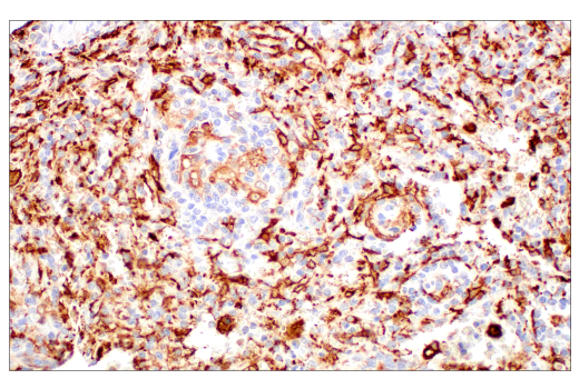
Immunohistochemical analysis of paraffin-embedded human colon adenocarcinoma using HO-1 (E8B7A) XP® Rabbit mAb.
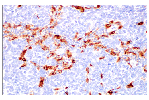
Immunohistochemical analysis of paraffin-embedded human neuroendocrine lung carcinoma using HO-1 (E8B7A) XP® Rabbit mAb.
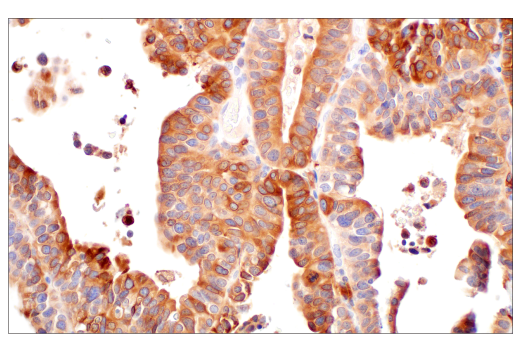
Immunohistochemical analysis of paraffin-embedded human ovarian serous carcinoma using HO-1 (E8B7A) XP® Rabbit mAb.

Revision 4
#26416
HO-1 (E8B7A) XP® Rabbit mAb
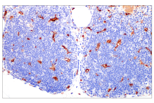
Immunohistochemical analysis of paraffin-embedded normal human spleen using HO-1 (E8B7A) XP® Rabbit mAb.
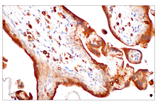
Immunohistochemical analysis of paraffin-embedded normal human placenta using HO-1 (E8B7A) XP® Rabbit mAb.
Immunohistochemical analysis of paraffin-embedded normal human thymus using HO-1 (E8B7A) XP® Rabbit mAb.
Revision 4
#26416
HO-1 (E8B7A) XP® Rabbit mAb
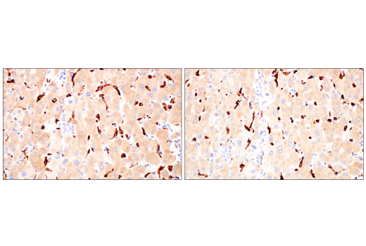
Immunohistochemical analysis of paraffin-embedded human hepatocellular carcinoma using HO-1 (E8B7A) XP® Rabbit mAb (left) or a HO-1 Rabbit mAb (right). These two antibodies detect unique, non-overlapping epitopes on human HO-1. The similar staining patterns obtained with both antibodies help to confirm the specificity of the staining.
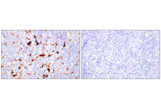
Immunohistochemical analysis of paraffin-embedded human B-cell non-Hodgkin lymphoma using HO-1 (E8B7A) XP® Rabbit mAb (left) compared to concentration-matched Rabbit (DA1E) mAb IgG XP® Isotype Control #3900 (right).
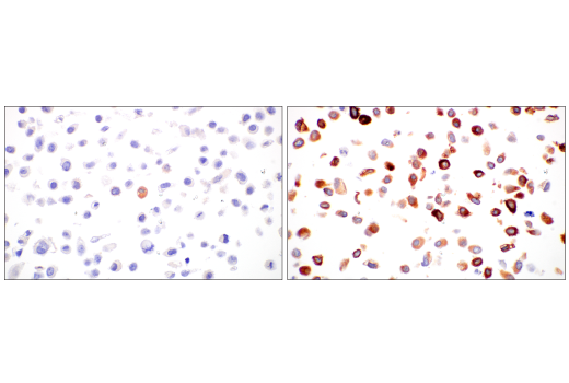
Immunohistochemical analysis of paraffin-embedded HeLa cell pellet, untreated (left) or treated with sodium arsenite (50 μM, 8 hr; right), using HO-1 (E8B7A) XP® Rabbit mAb.