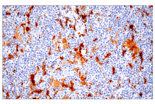
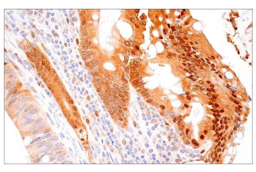
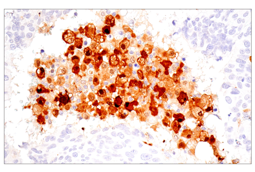
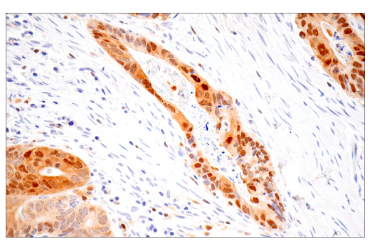
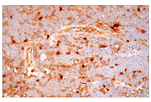
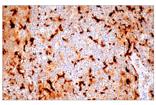
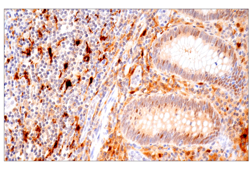
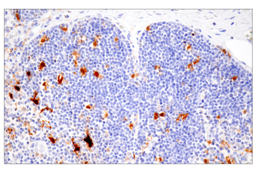
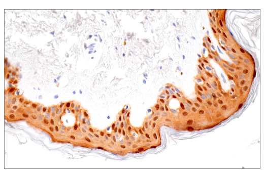
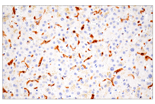
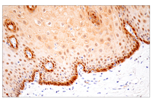
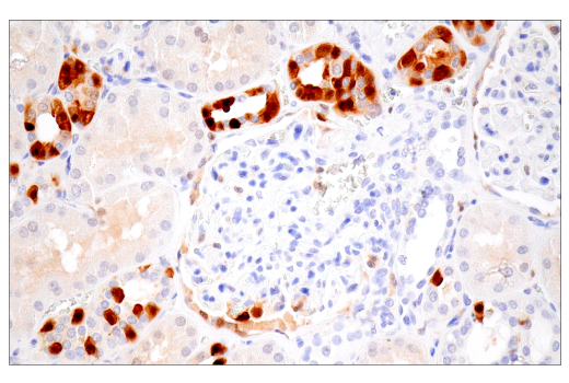
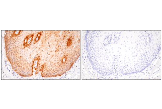
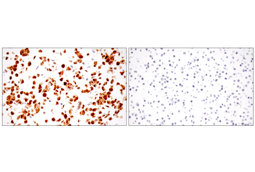
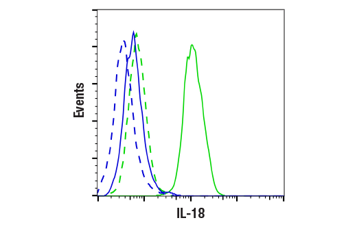
Revision 3

#67775Store at -20C

Cell Signaling Technology

Orders: 877-616-CELL (2355) [email protected]

Support: 877-678-TECH (8324)

Web: [email protected] cellsignal.com

3 Trask LaneDanversMassachusetts01923USA
For Research Use Only. Not for Use in Diagnostic Procedures.
Applications:

WB, IHC-P, FC-FP

REACTIVITY:

H

SENSITIVITY:

Endogenous

MW (kDa):

22

Source/Isotype:

Rabbit IgG

UniProt ID:

#Q14116

Entrez-Gene Id:

3606

Product Information

Product Usage Information

Application Dilution
Western Blotting 1:1000
Immunohistochemistry (Paraffin) 1:200 - 1:800
Flow Cytometry (Fixed/Permeabilized) 1:400 - 1:1600

Storage

Supplied in 10 mM sodium HEPES (pH 7.5), 150 mM NaCl, 100 µg/mL BSA, 50% glycerol, and less than 0.02% sodium azide. Store at –20°C. Do not aliquot the antibody.

For a carrier free (BSA and azide free) version of this product see product #33484.

Specificity / Sensitivity

IL-18 (E3G8R) Rabbit mAb recognizes endogenous levels of total IL-18 protein.

Species Reactivity:

Human

Source / Purification

Monoclonal antibody is produced by immunizing animals with human IL-18 recombinant protein.

Background

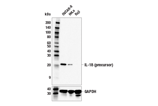
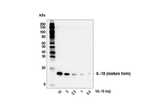
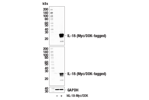
Interleukin-18 (IL-18), originally known as interferon-gamma inducing factor (IGIF), is part of the IL-superfamily of cytokines (1,2). This proinflammatory cytokine is synthesized as an inactive precursor which requires cleavage by Caspase-1 to become an active, mature molecule, which is secreted by monocytes and macrophages (3,4,6). It induces IFNγ production in T-helper-1 (Th1) cells and natural killer (NK) cells, and can regulate innate immunity via both Th1 and Th2 responses (4,5). Elevated IL-18 levels are associated with autoimmune disease, but may be balanced by binding to IL-18 binding protein (IL-18BP), which along with IL-18 neutralizing antibodies, are being examined in clinical trials (6,7).

  1. Okamura, H. et al. (1995) Nature 378, 88-91.
  2. Ushio, S. et al. (1996) J Immunol 156, 4274-9.
  3. Ghayur, T. et al. (1997) Nature 386, 619-23.
  4. Dinarello, C.A. (1999) Methods 19, 121-32.
  5. Nakanishi, K. et al. (2001) Annu Rev Immunol 19, 423-74.
  6. Novick, D. et al. (1999) Immunity 10, 127-36.
  7. Dinarello, C.A. et al. (2013) Front Immunol 4, 289.

Species Reactivity

Species reactivity is determined by testing in at least one approved application (e.g., western blot).

Western Blot Buffer

IMPORTANT: For western blots, incubate membrane with diluted primary antibody in 5% w/v BSA, 1X TBS, 0.1% Tween® 20 at 4°C with gentle shaking, overnight.

Applications Key

WB: Western Blotting IHC-P: Immunohistochemistry (Paraffin) FC-FP: Flow Cytometry (Fixed/Permeabilized)

Cross-Reactivity Key

H: human M: mouse R: rat Hm: hamster Mk: monkey Vir: virus Mi: mink C: chicken Dm: D. melanogaster X: Xenopus Z: zebrafish B: bovine Dg: dog Pg: pig Sc: S. cerevisiae Ce: C. elegans Hr: horse GP: Guinea Pig Rab: rabbit All: all species expected

Trademarks and Patents

Cell Signaling Technology is a trademark of Cell Signaling Technology, Inc.
Alexa Fluor is a registered trademark of Life Technologies Corporation.
All other trademarks are the property of their respective owners. Visit cellsignal.com/trademarks for more information.

Limited Uses

Except as otherwise expressly agreed in a writing signed by a legally authorized representative of CST, the following terms apply to Products provided by CST, its affiliates or its distributors. Any Customer's terms and conditions that are in addition to, or different from, those contained herein, unless separately accepted in writing by a legally authorized representative of CST, are rejected and are of no force or effect.

Products are labeled with For Research Use Only or a similar labeling statement and have not been approved, cleared, or licensed by the FDA or other regulatory foreign or domestic entity, for any purpose. Customer shall not use any Product for any diagnostic or therapeutic purpose, or otherwise in any manner that conflicts with its labeling statement. Products sold or licensed by CST are provided for Customer as the end-user and solely for research and development uses. Any use of Product for diagnostic, prophylactic or therapeutic purposes, or any purchase of Product for resale (alone or as a component) or other commercial purpose, requires a separate license from CST. Customer shall (a) not sell, license, loan, donate or otherwise transfer or make available any Product to any third party, whether alone or in combination with other materials, or use the Products to manufacture any commercial products, (b) not copy, modify, reverse engineer, decompile, disassemble or otherwise attempt to discover the underlying structure or technology of the Products, or use the Products for the purpose of developing any products or services that would compete with CST products or services, (c) not alter or remove from the Products any trademarks, trade names, logos, patent or copyright notices or markings, (d) use the Products solely in accordance with CST Product Terms of Sale and any applicable documentation, and (e) comply with any license, terms of service or similar agreement with respect to any third party products or services used by Customer in connection with the Products.

Revision 3
#67775

IL-18 (E3G8R) Rabbit mAb

Western Blotting Image 1: IL-18 (E3G8R) Rabbit mAb Expand Image
Western blot analysis of extracts from OVCAR-8, HeLa, and Raji cells using IL-18 (E3G8R) Rabbit mAb (upper) or GAPDH (D16H11) XP® Rabbit mAb #5174 (lower). Negative expression of IL-18 protein in Raji cells is consistent with the predicted expression pattern.
Western Blotting Image 2: IL-18 (E3G8R) Rabbit mAb Expand Image
Western blot analysis of recombinant human IL-18 protein (hIL-18) at indicated levels using IL-18 (E3G8R) Rabbit mAb.
Western Blotting Image 3: IL-18 (E3G8R) Rabbit mAb Expand Image
Western blot analysis of extracts from 293T cells, mock transfected (-) or transfected with a construct expressing Myc/DDK-tagged full-length human IL-18 protein (hIL-18-Myc/DDK; +), using IL-18 (E3G8R) Rabbit mAb (upper), Myc-Tag (71D10) Rabbit mAb #2278 (middle), or GAPDH (D16H11) XP® Rabbit mAb #5174 (lower).
Immunohistochemistry Image 1: IL-18 (E3G8R) Rabbit mAb Expand Image

Immunohistochemical analysis of paraffin-embedded human Hodgkin lymphoma using IL-18 (E3G8R) Rabbit mAb.

Immunohistochemistry Image 2: IL-18 (E3G8R) Rabbit mAb Expand Image
Immunohistochemical analysis of paraffin-embedded human colon carcinoma using IL-18 (E3G8R) Rabbit mAb.
Immunohistochemistry Image 3: IL-18 (E3G8R) Rabbit mAb Expand Image
Immunohistochemical analysis of paraffin-embedded human urothelial carcinoma using IL-18 (E3G8R) Rabbit mAb.
Immunohistochemistry Image 4: IL-18 (E3G8R) Rabbit mAb Expand Image

Immunohistochemical analysis of paraffin-embedded human adenocarcinoma of distal esophagus using IL-18 (E3G8R) Rabbit mAb.

Immunohistochemistry Image 5: IL-18 (E3G8R) Rabbit mAb Expand Image
Immunohistochemical analysis of paraffin-embedded human mantle cell lymphoma using IL-18 (E3G8R) Rabbit mAb.
Immunohistochemistry Image 6: IL-18 (E3G8R) Rabbit mAb Expand Image
Immunohistochemical analysis of paraffin-embedded human tonsil using IL-18 (E3G8R) Rabbit mAb.
Immunohistochemistry Image 7: IL-18 (E3G8R) Rabbit mAb Expand Image
Immunohistochemical analysis of paraffin-embedded human appendix using IL-18 (E3G8R) Rabbit mAb.
Immunohistochemistry Image 8: IL-18 (E3G8R) Rabbit mAb Expand Image
Immunohistochemical analysis of paraffin-embedded normal human thymus using IL-18 (E3G8R) Rabbit mAb.
Immunohistochemistry Image 9: IL-18 (E3G8R) Rabbit mAb Expand Image
Immunohistochemical analysis of paraffin-embedded normal human skin using IL-18 (E3G8R) Rabbit mAb.
Immunohistochemistry Image 10: IL-18 (E3G8R) Rabbit mAb Expand Image
Immunohistochemical analysis of paraffin-embedded normal human liver using IL-18 (E3G8R) Rabbit mAb.
Immunohistochemistry Image 11: IL-18 (E3G8R) Rabbit mAb Expand Image
Immunohistochemical analysis of paraffin-embedded normal human esophagus using IL-18 (E3G8R) Rabbit mAb.
Immunohistochemistry Image 12: IL-18 (E3G8R) Rabbit mAb Expand Image
Immunohistochemical analysis of paraffin-embedded normal human kidney using IL-18 (E3G8R) Rabbit mAb.
Immunohistochemistry Image 13: IL-18 (E3G8R) Rabbit mAb Expand Image
Immunohistochemical analysis of paraffin-embedded normal human esophagus using IL-18 (E3G8R) Rabbit mAb (left) compared to concentration-matched Rabbit (DA1E) mAb IgG XP® Isotype Control #3900 (right).
Immunohistochemistry Image 14: IL-18 (E3G8R) Rabbit mAb Expand Image
Immunohistochemical analysis of paraffin-embedded OVCAR-8 cell pellet (left, positive) or Raji cell pellet (right, negative) using IL-18 (E3G8R) Rabbit mAb.
Flow Cytometry Image 1: IL-18 (E3G8R) Rabbit mAb Expand Image
Flow cytometric analysis of Raji cells (blue, negative) and OVCAR-8 cells (green, positive) using IL-18 (E3G8R) Rabbit mAb (solid lines) or concentration-matched Rabbit (DA1E) mAb IgG XP® Isotype Control #3900 (dashed lines). Anti-rabbit IgG (H+L), F(ab')2 Fragment (Alexa Fluor® 488 Conjugate) #4412 was used as a secondary antibody.