Revision 7
#12242
Store at -20C
877-616-CELL (2355)
877-678-TECH (8324)
3 Trask Lane | Danvers | Massachusetts | 01923 | USA
For Research Use Only. Not for Use in Diagnostic Procedures.
Applications:
W, W-S, IHC-P
Reactivity:
H M
Sensitivity:
Endogenous
MW (kDa):
17,31
Source/Isotype:
Mouse IgG1
UniProt ID:
#P01584
Entrez-Gene Id:
3553
Product Usage Information
| Application | Dilution |
|---|---|
| Western Blotting | 1:1000 |
| Simple Western™ | 1:10 - 1:50 |
| Immunohistochemistry (Paraffin) | 1:50 - 1:200 |
Storage
For a carrier free (BSA and azide free) version of this product see product #27989.
Specificity/Sensitivity
IL-1β (3A6) Mouse mAb recognizes endogenous levels of total IL-1β protein.
Source / Purification
Monoclonal antibody is produced by immunizing animals with recombinant human IL-1β protein.
Background
Interleukin-1β (IL-1β), one of the major caspase-1 targets, is a multifunctional cytokine that is involved in a host of immune and proinflammatory responses (1). It is produced primarily by activated monocytes and macrophages. It signals through various adaptor proteins and kinases that lead to activation of numerous downstream targets (2-6). Human IL-1β is synthesized as a 31 kDa precursor. To gain activity, the precursor must be cleaved by caspase-1 between Asp116 and Ala117 to yield a 17 kDa mature form (7,8). Detection of the 17 kDa mature form of IL-1β is a good indicator of caspase-1 activity.
Background References
- Dinarello, C.A. (1998) Int Rev Immunol 16, 457-99.
- Burns, K. et al. (1998) J Biol Chem 273, 12203-9.
- Cao, Z. et al. (1996) Nature 383, 443-6.
- Cao, Z. et al. (1996) Science 271, 1128-31.
- Wesche, H. et al. (1997) Immunity 7, 837-47.
- Ninomiya-Tsuji, J. et al. (1999) Nature 398, 252-6.
- Thornberry, N.A. et al. (1992) Nature 356, 768-74.
- Cerretti, D.P. et al. (1992) Science 256, 97-100.
Species Reactivity
Species reactivity is determined by testing in at least one approved application (e.g., western blot).
Western Blot Buffer
IMPORTANT: For western blots, incubate membrane with diluted primary antibody in 5% w/v BSA, 1X TBS, 0.1% Tween® 20 at 4°C with gentle shaking, overnight.
Applications Key
W: Western Blotting IHC-P: Immunohistochemistry (Paraffin)
Cross-Reactivity Key
H: Human M: Mouse R: Rat Hm: Hamster Mk: Monkey Vir: Virus Mi: Mink C: Chicken Dm: D. melanogaster X: Xenopus Z: Zebrafish B: Bovine Dg: Dog Pg: Pig Sc: S. cerevisiae Ce: C. elegans Hr: Horse GP: Guinea Pig Rab: Rabbit G: Goat All: All Species Expected
Trademarks and Patents
Cell Signaling Technology is a trademark of Cell Signaling Technology, Inc.
All other trademarks are the property of their respective owners. Visit cellsignal.com/trademarks for more information.
Limited Uses
Except as otherwise expressly agreed in a writing signed by a legally authorized representative of CST, the following terms apply to Products provided by CST, its affiliates or its distributors. Any Customer's terms and conditions that are in addition to, or different from, those contained herein, unless separately accepted in writing by a legally authorized representative of CST, are rejected and are of no force or effect.
Products are labeled with For Research Use Only or a similar labeling statement and have not been approved, cleared, or licensed by the FDA or other regulatory foreign or domestic entity, for any purpose. Customer shall not use any Product for any diagnostic or therapeutic purpose, or otherwise in any manner that conflicts with its labeling statement. Products sold or licensed by CST are provided for Customer as the end-user and solely for research and development uses. Any use of Product for diagnostic, prophylactic or therapeutic purposes, or any purchase of Product for resale (alone or as a component) or other commercial purpose, requires a separate license from CST. Customer shall (a) not sell, license, loan, donate or otherwise transfer or make available any Product to any third party, whether alone or in combination with other materials, or use the Products to manufacture any commercial products, (b) not copy, modify, reverse engineer, decompile, disassemble or otherwise attempt to discover the underlying structure or technology of the Products, or use the Products for the purpose of developing any products or services that would compete with CST products or services, (c) not alter or remove from the Products any trademarks, trade names, logos, patent or copyright notices or markings, (d) use the Products solely in accordance with CST Product Terms of Sale and any applicable documentation, and (e) comply with any license, terms of service or similar agreement with respect to any third party products or services used by Customer in connection with the Products.
Revision 7
#12242
IL-1β (3A6) Mouse mAb
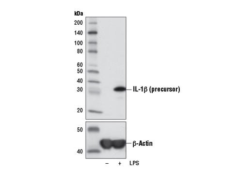
Western blot analysis of extracts from THP-1 cells, untreated (-) or LPS-treated (100 ng/mL, 3 hr; +), using IL-1β (3A6) Mouse mAb (upper) or β-Actin (D6A8) Rabbit mAb #8457 (lower).
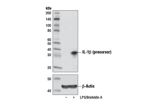
Western blot analysis of extracts from Raw 264.7 cells, untreated (-) or treated with Brefeldin A #9972 (300 ng/mL, last 3 hr of stimulation; +) and LPS (100 ng/mL, 6 hr; +), using IL-1β (3A6) Mouse mAb (upper) or β-Actin (D6A8) Rabbit mAb #8457 (lower).
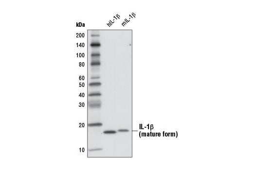
Western blot analysis of 1 ng recombinant Human Interleukin-1β (hIL-1β) #8900 and 1 ng recombinant Mouse Interleukin-1β (mIL-1β) #5204 using IL-1β (3A6) Mouse mAb.
Revision 7
#12242
IL-1β (3A6) Mouse mAb
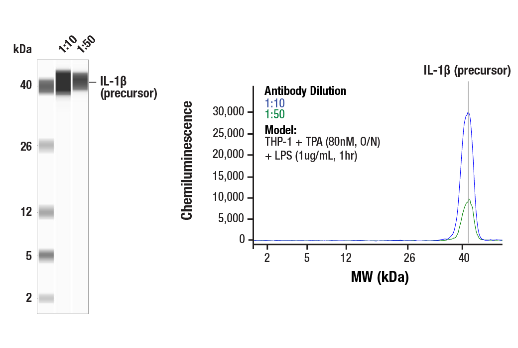
Simple Western™ analysis of lysates (1.0 mg/mL) from THP-1 cells treated with TPA (80nM, O/N) and LPS (1ug/ml, 1 hr) using IL-1β (3A6) Mouse mAb #12242. The virtual lane view (left) shows the target band (as indicated) at 1:10 and 1:50 dilutions of primary antibody. The corresponding electropherogram view (right) plots chemiluminescence by molecular weight along the capillary at 1:10 (blue line) and 1:50 (green line) dilutions of primary antibody. This experiment was performed under reducing conditions on the Jess™ Simple Western instrument from ProteinSimple, a BioTechne brand, using the 2-40 kDa separation module.
Immunohistochemical analysis of paraffin-embedded human large intestine (chronic colitis of the colon) using IL-1-β (3A6) Mouse mAb.
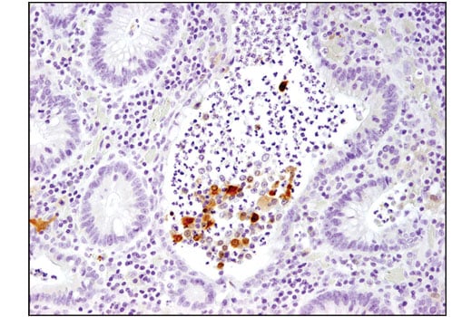
Immunohistochemical analysis of paraffin-embedded human large intestine (ulcerative chronic colitis of the rectum) using IL-1-β (3A6) Mouse mAb.
Revision 7
#12242
IL-1β (3A6) Mouse mAb
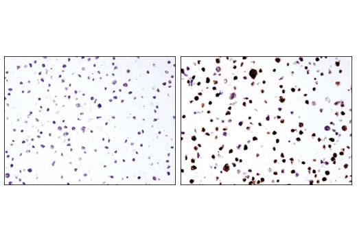
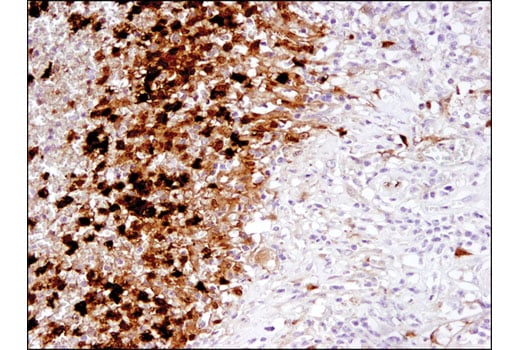
Immunohistochemical analysis of paraffin embedded THP-1 cell pellets, control (left) or LPS-treated (right) using IL-1-β (3A6) Mouse mAb.