| Product Includes | Product # | Quantity | Mol. Wt | Isotype/Source |
|---|---|---|---|---|
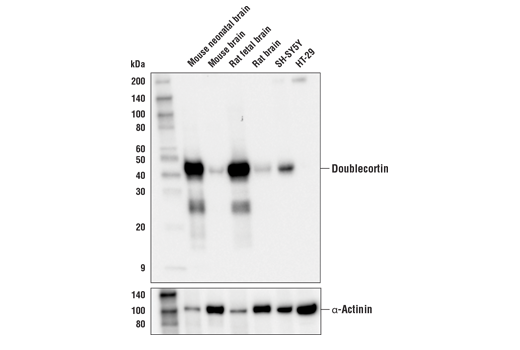
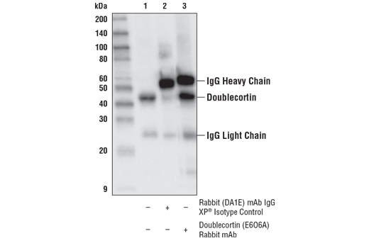
| Doublecortin (E6O6A) Rabbit mAb | 91954 | 20 µl | 45 kDa | Rabbit IgG |
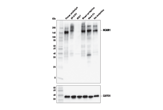
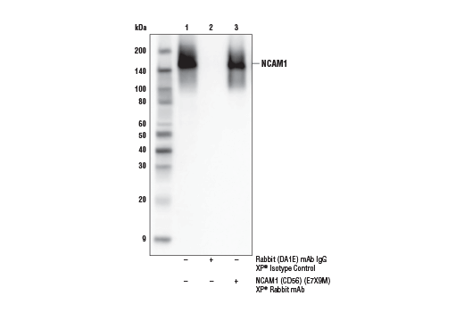
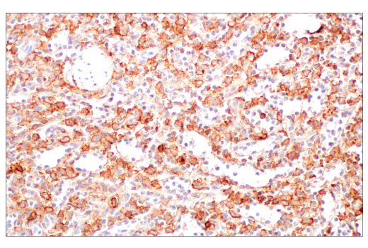
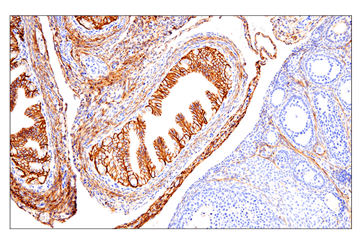
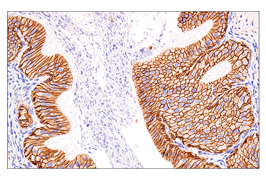
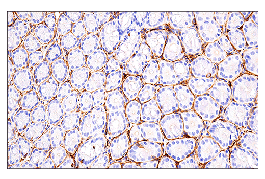
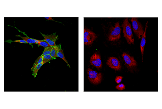
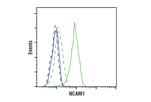
| NCAM1 (CD56) (E7X9M) XP® Rabbit mAb | 99746 | 20 µl | 120 to 220 kDa | Rabbit IgG |
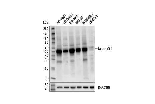
| NeuroD1 (D90G12) Rabbit mAb | 7019 | 20 µl | 49 kDa | Rabbit IgG |
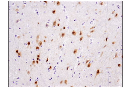
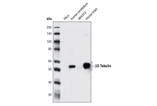
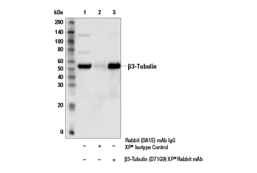
| β3-Tubulin (D71G9) XP® Rabbit mAb | 5568 | 20 µl | 55 kDa | Rabbit IgG |
| TBR1 (D6C6X) Rabbit mAb | 49661 | 20 µl | 74 kDa | Rabbit IgG |
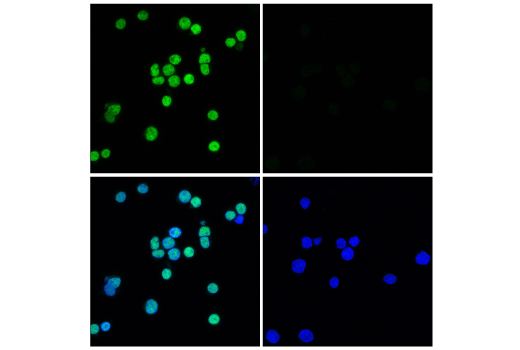
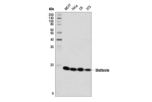
| Stathmin (D1Y5A) Rabbit mAb | 13655 | 20 µl | 19 kDa | Rabbit IgG |
| Anti-rabbit IgG, HRP-linked Antibody | 7074 | 100 µl | Goat |
Please visit cellsignal.com for individual component applications, species cross-reactivity, dilutions, protocols, and additional product information.
Description
The Immature Neuron Marker Antibody Sampler Kit provides an economical means for detecting immature neuron proteins by western blot. The kit includes enough antibodies to perform two western blot experiments with each primary antibody.
Storage
Background
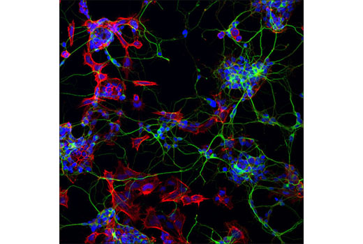
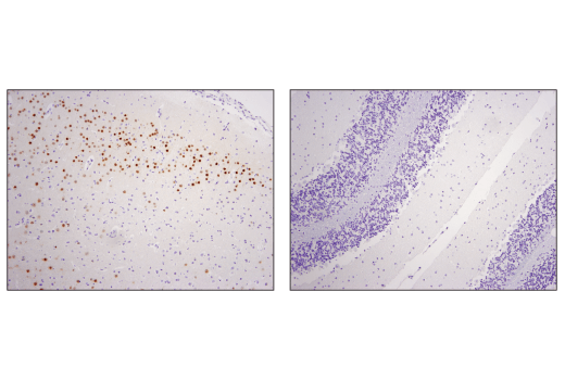
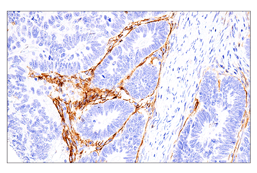
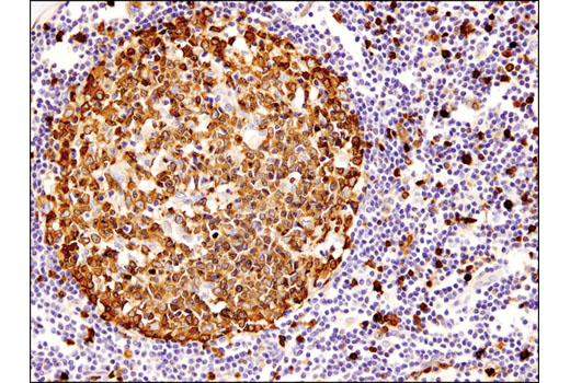
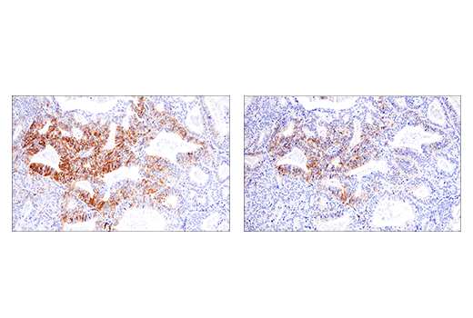
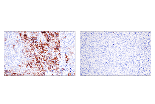
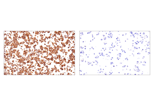
The antibodies in this kit serve to characterize and identify immature neurons. During development, radial glia (RG) cells located in the ventricular zone (VZ) of the brain divide asymmetrically, each producing a neuronal and RG daughter cell. The daughter RG cell is also known as a neural progenitor cell (NPC) or an intermediate progenitor cell (IPC). Newly formed IPCs migrate to the subventricular zone (SVZ) where they divide symmetrically, each giving rise to two post-mitotic neurons that can then migrate to their final destination. In adulthood, NPCs reside within the subgranular zone (SGZ) of the dentate gyrus, and the adult SVZ, which surrounds the lateral ventricles of the cerebral cortex. NPCs within the SGZ and SVZ divide and give rise to immature neurons (1). The cytoskeleton of these cells plays an important role in generating neuronal processes. The cytoskeleton consists of three types of cytosolic fibers: actin microfilaments, intermediate filaments, and microtubules. β3-tubulin is one of six β-tubulin isoforms that make up the building blocks of microtubules (2). Stathmin is a tubulin binding protein that regulates microtubule dynamics in a phosphorylation dependent manner. Stathmin is heavily expressed during neuronal development, mediating differentiation and synaptic plasticity (3,4). Doublecortin is a microtubule-associated protein that facilitates neurite outgrowth and cell migration (5). The dual expression of doublecortin and NCAM (neural cell adhesion molecule, CD56), combined with the lack of expression of mature neuronal markers, is evidence of an immature neuronal phenotype (6). NCAM mediates neuronal attachment, neurite extension, and cell to cell interactions through homo and heterophilic interactions. Polysialic acid (PSA) post-translational modification of NCAM disrupts cell to cell adhesion, promoting axonal growth, cell migration, and synaptic plasticity during neurogenesis (7-9).
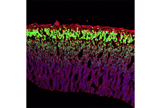
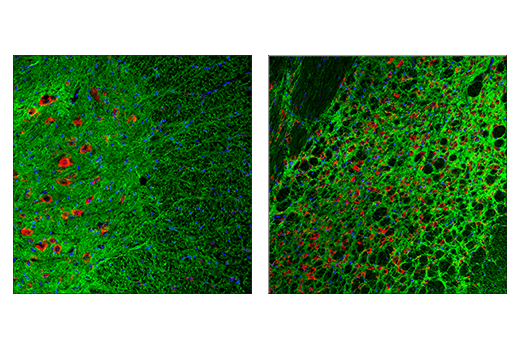
Transcription factors also play a key role in immature neuron growth and differentiation. NeuroD1 is a member of the basic helix-loop-helix (bHLH) family of transcription factors. These proteins function by forming heterodimers with E-proteins and binding to the canonical E-box sequence CANNTG (10,11). Neuronal activity results in CaMKII-mediated phosphorylation of NeuroD1 at Ser336, which is necessary for the formation and growth of dendrites (12,13). T-box, brain, 1 (TBR1) is a transcription factor important in vertebrate embryo development. As a member of the T-Box family of transcription factors, TBR1 is expressed in postmitotic glutamatergic projection neurons (14). During cortical neurogenesis, sequential expression of transcription factors Pax6, TBR2, and TBR1 regulates discrete steps in projection neuron differentiation (15).
- Martínez-Cerdeño, V. and Noctor, S.C. (2018) Front Neuroanat 12, 104.
- Jiang, Y.Q. and Oblinger, M.M. (1992) J Cell Sci 103 (Pt 3), 643-51.
- Chauvin, S. and Sobel, A. (2015) Prog Neurobiol 126, 1-18.
- Uchida, S. et al. (2014) Nat Commun 5, 4389.
- Reiner, O. et al. (2004) Cell Cycle 3, 747-51.
- Coviello, S. et al. (2022) Front Neuroanat 16, 851432.
- Seidenfaden, R. et al. (2003) Mol Cell Biol 23, 5908-18.
- Bonfanti, L. and Seki, T. (2021) Cells 10, 2542.
- Wędzony, K. et al. (2013) Pharmacol Rep 65, 1471-8.
- Schonhoff, S.E. et al. (2004) Endocrinology 145, 2639-44.
- Sharma, A. et al. (1999) Mol Cell Biol 19, 704-13.
- Chae, J.H. et al. (2004) Mol Cells 18, 271-88.
- Gaudillière, B. et al. (2004) Neuron 41, 229-41.
- Hevner, R.F. et al. (2001) Neuron 29, 353-66.
- Englund, C. et al. (2005) J Neurosci 25, 247-51.
Background References
Trademarks and Patents
Limited Uses
Except as otherwise expressly agreed in a writing signed by a legally authorized representative of CST, the following terms apply to Products provided by CST, its affiliates or its distributors. Any Customer's terms and conditions that are in addition to, or different from, those contained herein, unless separately accepted in writing by a legally authorized representative of CST, are rejected and are of no force or effect.
Products are labeled with For Research Use Only or a similar labeling statement and have not been approved, cleared, or licensed by the FDA or other regulatory foreign or domestic entity, for any purpose. Customer shall not use any Product for any diagnostic or therapeutic purpose, or otherwise in any manner that conflicts with its labeling statement. Products sold or licensed by CST are provided for Customer as the end-user and solely for research and development uses. Any use of Product for diagnostic, prophylactic or therapeutic purposes, or any purchase of Product for resale (alone or as a component) or other commercial purpose, requires a separate license from CST. Customer shall (a) not sell, license, loan, donate or otherwise transfer or make available any Product to any third party, whether alone or in combination with other materials, or use the Products to manufacture any commercial products, (b) not copy, modify, reverse engineer, decompile, disassemble or otherwise attempt to discover the underlying structure or technology of the Products, or use the Products for the purpose of developing any products or services that would compete with CST products or services, (c) not alter or remove from the Products any trademarks, trade names, logos, patent or copyright notices or markings, (d) use the Products solely in accordance with CST Product Terms of Sale and any applicable documentation, and (e) comply with any license, terms of service or similar agreement with respect to any third party products or services used by Customer in connection with the Products.