Revision 6
#98344
Store at -20C
877-616-CELL (2355)
877-678-TECH (8324)
3 Trask Lane | Danvers | Massachusetts | 01923 | USA
For Research Use Only. Not for Use in Diagnostic Procedures.
Applications:
W, W-S, IP, IF-F, IF-IC, FC-FP, ChIP
Reactivity:
H M R
Sensitivity:
Endogenous
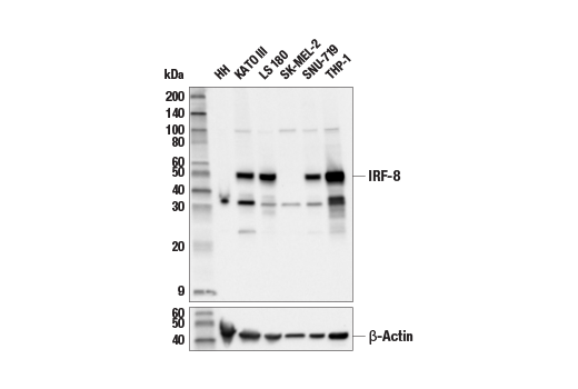
MW (kDa):
50
Source/Isotype:
Rabbit IgG
UniProt ID:
#Q02556
Entrez-Gene Id:
3394
Product Usage Information
| Application | Dilution |
|---|---|
| Western Blotting | 1:1000 |
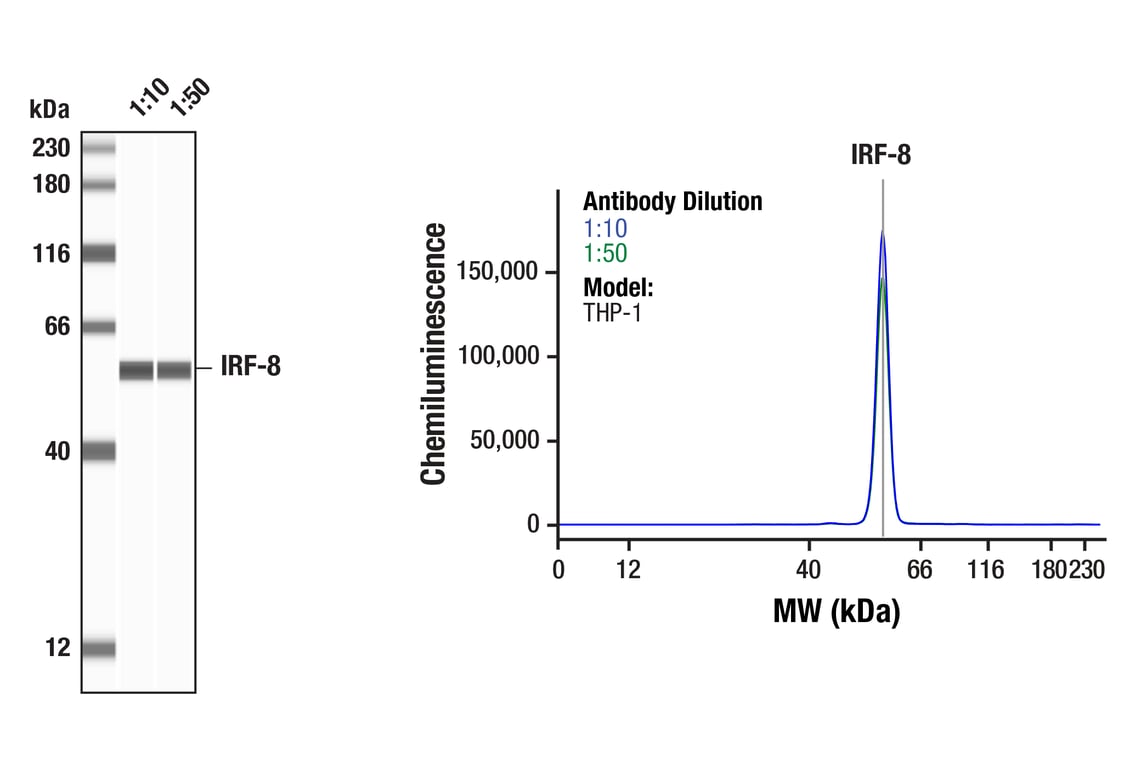
| Simple Western™ | 1:10 - 1:50 |
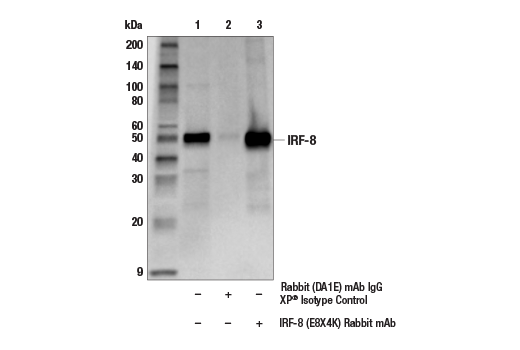
| Immunoprecipitation | 1:100 |
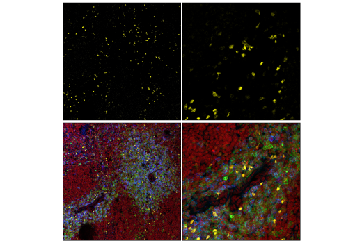
| Immunofluorescence (Frozen) | 1:100 |
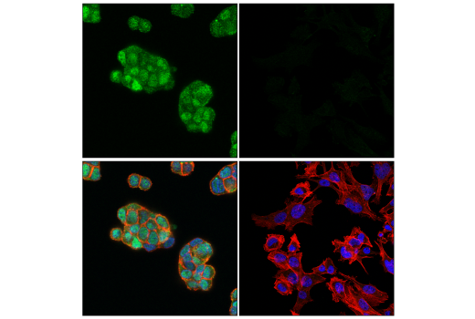
| Immunofluorescence (Immunocytochemistry) | 1:800 |
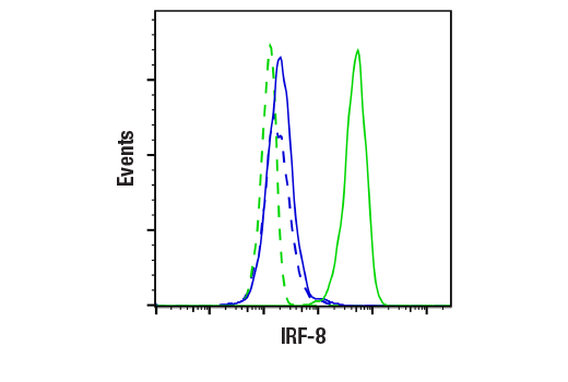
| Flow Cytometry (Fixed/Permeabilized) | 1:200 - 1:800 |
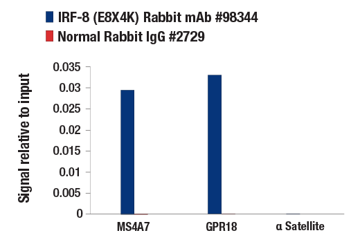
| Chromatin IP | 1:50 |
Storage
For a carrier free (BSA and azide free) version of this product see product #81517.
Specificity/Sensitivity
Source / Purification
Background
IRF-8/ICSCP is expressed predominately in hematopoietic cells and is further increased upon treatment with interferon (3,4). IRF-8 can function as a transcription repressor of ICS-containing promoters (4). Expression of IRF-8 can lead to the downregulation of the anti-apoptotic protein Bcl-2 (5). Originally described as being induced by IFN-γ, IRF-8 expression is also elevated by IRF-α as well as IL-12 in NK and T cells (6). IRF-8 deficient mice have enhanced susceptibility to various pathogens and impaired production of interferons, as well as deregulated hematopoiesis that resembles chronic myelogenous leukemia (7,8). IRF-8 also regulates bone metabolism by suppressing osteoclast formation (9).
Background References
- Taniguchi, T. et al. (2001) Annu Rev Immunol 19, 623-55.
- Honda, K. and Taniguchi, T. (2006) Nat Rev Immunol 6, 644-58.
- Driggers, P.H. et al. (1990) Proc Natl Acad Sci U S A 87, 3743-7.
- Weisz, A. et al. (1992) J Biol Chem 267, 25589-96.
- Burchert, A. et al. (2004) Blood 103, 3480-9.
- Lehtonen, A. et al. (2003) Cytokine 24, 81-90.
- Holtschke, T. et al. (1996) Cell 87, 307-17.
- Fehr, T. et al. (1997) J Exp Med 185, 921-31.
- Zhao, B. et al. (2009) Nat Med 15, 1066-71.
Species Reactivity
Species reactivity is determined by testing in at least one approved application (e.g., western blot).
Western Blot Buffer
IMPORTANT: For western blots, incubate membrane with diluted primary antibody in 5% w/v BSA, 1X TBS, 0.1% Tween® 20 at 4°C with gentle shaking, overnight.
Applications Key
W: Western Blotting W-S: Simple Western™ IP: Immunoprecipitation IF-F: Immunofluorescence (Frozen) IF-IC: Immunofluorescence (Immunocytochemistry) FC-FP: Flow Cytometry (Fixed/Permeabilized) ChIP: Chromatin IP
Cross-Reactivity Key
H: Human M: Mouse R: Rat
Trademarks and Patents
Cell Signaling Technology is a trademark of Cell Signaling Technology, Inc.
Alexa Fluor is a registered trademark of Life Technologies Corporation.
All other trademarks are the property of their respective owners. Visit cellsignal.com/trademarks for more information.
Limited Uses
Except as otherwise expressly agreed in a writing signed by a legally authorized representative of CST, the following terms apply to Products provided by CST, its affiliates or its distributors. Any Customer's terms and conditions that are in addition to, or different from, those contained herein, unless separately accepted in writing by a legally authorized representative of CST, are rejected and are of no force or effect.
Products are labeled with For Research Use Only or a similar labeling statement and have not been approved, cleared, or licensed by the FDA or other regulatory foreign or domestic entity, for any purpose. Customer shall not use any Product for any diagnostic or therapeutic purpose, or otherwise in any manner that conflicts with its labeling statement. Products sold or licensed by CST are provided for Customer as the end-user and solely for research and development uses. Any use of Product for diagnostic, prophylactic or therapeutic purposes, or any purchase of Product for resale (alone or as a component) or other commercial purpose, requires a separate license from CST. Customer shall (a) not sell, license, loan, donate or otherwise transfer or make available any Product to any third party, whether alone or in combination with other materials, or use the Products to manufacture any commercial products, (b) not copy, modify, reverse engineer, decompile, disassemble or otherwise attempt to discover the underlying structure or technology of the Products, or use the Products for the purpose of developing any products or services that would compete with CST products or services, (c) not alter or remove from the Products any trademarks, trade names, logos, patent or copyright notices or markings, (d) use the Products solely in accordance with CST Product Terms of Sale and any applicable documentation, and (e) comply with any license, terms of service or similar agreement with respect to any third party products or services used by Customer in connection with the Products.
Revision 6
#98344
IRF-8 (E8X4K) Rabbit mAb
Revision 6
#98344
IRF-8 (E8X4K) Rabbit mAb
Revision 6
#98344
IRF-8 (E8X4K) Rabbit mAb