| Product Includes | Product # | Quantity | Mol. Wt | Isotype/Source |
|---|---|---|---|---|
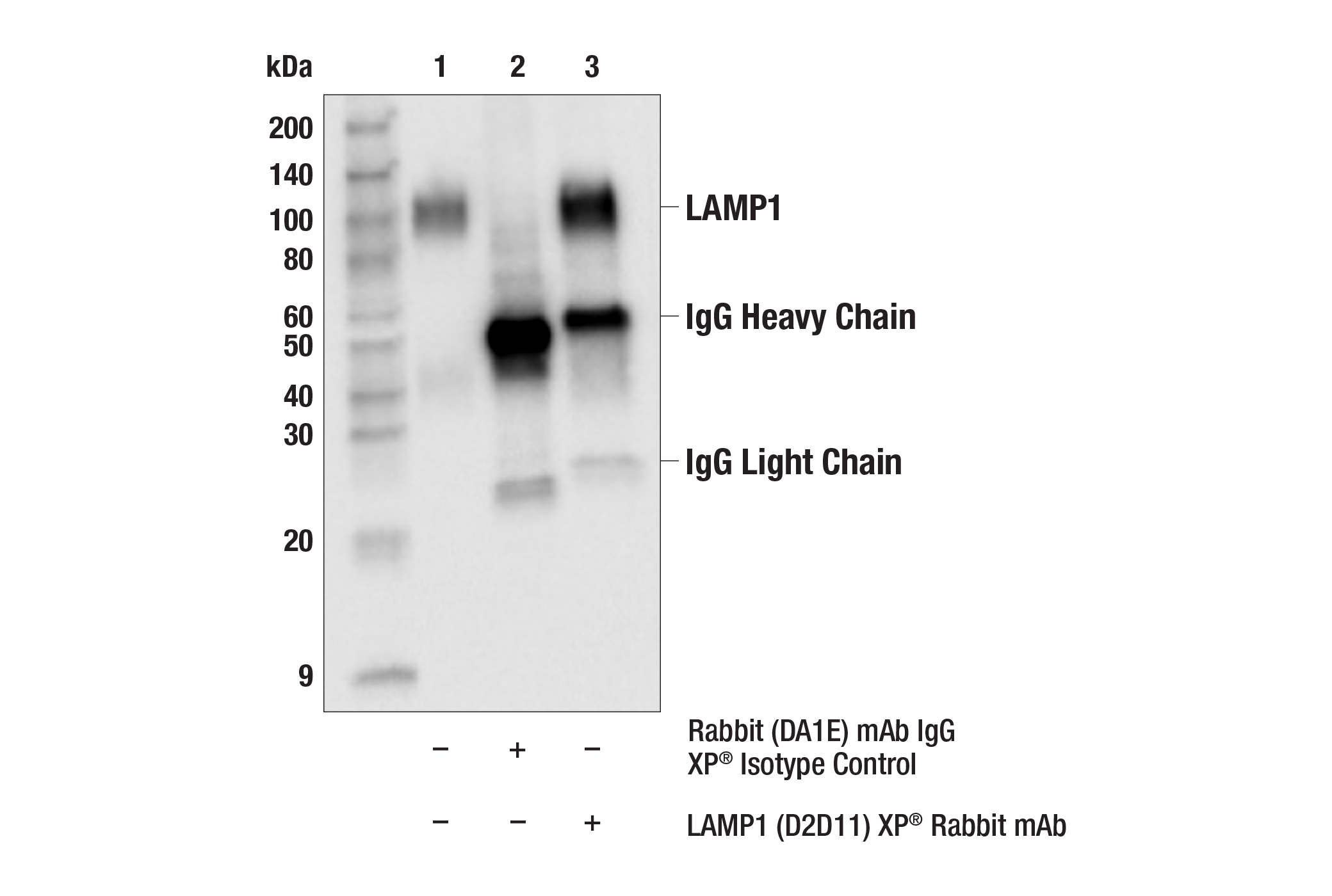
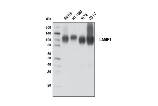
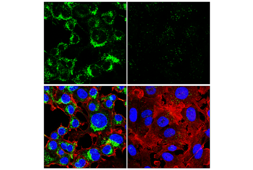
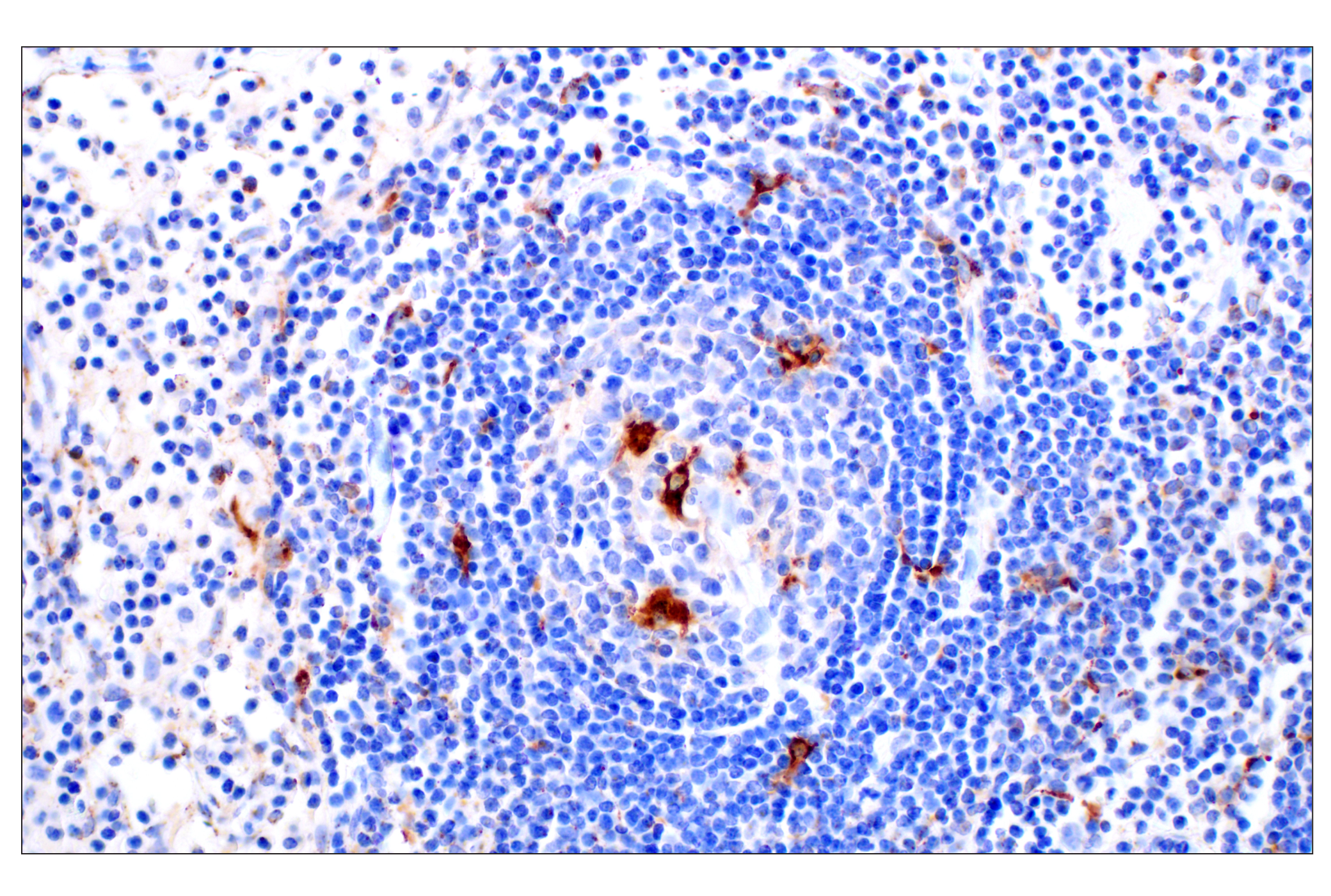
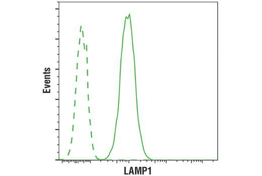
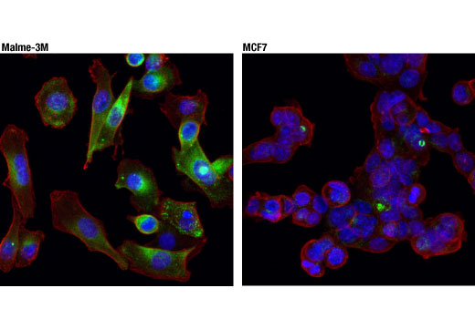
| LAMP1 (D2D11) XP® Rabbit mAb | 9091 | 20 µl | 42 (non-glycosylated), 90-120 (glycosylated) kDa | Rabbit IgG |
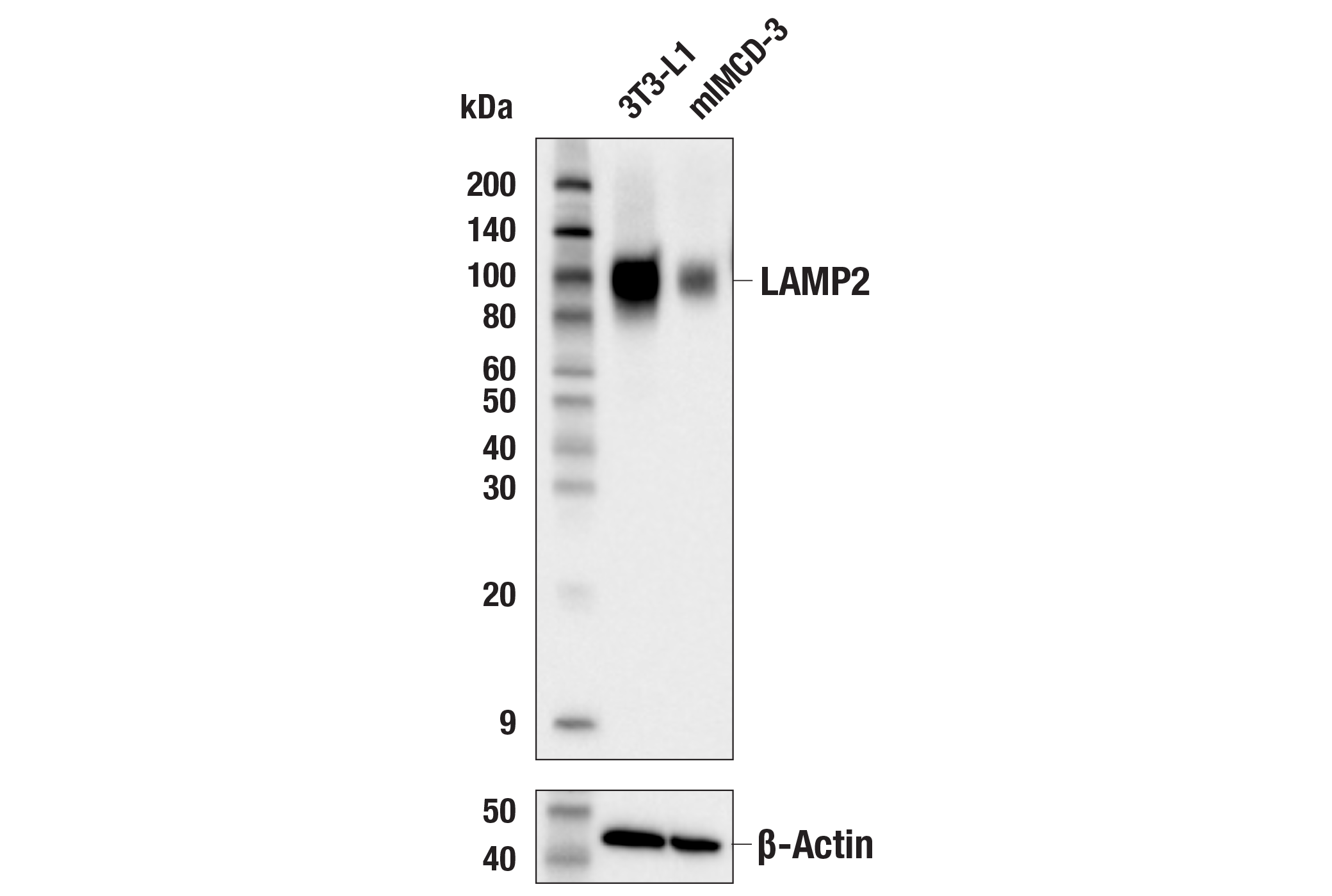
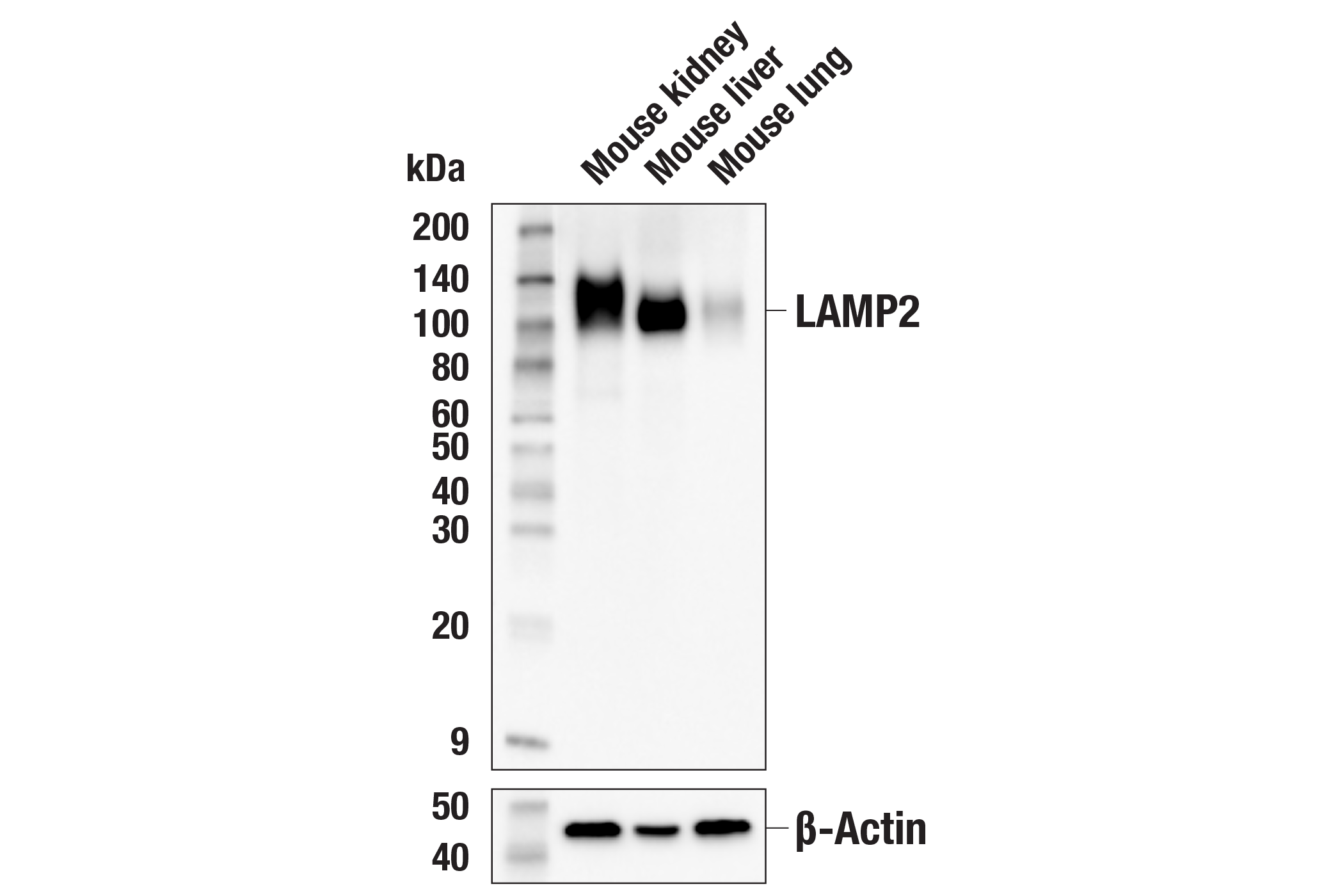
| LAMP2 (E6A6S) Rabbit mAb | 34141 | 20 µl | 100-130 kDa | Rabbit IgG |
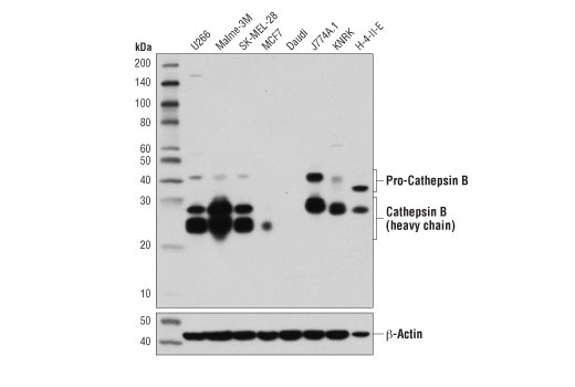
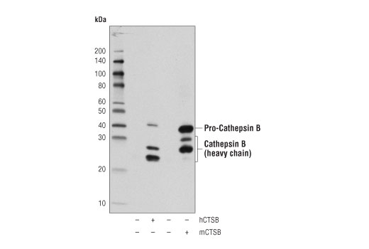
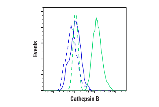
| Cathepsin B (D1C7Y) XP® Rabbit mAb | 31718 | 20 µl | 44, 27, 24 kDa | Rabbit IgG |
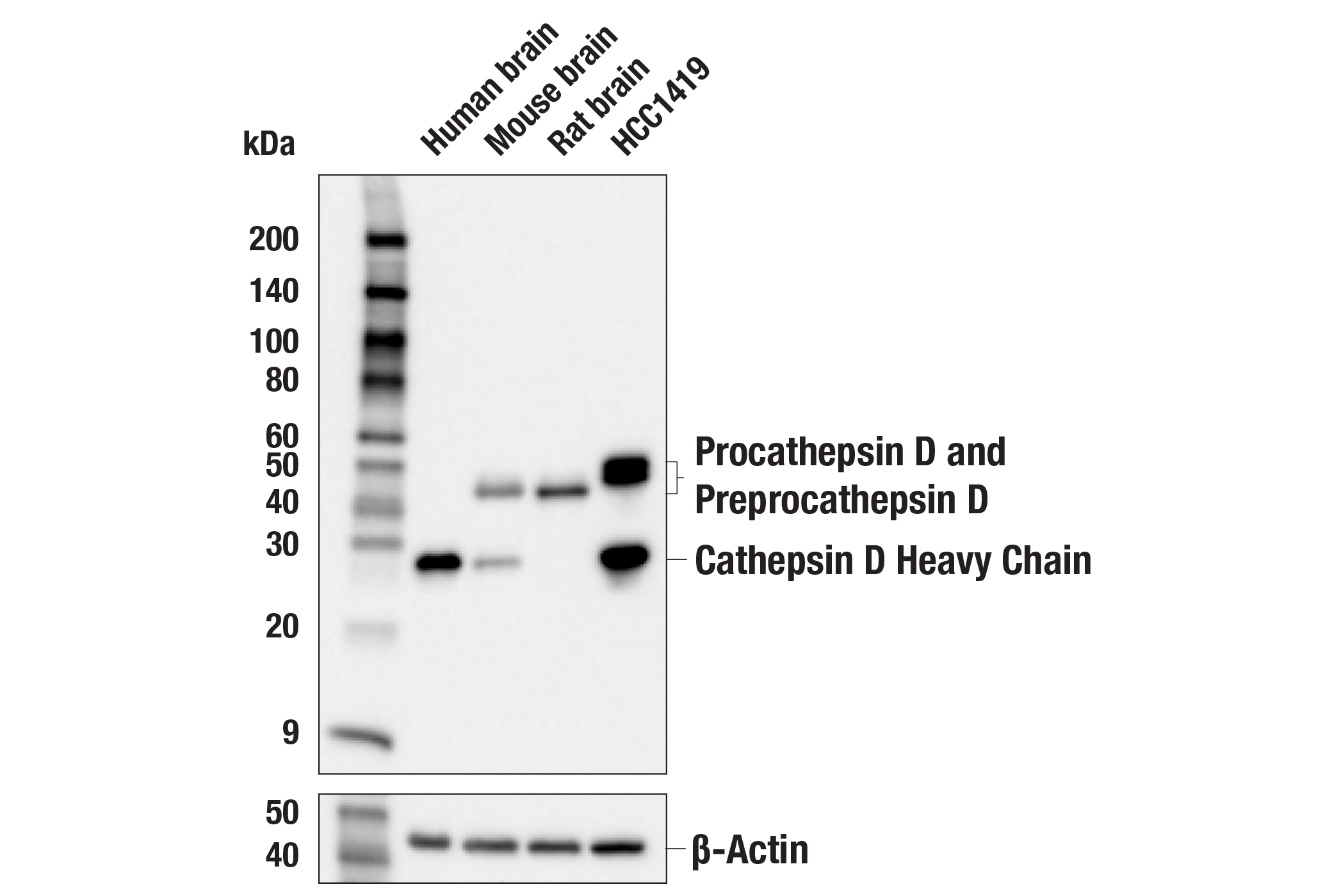
| Cathepsin D (E5V4H) Rabbit mAb | 74089 | 20 µl | 46, 43, 28 kDa | Rabbit IgG |
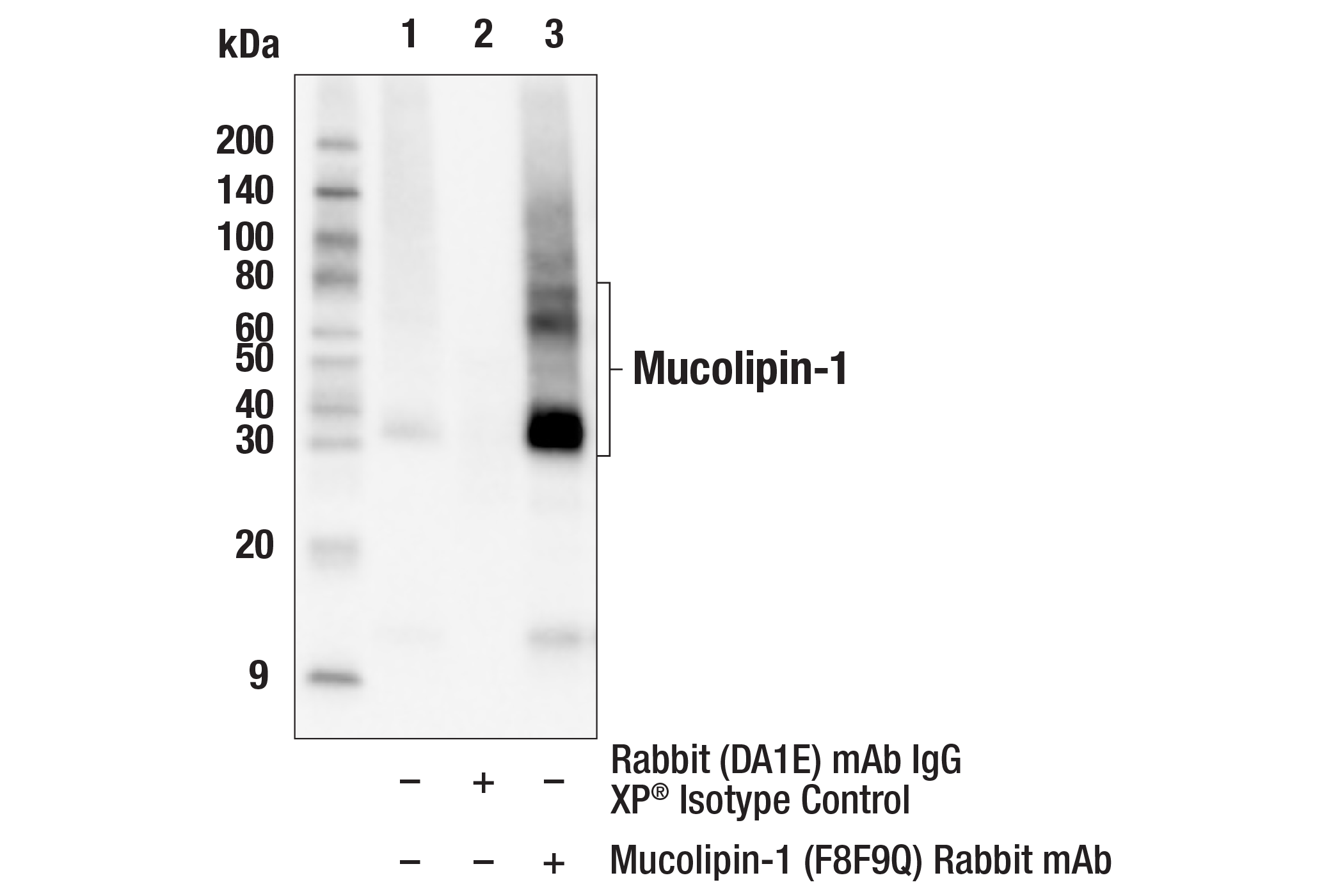
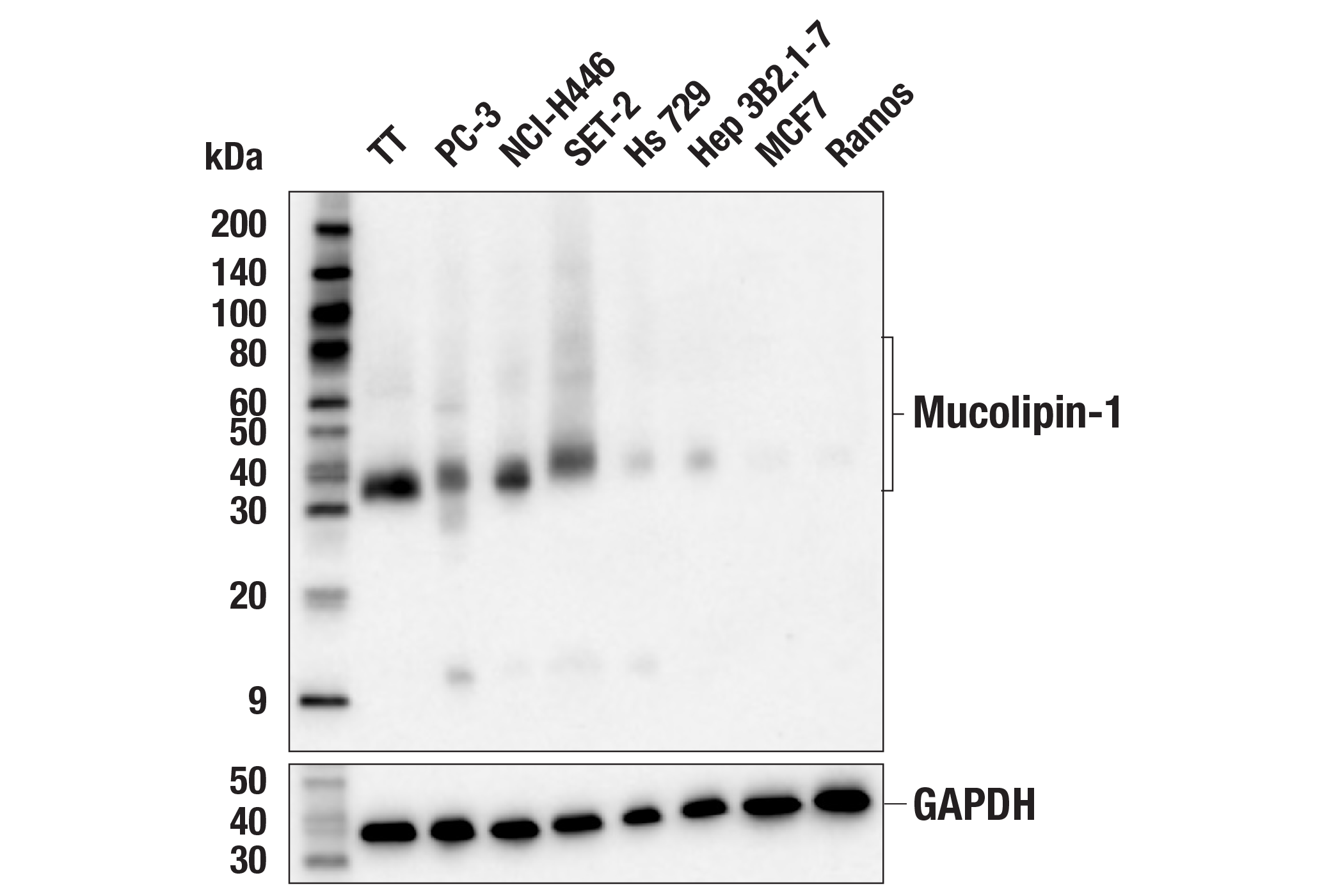
| Mucolipin-1 (F8F9Q) Rabbit mAb | 27748 | 20 µl | 35-40, 65 kDa | Rabbit IgG |
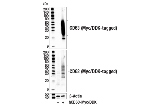
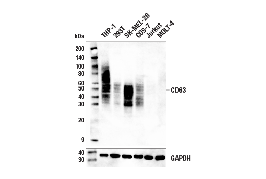
| CD63 (E1W3T) Rabbit mAb | 52090 | 20 µl | 25-60 kDa | Rabbit IgG |
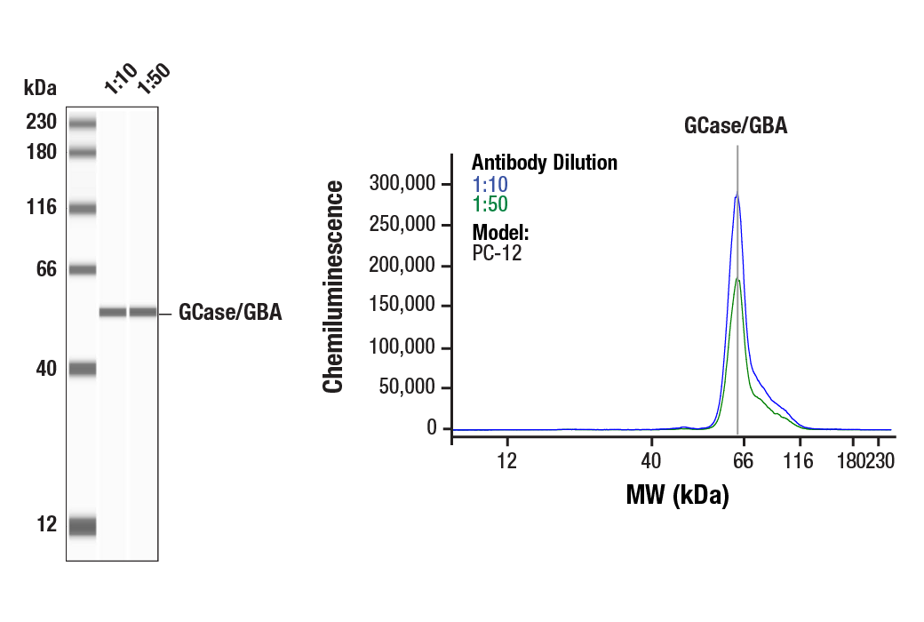
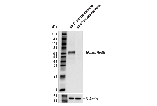
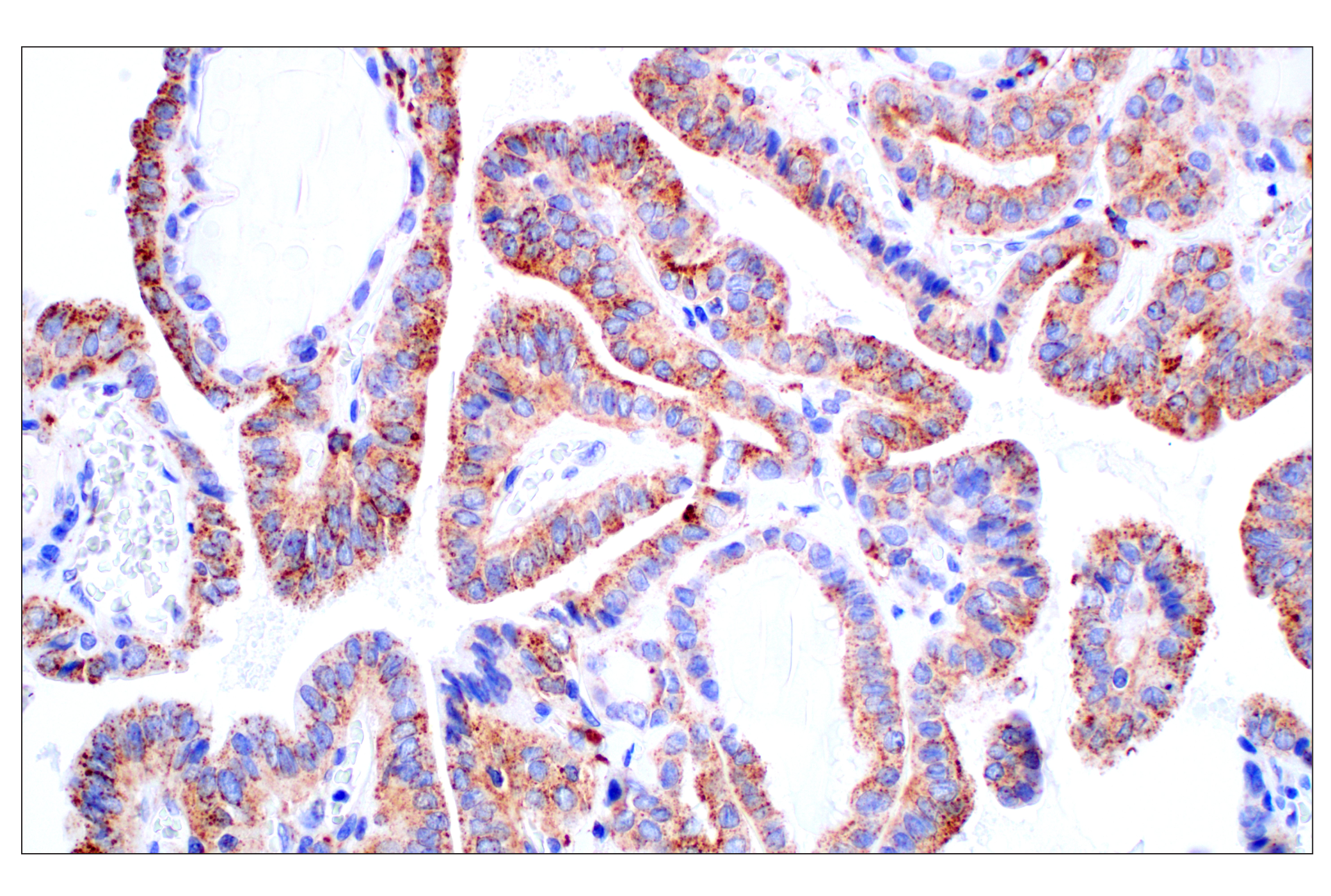
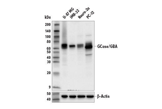
| GCase/GBA (E2R1L) Rabbit mAb | 88162 | 20 µl | 65 kDa | Rabbit IgG |
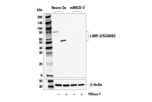
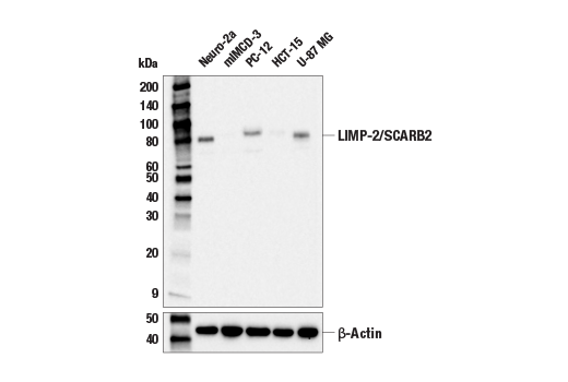
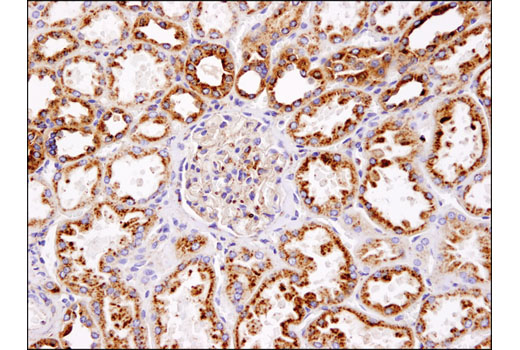
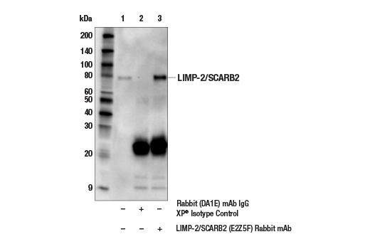
| LIMP-2/SCARB2 (E2Z5F) Rabbit mAb | 27960 | 20 µl | 54, 80 kDa | Rabbit IgG |
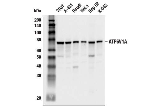
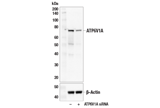
| ATP6V1A (E5N9E) Rabbit mAb | 39517 | 20 µl | 75 kDa | Rabbit IgG |
| Anti-rabbit IgG, HRP-linked Antibody | 7074 | 100 µl | Goat |
Please visit cellsignal.com for individual component applications, species cross-reactivity, dilutions, protocols, and additional product information.
Description
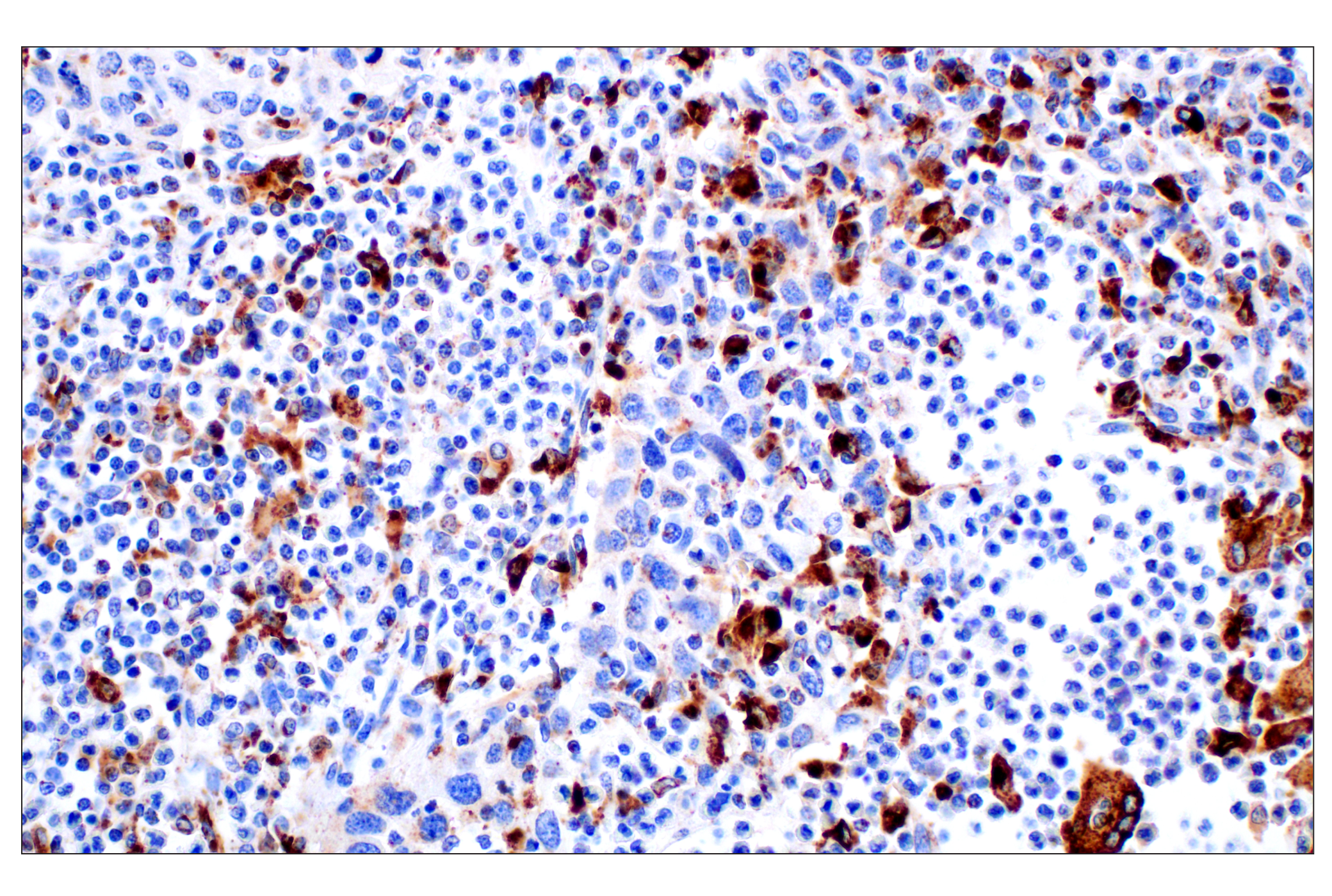
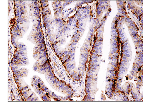
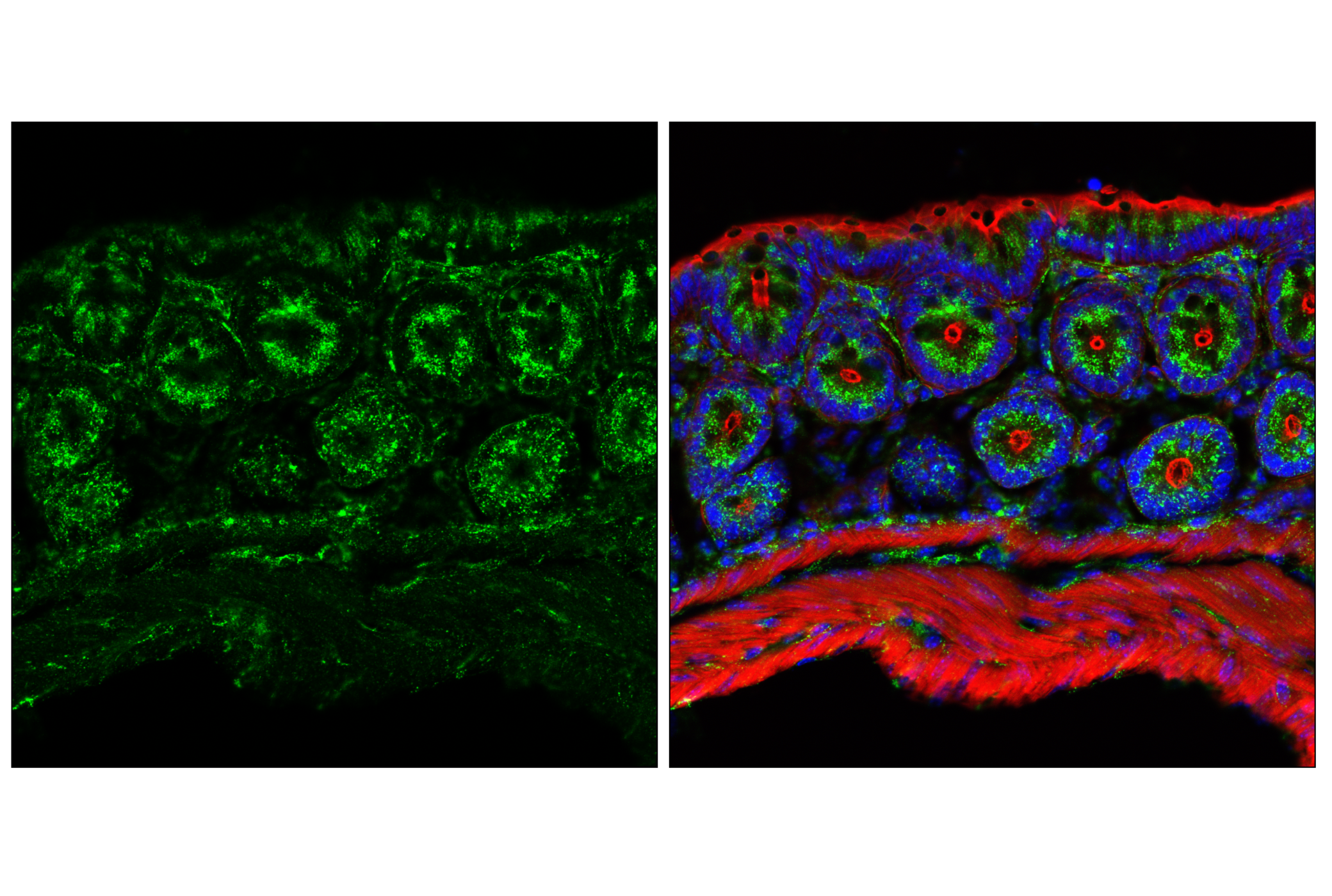
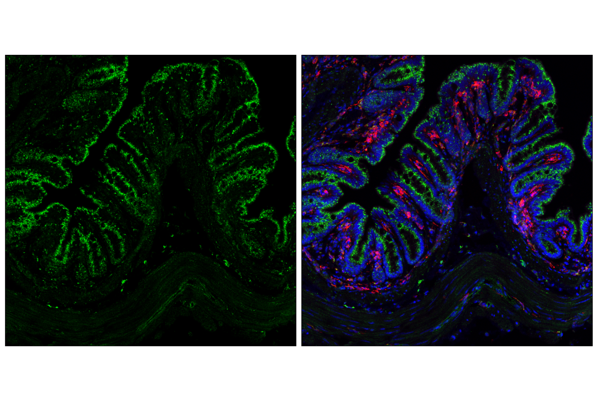
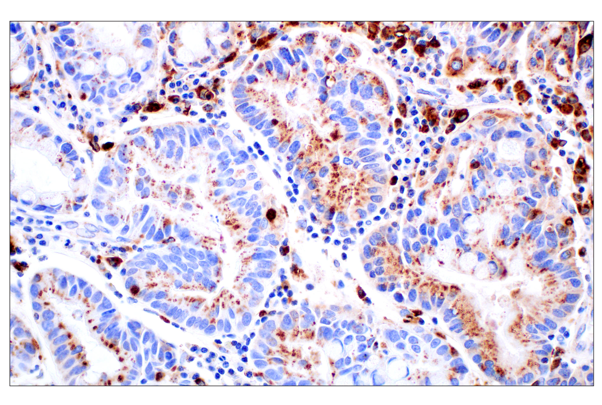
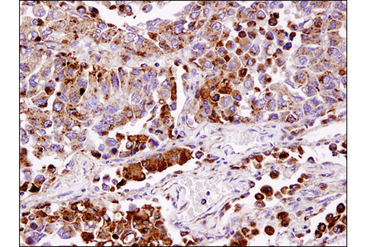
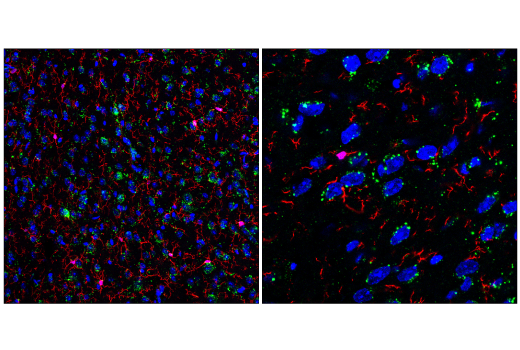
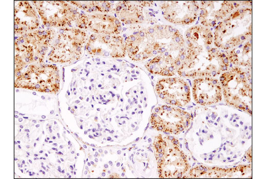
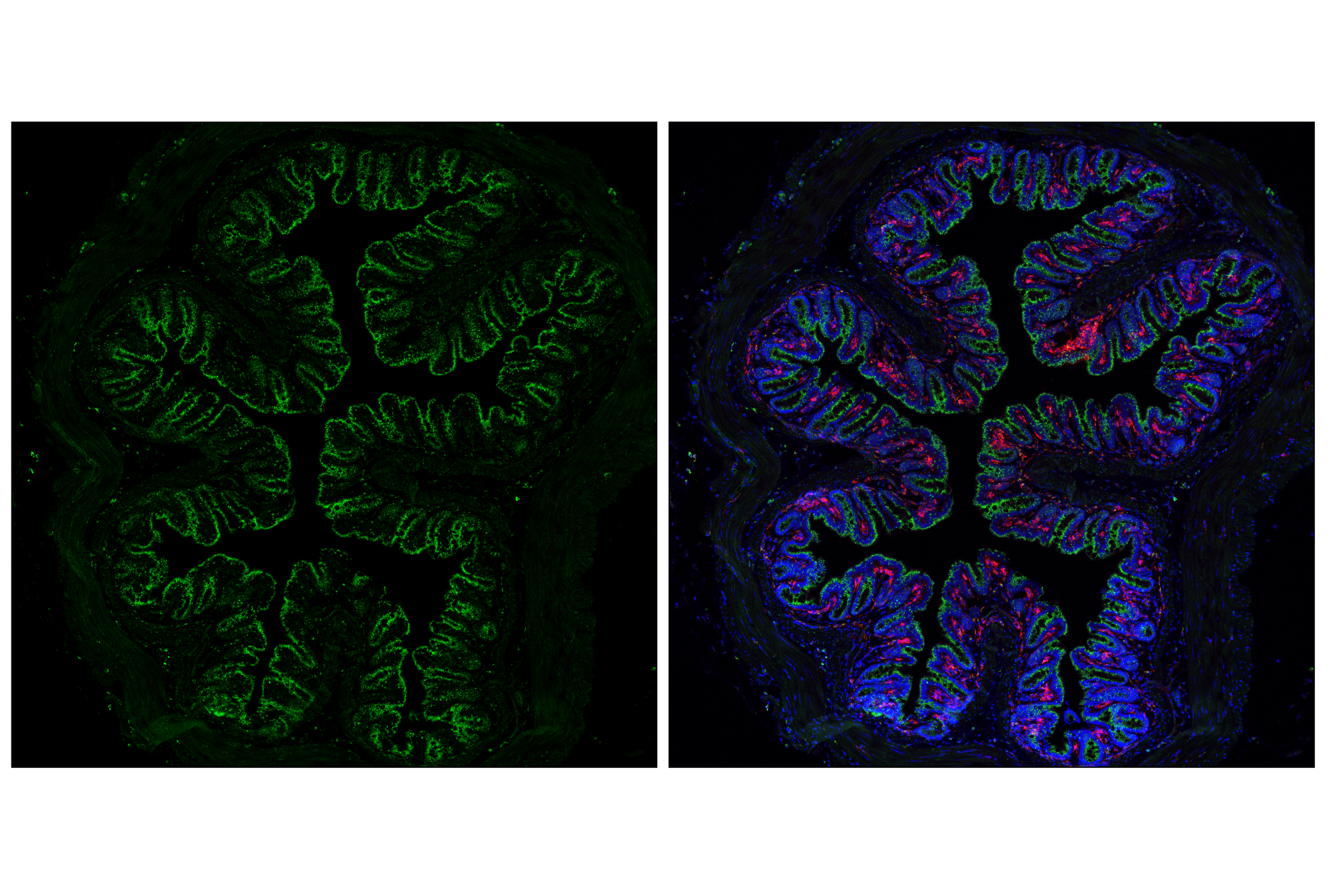
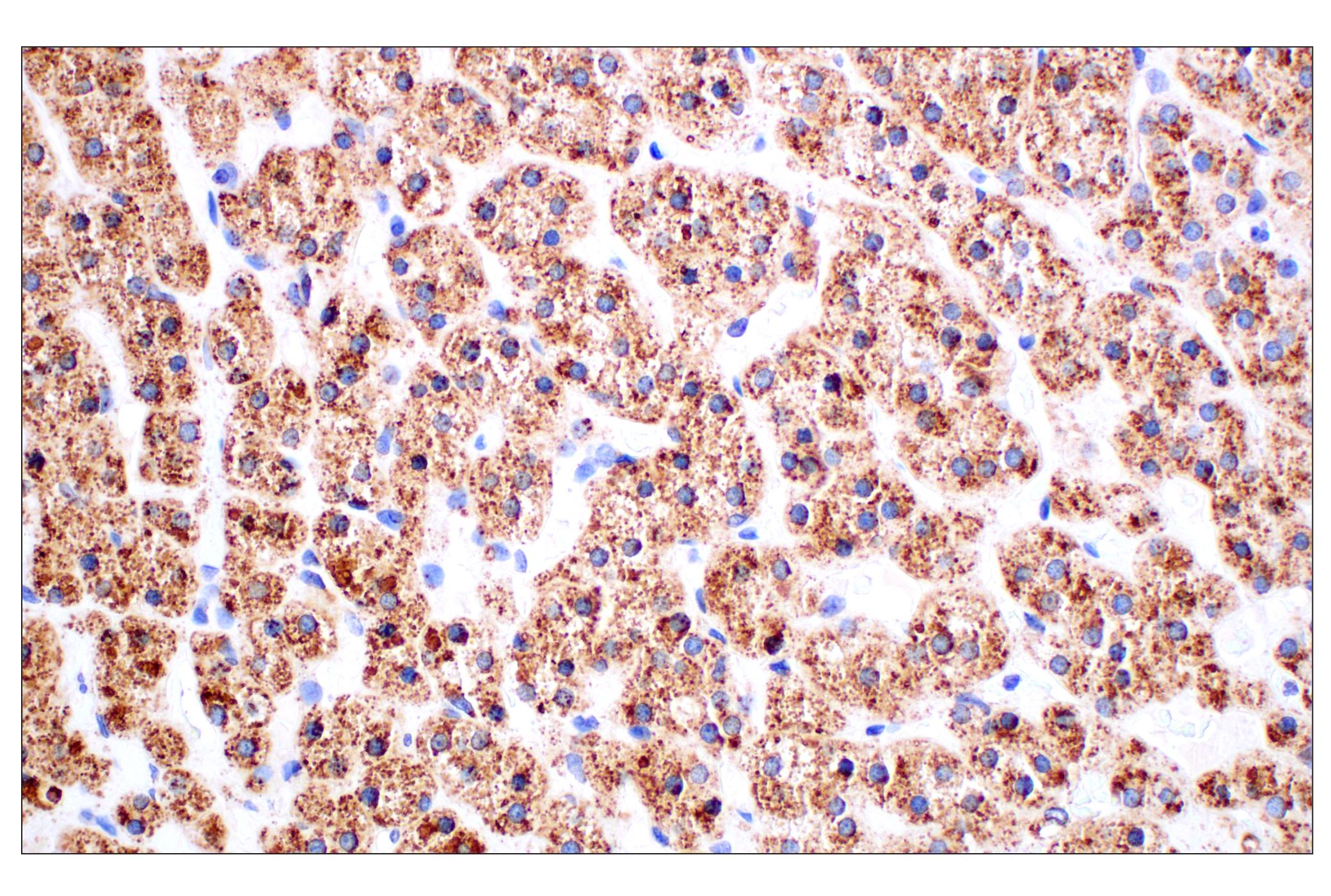
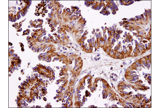
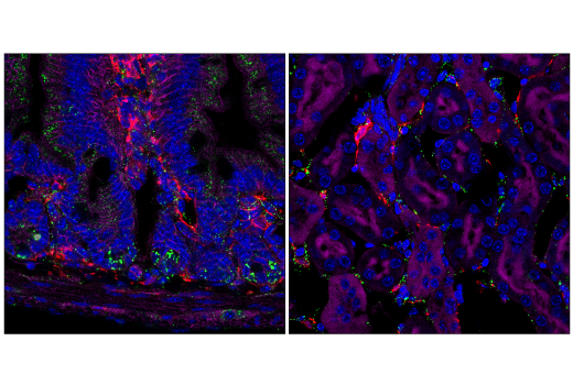
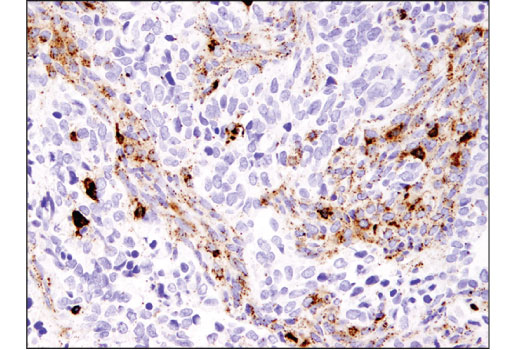
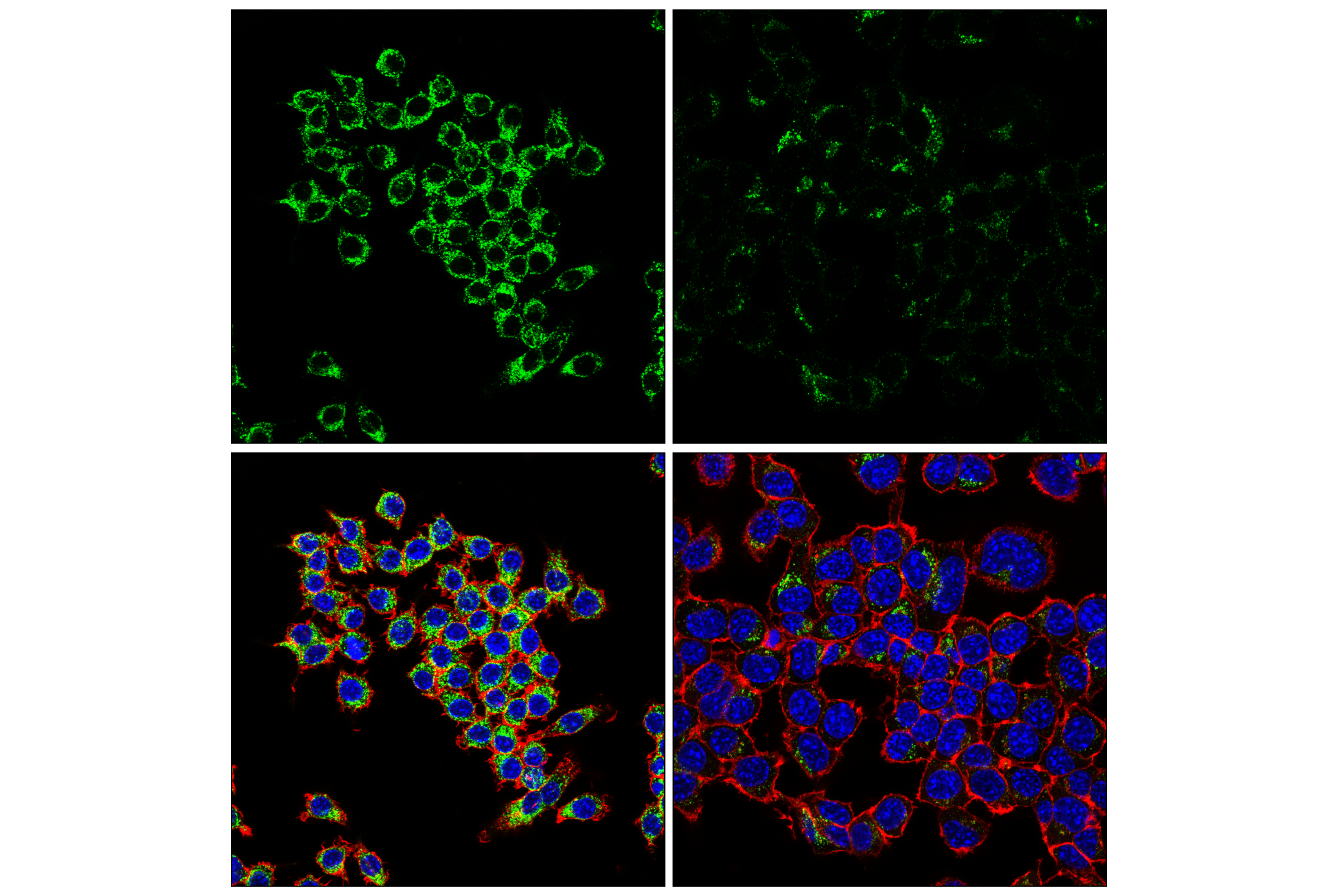
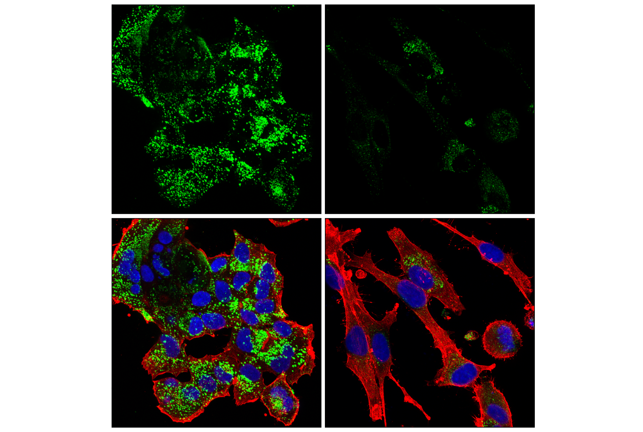
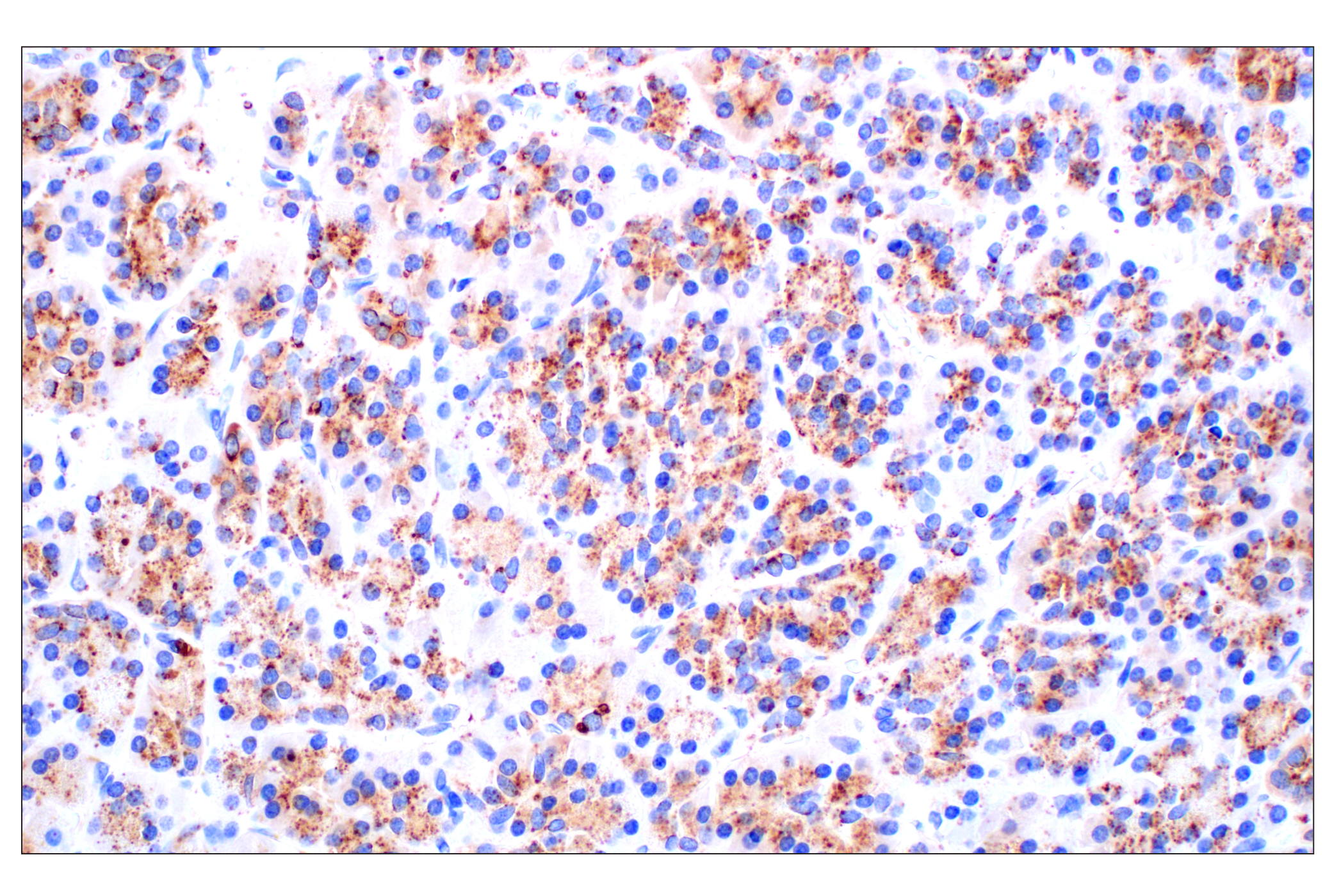
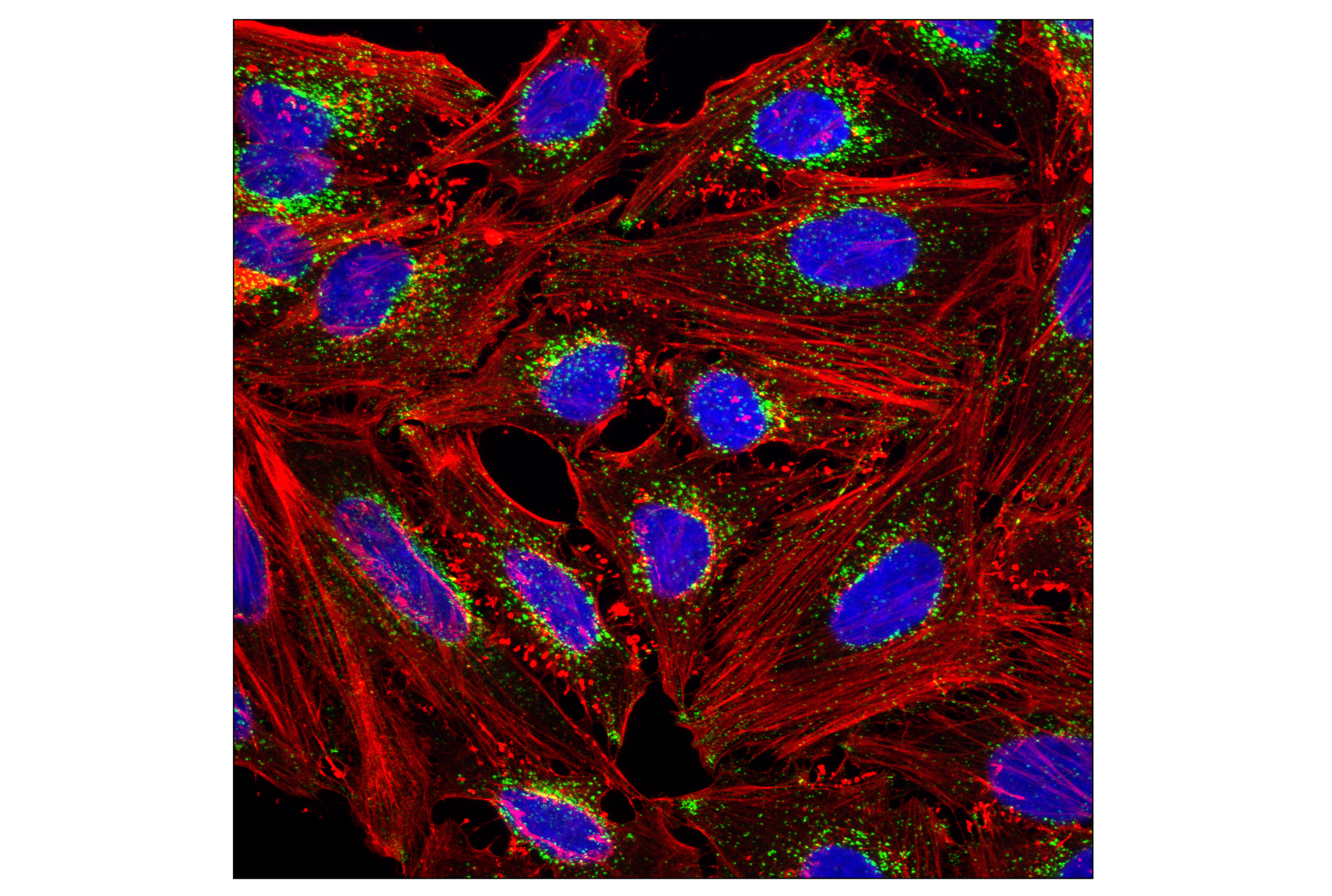
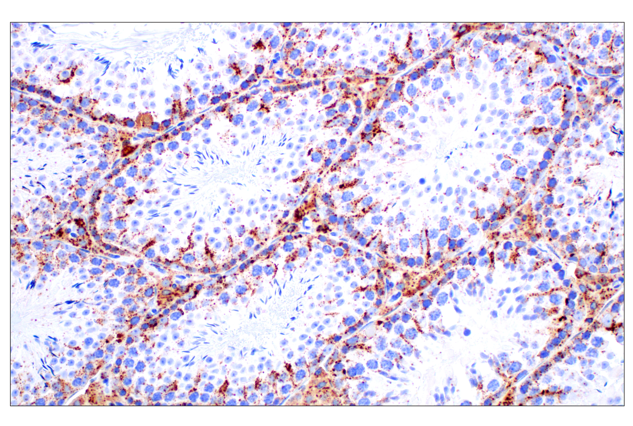
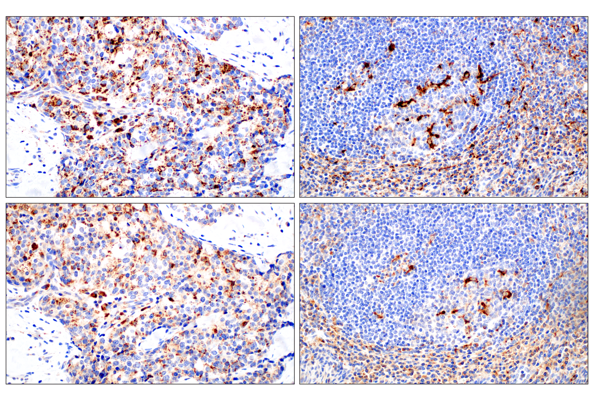
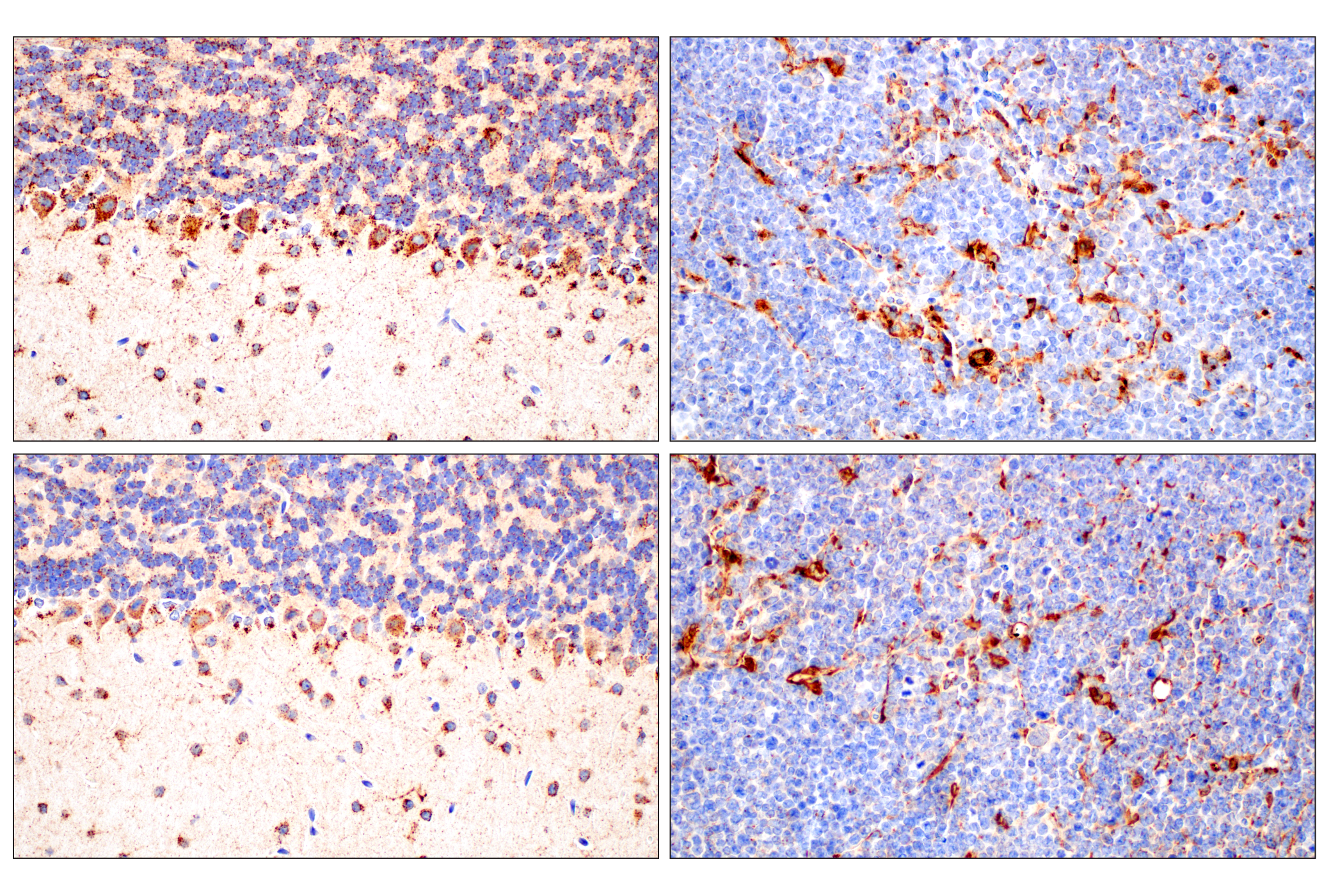
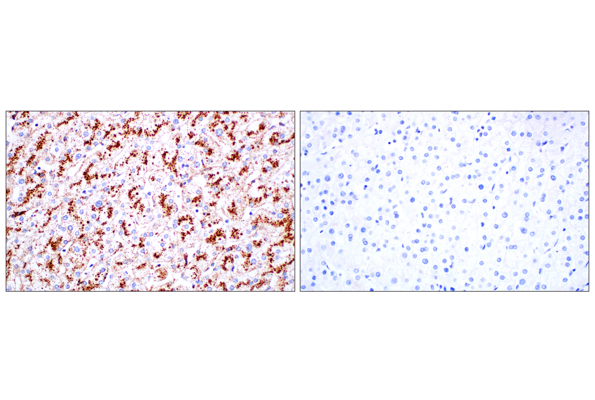
The Lysosomal Marker Antibody Sampler Kit provides an economical means of detecting proteins localized to lysosomes. The kit includes enough antibodies to perform two western blot experiments with each primary antibody.
Storage
Background
Lysosomes are single-membrane bound organelles of eukaryotic cells that are critically involved in the digestion of macromolecules mainly delivered by endocytosis and autophagy (1). Degradation occurs through hydrolytic enzymes requiring a low-pH environment (~4.5–5.0). Lysosome-associated membrane proteins play a critical role in lysosomal function (2). Lysosome-associated membrane protein 1 and 2 (LAMP1 and LAMP2) are two abundant lysosomal membrane proteins involved in regulating lysosomal motility during lysosome-phagosome fusion and cholesterol trafficking (3,4). CD63/LAMP3, a member of the tetraspanin family, is expressed on the surface of late endosomes and lysosomes and is also enriched in secreted exosomes (5). Lysosomal integral membrane protein type 2 (LIMP-2, also known as SCARB2) functions in cholesterol transport from the lysosome to the membrane, an important function as cholesterol contributes to various biophysical properties of cell membranes and may contribute to various diseases (6). Mucolipin-1 (MCOLN1, TRPML1), a member of the transient receptor potential ion channel superfamily, is a lysosomal/late endosome Ca2+ efflux channel that is important in autophagy and lysosomal biogenesis (7).
The acidic environment of the lysosome is maintained by H+ ATPase (V-ATPase), a large heteromultimeric proton pump located on the lysosomal transmembrane. Vacuolar ATPase enzymes are large, multimeric protein complexes with component proteins found in either the V1 peripheral domain or the V0 integral domain (8). The cytoplasmic V1 domain contains a hexamer of A and B catalytic subunits and several other protein subunits required for ATPase assembly and ATP hydrolysis. The integral V0 V-ATPase domain exhibits protein translocase activity and is responsible for proton transport across the membrane. The V-ATPase subunits ATP6V0c, ATP6V0d1, ATP6V1A, ATP6V1B2, and ATP6V1D interact with the Ragulator protein complex and are essential for amino acid induced activation of mTORC1 on the surface of lysosomes (9). Additionally, ATP6V1A has been shown to interact with SARS-CoV-2 M protein and facilitate viral infection (10,11).
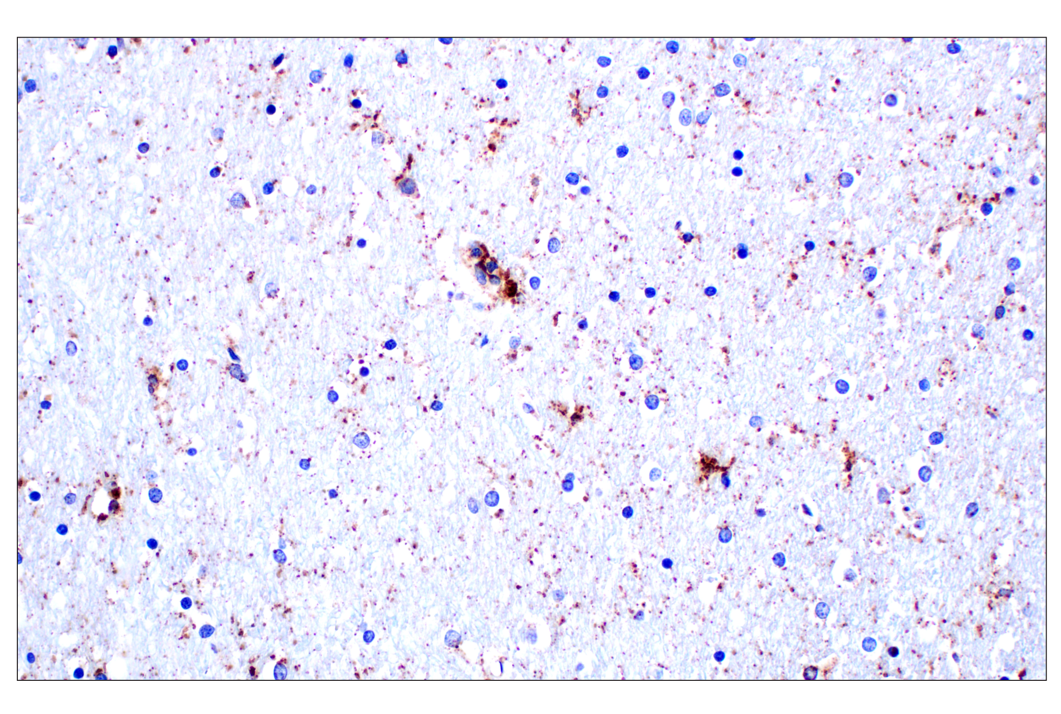
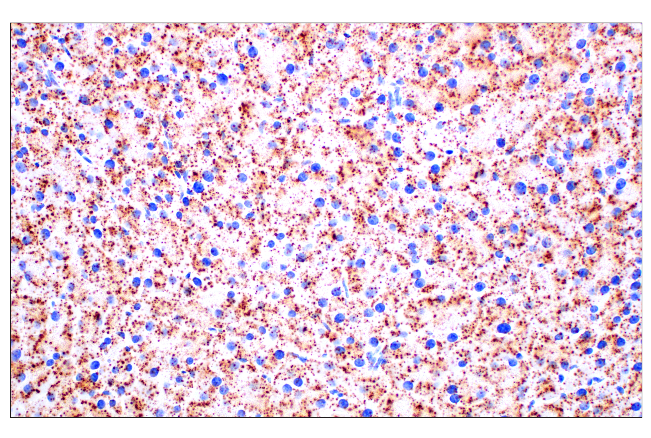
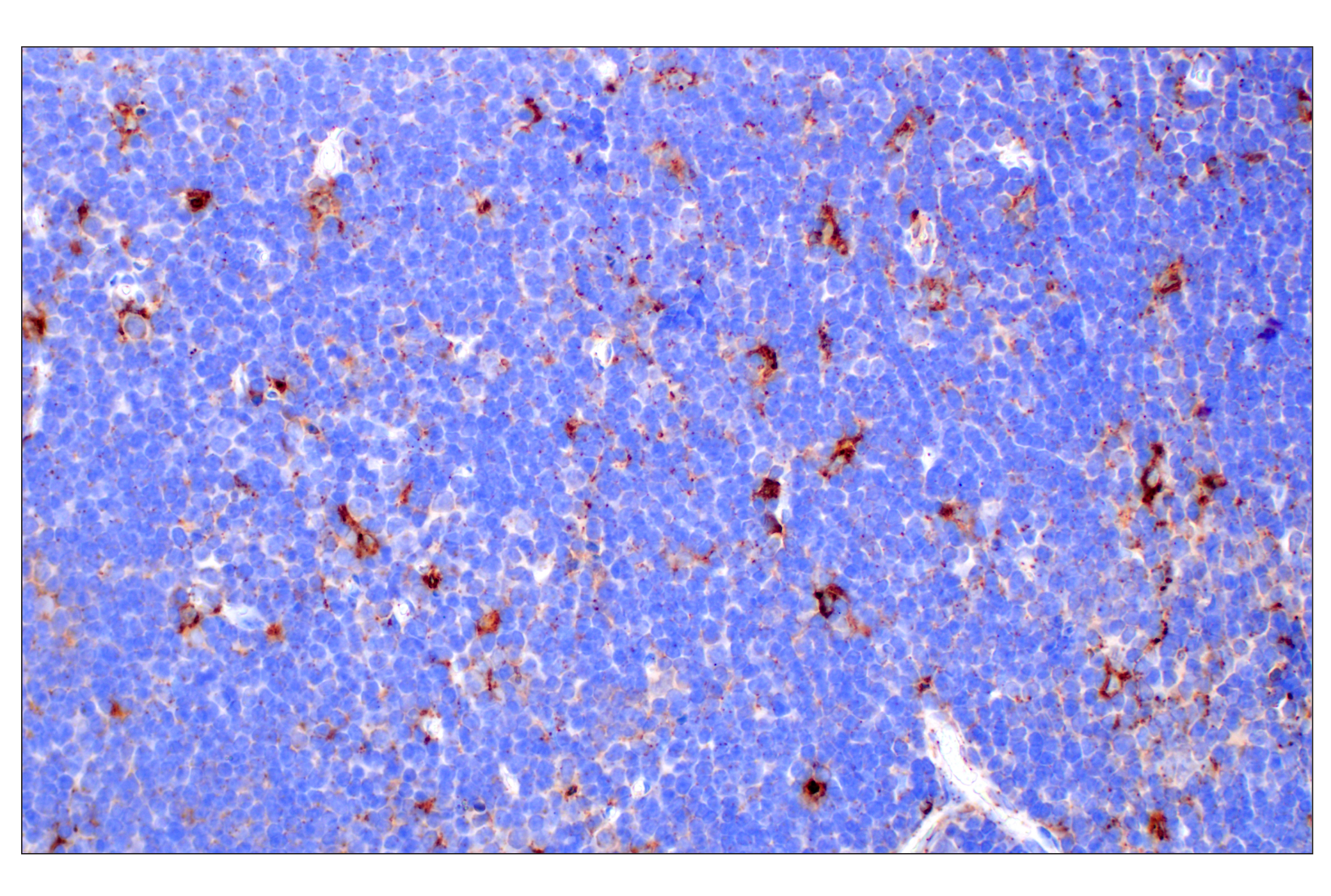
Cathepsins are the most abundant lysosomal proteases that can be categorized into groups of cysteine, serine, and aspartic proteases (12). Cathepsin B (CTSB), a cysteine protease, has been associated with multiple sclerosis (13), rheumatoid arthritis (14), and pancreatitis (15). Expression can correlate with poor prognosis for a variety of forms of cancer (16). Cathepsin D (CTSD) is a lysosomal aspartyl protease that plays a role in neuronal degradation and malignant transformation, particularly in breast cancer (17,18).
β-glucocerebrosidase (GCase) is a lysosomal enzyme that catalyzes the hydrolysis of glucocerebroside into free ceramide and glucose (19). GBA mutations are the most common genetic risk factor for Parkinson’s disease (20).
- Schröder, B.A. et al. (2010) Proteomics 10, 4053-76.
- Eskelinen, E.L. et al. (2003) Trends Cell Biol 13, 137-45.
- Huynh, K.K. et al. (2007) EMBO J 26, 313-24.
- Eskelinen, E.L. et al. (2004) Mol Biol Cell 15, 3132-45.
- Escola, J.M. et al. (1998) J Biol Chem 273, 20121-7.
- Heybrock, S. et al. (2019) Nat Commun 10, 3521.
- Cheng, X. et al. (2010) FEBS Lett 584, 2013-21.
- Jefferies, K.C. et al. (2008) Arch Biochem Biophys 476, 33-42.
- Zoncu, R. et al. (2011) Science 334, 678-83.
- Gordon, D.E. et al. (2020) Nature 583, 459-468.
- Daniloski, Z. et al. (2021) Cell 184, 92-105.e16.
- Yadati, T. et al. (2020) Cells 9, 1679. doi: 10.3390/cells9071679.
- Bever, C.T. et al. (1994) Neurology 44, 745-8.
- Hashimoto, Y. et al. (2001) Biochem Biophys Res Commun 283, 334-9.
- Halangk, W. et al. (2000) J Clin Invest 106, 773-81.
- Mijanović, O. et al. (2019) Cancer Lett 449, 207-214.
- Nomura, T. and Katunuma, N. (2005) J Med Invest 52, 1-9.
- Nakanishi, H. (2003) Ageing Res Rev 2, 367-81.
- Ho, M.W. et al. (1973) Biochem J 131, 173-6.
- Sidransky, E. and Lopez, G. (2012) Lancet Neurol 11, 986-98.
Background References
Trademarks and Patents
Limited Uses
Except as otherwise expressly agreed in a writing signed by a legally authorized representative of CST, the following terms apply to Products provided by CST, its affiliates or its distributors. Any Customer's terms and conditions that are in addition to, or different from, those contained herein, unless separately accepted in writing by a legally authorized representative of CST, are rejected and are of no force or effect.
Products are labeled with For Research Use Only or a similar labeling statement and have not been approved, cleared, or licensed by the FDA or other regulatory foreign or domestic entity, for any purpose. Customer shall not use any Product for any diagnostic or therapeutic purpose, or otherwise in any manner that conflicts with its labeling statement. Products sold or licensed by CST are provided for Customer as the end-user and solely for research and development uses. Any use of Product for diagnostic, prophylactic or therapeutic purposes, or any purchase of Product for resale (alone or as a component) or other commercial purpose, requires a separate license from CST. Customer shall (a) not sell, license, loan, donate or otherwise transfer or make available any Product to any third party, whether alone or in combination with other materials, or use the Products to manufacture any commercial products, (b) not copy, modify, reverse engineer, decompile, disassemble or otherwise attempt to discover the underlying structure or technology of the Products, or use the Products for the purpose of developing any products or services that would compete with CST products or services, (c) not alter or remove from the Products any trademarks, trade names, logos, patent or copyright notices or markings, (d) use the Products solely in accordance with CST Product Terms of Sale and any applicable documentation, and (e) comply with any license, terms of service or similar agreement with respect to any third party products or services used by Customer in connection with the Products.