Revision 1
#89884
Store at -20C
Mismatch DNA Repair (MMR) Antibody Sampler Kit
1 Kit
(5 x 20 microliters)
877-616-CELL (2355)
877-678-TECH (8324)
3 Trask Lane | Danvers | Massachusetts | 01923 | USA
For Research Use Only. Not for Use in Diagnostic Procedures.
| Product Includes | Product # | Quantity | Mol. Wt | Isotype/Source |
|---|---|---|---|---|
| MSH2 (D24B5) XP® Rabbit mAb | 2017 | 20 µl | 100 kDa | Rabbit IgG |
| MLH1 (D38G9) Rabbit mAb | 4256 | 20 µl | 84 kDa | Rabbit IgG |
| MSH6 (D60G2) XP® Rabbit mAb | 5424 | 20 µl | 160 kDa | Rabbit IgG |
| PMS2 (E9U4P) Rabbit mAb | 27884 | 20 µl | 110 kDa | Rabbit IgG |
| EXO1 (E6P2B) Rabbit mAb | 63862 | 20 µl | 120 kDa | Rabbit IgG |
| Anti-rabbit IgG, HRP-linked Antibody | 7074 | 100 µl | Goat |
Please visit cellsignal.com for individual component applications, species cross-reactivity, dilutions, protocols, and additional product information.
Description
The Mismatch DNA Repair (MMR) Antibody Sampler Kit provides an economical means of detecting proteins involved in MMR. The kit includes enough antibodies to perform two western blot experiments with each primary antibody.
Storage
Background
DNA mismatch repair (MMR), a conserved process for detecting and correcting errors made during DNA synthesis, is crucial to the maintenance of genomic integrity (1). In prokaryotes, a MutS homodimer recruits a MutL homodimer to sites of DNA mismatches. In eukaryotes, six MutS homologues (MSH1-6) and four MutL homologues (MLH1, PMS2, PMS1, and MLH3) have been identified. Heterodimers composed of two MutL homologues detect distinct DNA mismatch lesions, and heterodimers composed of two MutS homologues perform the repair (2). Other factors required for MMR in eukaryotes are EXO1, PCNA, RFC, RPA, DNA polymerases, and DNA ligases (1).
Microsatellite instability (MSI) is a predisposition to genetic mutation resulting from MMR deficiency (dMMR). High MSI (MSI-H) arising from dMMR results in Lynch syndrome, also known as hereditary non-polyposis colorectal cancer (HNPCC). Lynch syndrome is associated with colon cancer, as well as other human cancers (3). MSI and dMMR are strongly associated with tumor responsiveness to immune checkpoint blockade (4,5). MSI status can be determined through PCR amplification of microsatellite markers and/or immunohistochemical detection of MMR proteins MLH1, PMS2, MSH2, and MSH6. The absence of expression of any of these MMR proteins indicates dMMR (3).
Trademarks and Patents
Cell Signaling Technology is a trademark of Cell Signaling Technology, Inc.
XP is a registered trademark of Cell Signaling Technology, Inc.
All other trademarks are the property of their respective owners. Visit cellsignal.com/trademarks for more information.
Limited Uses
Except as otherwise expressly agreed in a writing signed by a legally authorized representative of CST, the following terms apply to Products provided by CST, its affiliates or its distributors. Any Customer's terms and conditions that are in addition to, or different from, those contained herein, unless separately accepted in writing by a legally authorized representative of CST, are rejected and are of no force or effect.
Products are labeled with For Research Use Only or a similar labeling statement and have not been approved, cleared, or licensed by the FDA or other regulatory foreign or domestic entity, for any purpose. Customer shall not use any Product for any diagnostic or therapeutic purpose, or otherwise in any manner that conflicts with its labeling statement. Products sold or licensed by CST are provided for Customer as the end-user and solely for research and development uses. Any use of Product for diagnostic, prophylactic or therapeutic purposes, or any purchase of Product for resale (alone or as a component) or other commercial purpose, requires a separate license from CST. Customer shall (a) not sell, license, loan, donate or otherwise transfer or make available any Product to any third party, whether alone or in combination with other materials, or use the Products to manufacture any commercial products, (b) not copy, modify, reverse engineer, decompile, disassemble or otherwise attempt to discover the underlying structure or technology of the Products, or use the Products for the purpose of developing any products or services that would compete with CST products or services, (c) not alter or remove from the Products any trademarks, trade names, logos, patent or copyright notices or markings, (d) use the Products solely in accordance with CST Product Terms of Sale and any applicable documentation, and (e) comply with any license, terms of service or similar agreement with respect to any third party products or services used by Customer in connection with the Products.
Revision 1
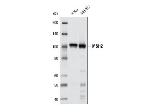
Western blot analysis of extracts of HeLa and NIH/3T3 cells using MSH2 (D24B5) XP® Rabbit mAb.
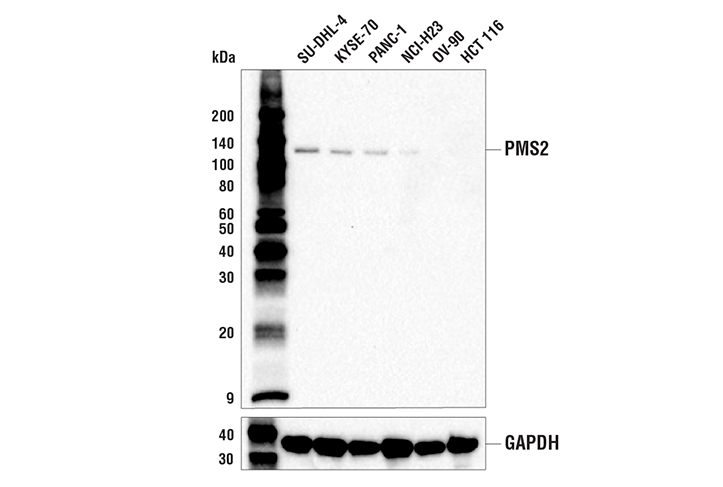
Western blot analysis of extracts from various human cell lines using PMS2 (E9U4P) Rabbit mAb (upper) and GAPDH (D16H11) XP® Rabbit mAb #5174 (lower). Low expression of PMS2 protein in OV-90 and HCT 116 cells is consistent with the predicted expression pattern.
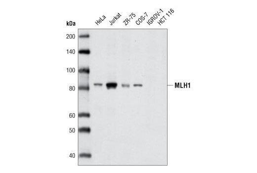
Western blot analysis of extracts from various cell types using MLH1 (D38G9) Rabbit mAb.
Revision 1
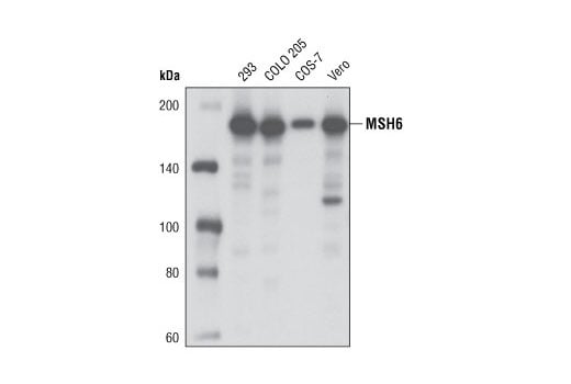
Western blot analysis of extracts from various cell types using MSH6 (D60G2) XP® Rabbit mAb.
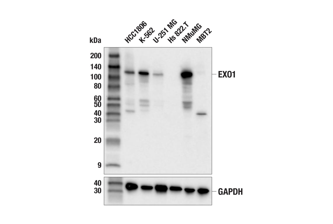
Western blot analysis of extracts from various cell lines using EXO1 (E6P2B) Rabbit mAb (upper) or GAPDH (D16H11) XP® Rabbit mAb #5174 (lower). Low/negative expression of EXO1 protein in Hs 822.T and MBT2 cells is consistent with the predicted expression pattern.
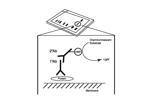
After the primary antibody is bound to the target protein, a complex with HRP-linked secondary antibody is formed. The LumiGLO® is added and emits light during enzyme catalyzed decomposition.
Revision 1
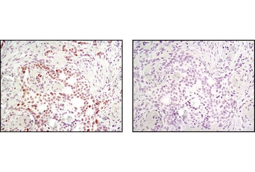
Immunohistochemical analysis of paraffin-embedded human breast carcinoma using MSH2 (D24B5) XP® Rabbit mAb in the presence of control peptide (left) or antigen-specific peptide (right).
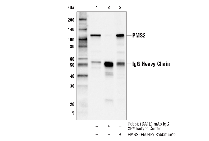
Immunoprecipitation of PMS2 protein from KYSE-70 cell extracts. Lane 1 is 10% input, lane 2 is Rabbit (DA1E) mAb IgG XP® Isotype Control #3900, and lane 3 is PMS2 (E9U4P) Rabbit mAb. Western blot analysis was performed using a PMS2 rabbit monoclonal antibody which detects an epitope distinct from that detected by PMS2 (E9U4P) Rabbit mAb. Anti-rabbit IgG, HRP-linked Antibody #7074 was used as the secondary antibody.
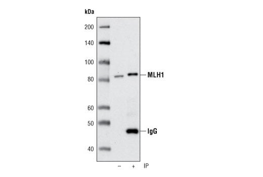
Immunoprecipitation of MLH1 from HeLa cells using MLH1 (D38G9) Rabbit mAb. Western blot was performed using the same antibody. IP () is 5% input.
Revision 1
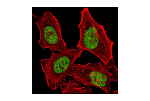
Confocal immunofluorescent analysis of 293 cells using MSH6 (D60G2) XP® Rabbit mAb (green). Actin filaments were labeled with DY-554 phalloidin (red).
Confocal immunofluorescent analysis of HeLa cells using MSH2 (D24B5) XP® Rabbit mAb (green). Actin filaments have been labeled with DY-554 phalloidin (red).
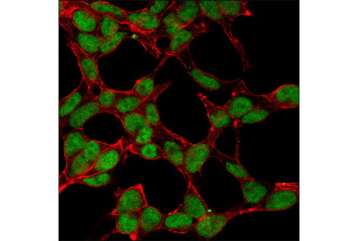
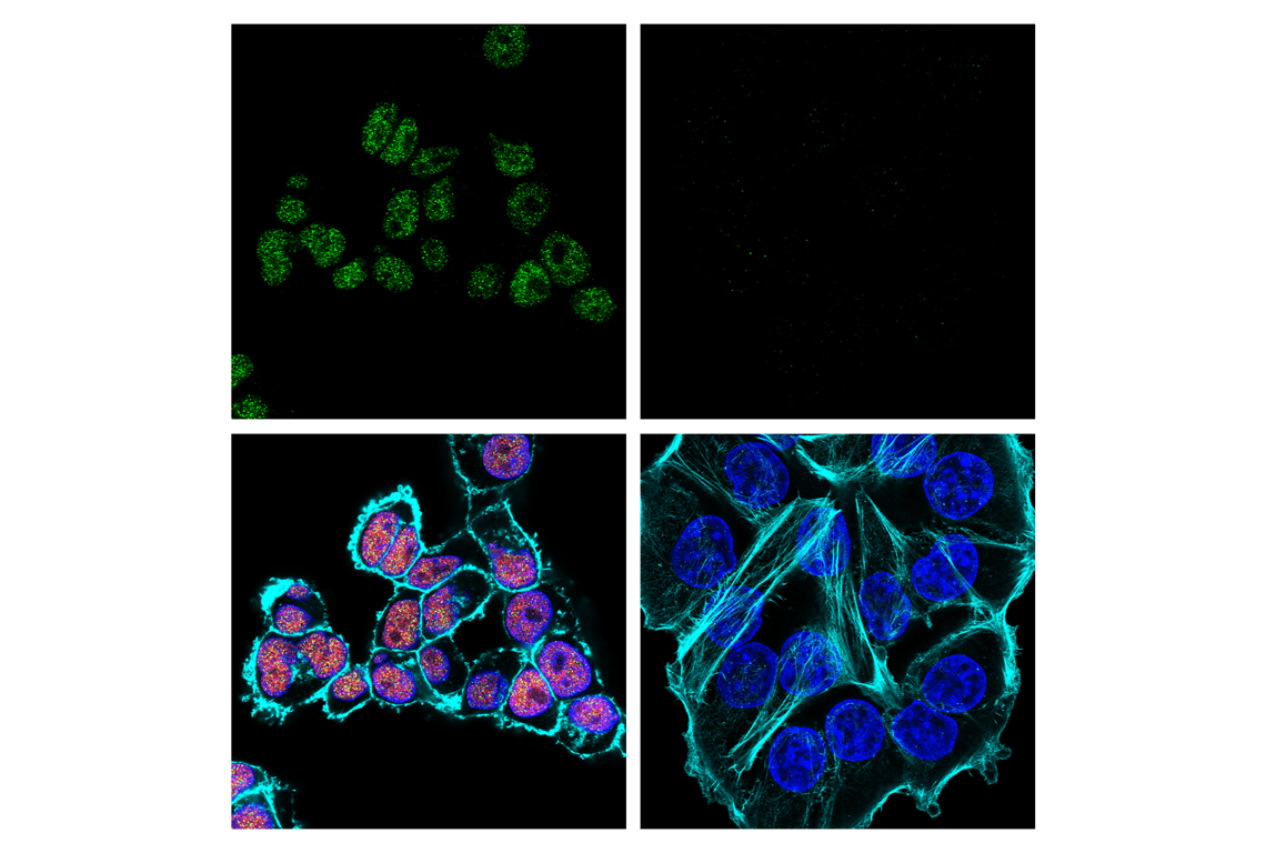
Confocal immunofluorescent analysis of KYSE-70 cells (left, positive) and HCT 116 cells (right, negative) using PMS2 (E9U4P) Rabbit mAb (green) and MLH1 (4C9C7) Mouse mAb #3515 (red). PMS2 forms a heterodimer with MLH1 (6). Actin filaments were labeled with DyLight™ 554 Phalloidin #13054 (cyan pseudocolor). Blue = DAPI #4083 (fluorescent DNA dye).
Revision 1
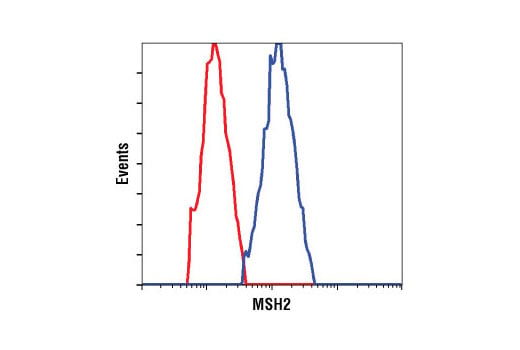
Flow cytometric analysis of HeLa cells using MSH2 (D24B5) XP® Rabbit mAb (blue) compared to a nonspecific negative control antibody (red).