Revision 1
#96128
Store at -20C
Mouse Reactive Exosome Marker Antibody Sampler Kit
1 Kit
(6 x 20 microliters)
877-616-CELL (2355)
877-678-TECH (8324)
3 Trask Lane | Danvers | Massachusetts | 01923 | USA
For Research Use Only. Not for Use in Diagnostic Procedures.
| Product Includes | Product # | Quantity | Mol. Wt | Isotype/Source |
|---|---|---|---|---|
| CD9 (E8L5J) Rabbit mAb | 98327 | 20 µl | 22-27 kDa | Rabbit IgG |
| CD81 (D5O2Q) Rabbit mAb | 10037 | 20 µl | 22 kDa | Rabbit IgG |
| TSG101 (E6V1X) Rabbit mAb | 72312 | 20 µl | 50 kDa | Rabbit IgG |
| Alix (E6P9B) Rabbit mAb | 92880 | 20 µl | 90-100 kDa | Rabbit IgG |
| Flotillin-1 (D2V7J) XP® Rabbit mAb | 18634 | 20 µl | 49 kDa | Rabbit IgG |
| HSP70 Antibody | 4872 | 20 µl | 72, 73 kDa | Rabbit |
| Anti-rabbit IgG, HRP-linked Antibody | 7074 | 100 µl | Goat |
Please visit cellsignal.com for individual component applications, species cross-reactivity, dilutions, protocols, and additional product information.
Description
The Mouse Reactive Exosome Marker Antibody Sampler Kit provides an economical means of analyzing proteins that can be present on exosomes. The kit includes enough antibodies to perform two western blot experiments with each primary antibody.
Storage
Background
Exosomes are small (30-150 nm) membrane-bound vesicles that are secreted by various cell types under normal and pathological conditions (1,2). They originate from intracellular multivesicular endosomes upon fusion with the plasma membrane. Exosomes have emerged as an important mechanism of intercellular communication facilitating the transfer of membrane and cytosolic proteins, lipids, and RNA.
A variety of methods have been described to isolate exosomes and understand their composition (3-7). Heterogeneity in exosome composition can be attributed to the cells of origin as well as the isolation methods. However, there are protein markers that appear with high frequency. Tetraspanins are a family of cell surface glycoproteins with four transmembrane domains often found in exosomes (8). Tetraspanins CD9, CD81, and CD63 appear in exosomes and have been the target of immune-affinity approaches of exosome isolation. Flotillin-1 is a lipid raft-associated integral membrane protein that is incorporated into exosomes (9). Exosomes also contain proteins involved in endosomal membrane trafficking, collectively known as the ESCRT (endosomal sorting complex required for transport) pathway. Alix regulates cellular processes, such as endocytic membrane trafficking and cell adhesion through interactions with ESCRT proteins including endophilins, and CIN85 (Cbl-interacting protein of 85 kDa), and plays a role in exosome biogenesis (10-12). Syntenin-1 (MDA9, SDCBP) is a member of the PDZ family of proteins that functions as a scaffold adaptor protein regulating numerous signal transduction pathways (13). Syntenin-1 interacts with Alix to regulate exosome biogenesis (12). Tumor susceptibility gene 101 (TSG101) is a fundamental component of the ESCRT complex I involved in regulating the trafficking of proteins throughout the endosomal compartment (14). TSG101 is involved in regulating diverse biological processes, such as cell proliferation, viral budding and release, and exosome biosynthesis (15,16). The heat shock protein HSP70 is a molecular chaperone involved in protein folding that can be induced upon environmental stress (17). HSP70 may also be secreted through exosomes (18).
Background References
- Raposo, G. and Stoorvogel, W. (2013) J Cell Biol 200, 373-83.
- van Niel, G. et al. (2018) Nat Rev Mol Cell Biol 19, 213-228.
- Jeppesen, D.K. et al. (2019) Cell 177, 428-445.e18.
- Kowal, J. et al. (2016) Proc Natl Acad Sci U S A 113, E968-77.
- Sidhom, K. et al. (2020) Int J Mol Sci 21, 6466. doi: 10.3390/ijms21186466.
- Patel, G.K. et al. (2019) Sci Rep 9, 5335.
- Tauro, B.J. et al. (2012) Methods 56, 293-304.
- Hemler, M.E. (2005) Nat Rev Mol Cell Biol 6, 801-11.
- de Gassart, A. et al. (2003) Blood 102, 4336-44.
- Katoh, K. et al. (2003) J Biol Chem 278, 39104-13.
- Chatellard-Causse, C. et al. (2002) J Biol Chem 277, 29108-15.
- Baietti, M.F. et al. (2012) Nat Cell Biol 14, 677-85.
- Pradhan, A.K. et al. (2020) Cancer Metastasis Rev 39, 769-781.
- Katzmann, D.J. et al. (2001) Cell 106, 145-55.
- Garrus, J.E. et al. (2001) Cell 107, 55-65.
- Zhong, Q. et al. (1998) Cancer Res 58, 2699-702.
- Nollen, E.A. and Morimoto, R.I. (2002) J Cell Sci 115, 2809-16.
- Zhan, R. et al. (2009) Biochem Biophys Res Commun 387, 229-33.
Trademarks and Patents
Cell Signaling Technology is a trademark of Cell Signaling Technology, Inc.
XP is a registered trademark of Cell Signaling Technology, Inc.
All other trademarks are the property of their respective owners. Visit cellsignal.com/trademarks for more information.
Limited Uses
Except as otherwise expressly agreed in a writing signed by a legally authorized representative of CST, the following terms apply to Products provided by CST, its affiliates or its distributors. Any Customer's terms and conditions that are in addition to, or different from, those contained herein, unless separately accepted in writing by a legally authorized representative of CST, are rejected and are of no force or effect.
Products are labeled with For Research Use Only or a similar labeling statement and have not been approved, cleared, or licensed by the FDA or other regulatory foreign or domestic entity, for any purpose. Customer shall not use any Product for any diagnostic or therapeutic purpose, or otherwise in any manner that conflicts with its labeling statement. Products sold or licensed by CST are provided for Customer as the end-user and solely for research and development uses. Any use of Product for diagnostic, prophylactic or therapeutic purposes, or any purchase of Product for resale (alone or as a component) or other commercial purpose, requires a separate license from CST. Customer shall (a) not sell, license, loan, donate or otherwise transfer or make available any Product to any third party, whether alone or in combination with other materials, or use the Products to manufacture any commercial products, (b) not copy, modify, reverse engineer, decompile, disassemble or otherwise attempt to discover the underlying structure or technology of the Products, or use the Products for the purpose of developing any products or services that would compete with CST products or services, (c) not alter or remove from the Products any trademarks, trade names, logos, patent or copyright notices or markings, (d) use the Products solely in accordance with CST Product Terms of Sale and any applicable documentation, and (e) comply with any license, terms of service or similar agreement with respect to any third party products or services used by Customer in connection with the Products.
Revision 1
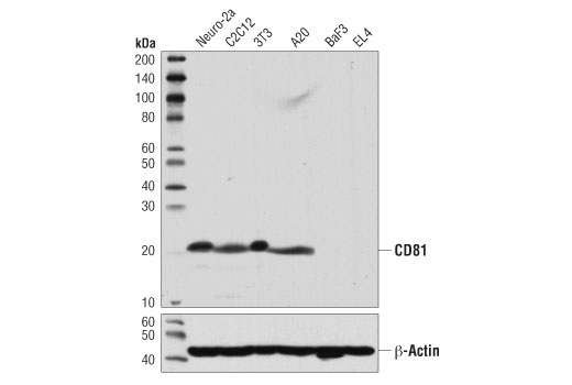
Western blot analysis of extracts from various cell lines using CD81 (D5O2Q) Rabbit mAb (upper) or β-Actin (D6A8) Rabbit mAb #8457 (lower).
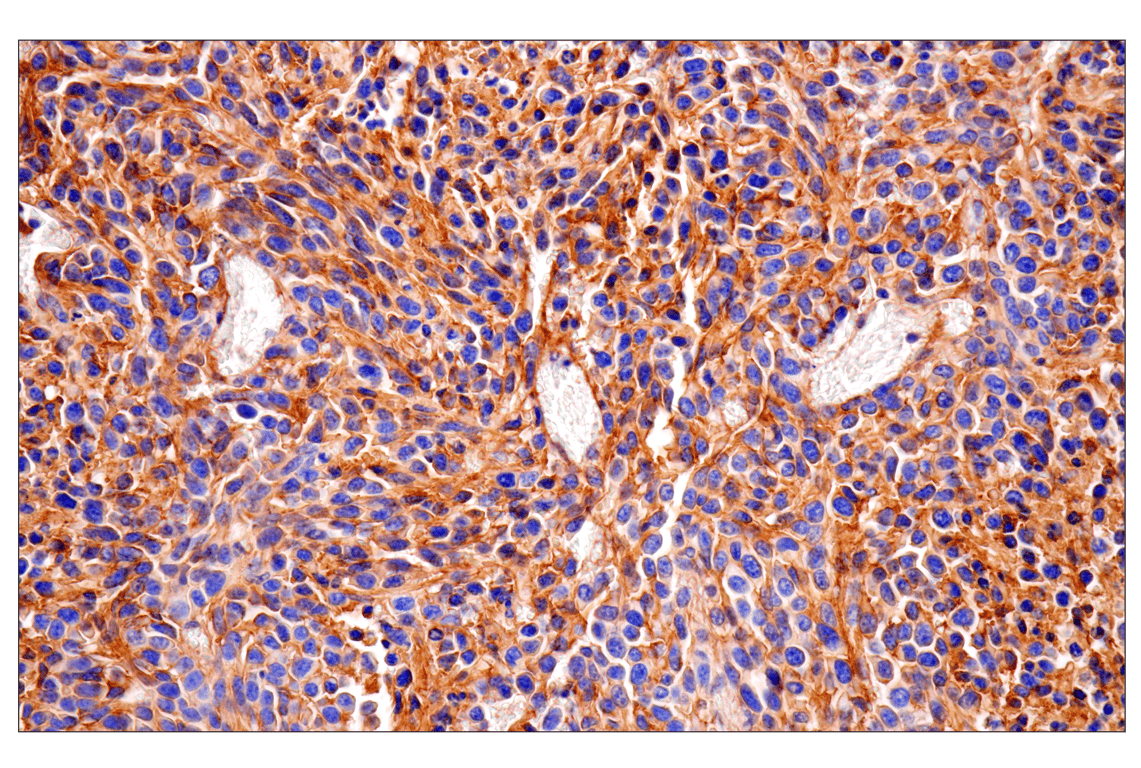
Immunohistochemical analysis of paraffin-embedded CT26.WT syngeneic tumor using CD81 (D5O2Q) Rabbit mAb.
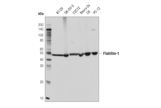
Western blot analysis of extracts from various cell lines using Flotillin-1 (D2V7J) XP® Rabbit mAb.
Revision 1
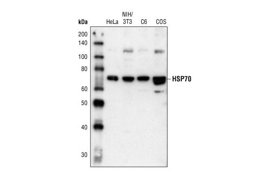
Western blot analysis of extracts from HeLa, NIH/3T3, C6 and COS cells, using HSP70 Antibody.
After the primary antibody is bound to the target protein, a complex with HRP-linked secondary antibody is formed. The LumiGLO® is added and emits light during enzyme catalyzed decomposition.
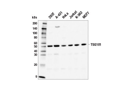
Western blot analysis of extracts from various cell lines using TSG101 (E6V1X) Rabbit mAb.
Revision 1
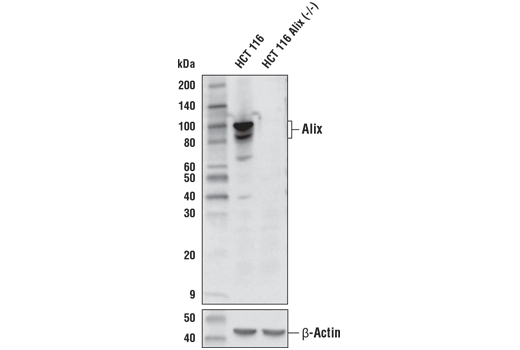
Western blot analysis of extracts from HCT 116 and HCT 116 Alix knockout (-/-) cells using Alix (E6P9B) Rabbit mAb (upper) or β-Actin (D6A8) Rabbit mAb #8457 (lower).
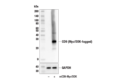
Western blot analysis of extracts from 293T cells, mock transfected (-) or transfected with a construct expressing Myc/DDK-tagged full-length mouse CD9 protein (mCD9-Myc/DDK; +), using CD9 (E8L5J) Rabbit mAb (upper) and GAPDH (D16H11) XP® Rabbit mAb #5174 (lower).
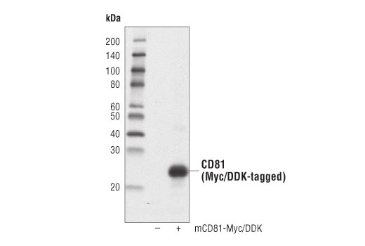
Western blot analysis of extracts from 293T cells, untransfected (-) or transfected with a construct expressing Myc/DDK-tagged full-length mouse CD81 (mCD81-Myc/DDK, +), using CD81 (D5O2Q) Rabbit mAb.
Revision 1
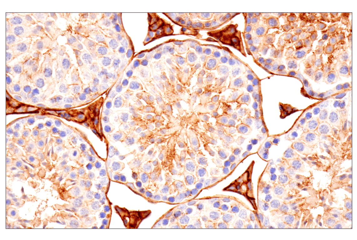
Immunohistochemical analysis of paraffin-embedded mouse testis using CD81 (D5O2Q) Rabbit mAb.
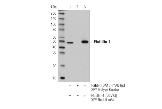
Immunoprecipitation of flotillin-1 protein from BT-20 cell extracts. Lane 1 is 10% input, lane 2 is Rabbit (DA1E) mAb IgG XP® Isotype Control #3900, and lane 3 is Flotillin-1 (D2V7J) XP® Rabbit mAb. Western blot analysis was performed using Flotillin-1 (D2V7J) XP® Rabbit mAb.
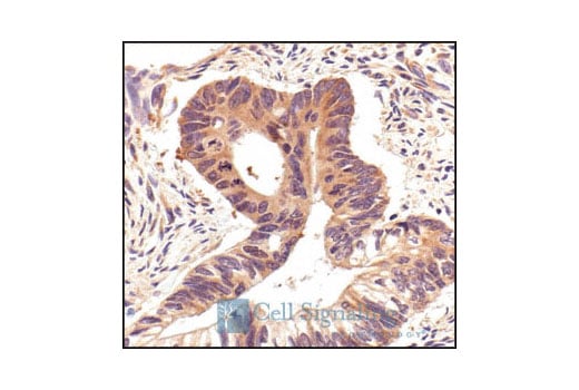
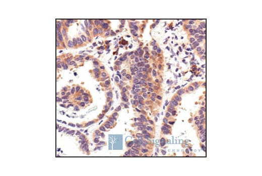
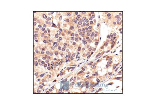
Immunohistochemical analysis of paraffin-embedded human breast carcinoma, using HSP70 Antibody.
Revision 1
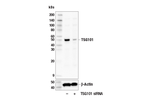
Western blot analysis of extracts from 293T cells, transfected with control siRNA (-) or TSG101 siRNA (+), using TSG101 (E6V1X) Rabbit mAb (upper) or β-Actin (D6A8) Rabbit mAb #8457 (lower).
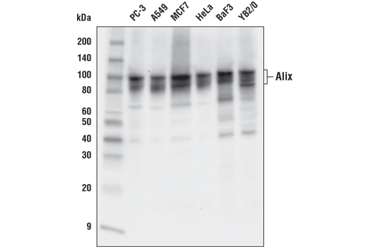
Western blot analysis of extracts from various cell lines using Alix (E6P9B) Rabbit mAb.
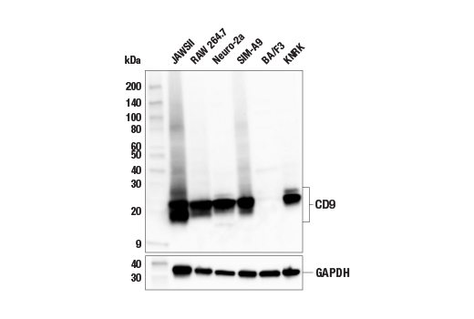
Western blot analysis of extracts from various cell lines using CD9 (E8L5J) Rabbit mAb (upper) and GAPDH (D16H11) XP® Rabbit mAb #5174 (lower). Absence of signal in BA/F3 cells is predicted from RNAseq data and confirms the specificity of the antibody.
Revision 1
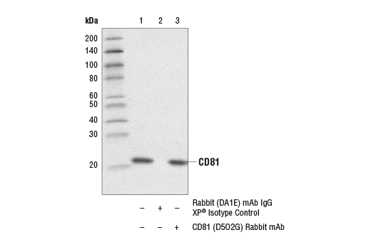
Immunoprecipitation of CD81 from extracts of C2C12 cells. Lane 1 is 10% input, Lane 2 is Rabbit (DA1E) mAb IgG XP® Isotype Control #3900, and lane 3 is CD81 (D5O2Q) Rabbit mAb. Western blot analysis was performed using CD81 (D5O2Q) Rabbit mAb.
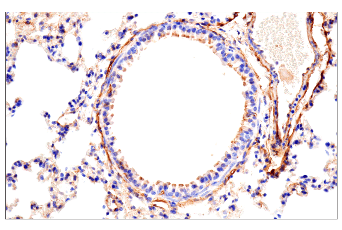
Immunohistochemical analysis of paraffin-embedded mouse lung using CD81 (D5O2Q) Rabbit mAb.
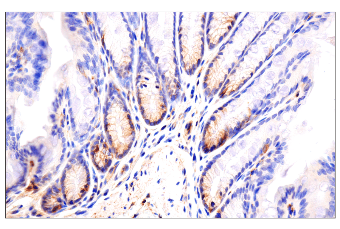
Immunohistochemical analysis of paraffin-embedded human colon carcinoma, using HSP70 Antibody.
Revision 1
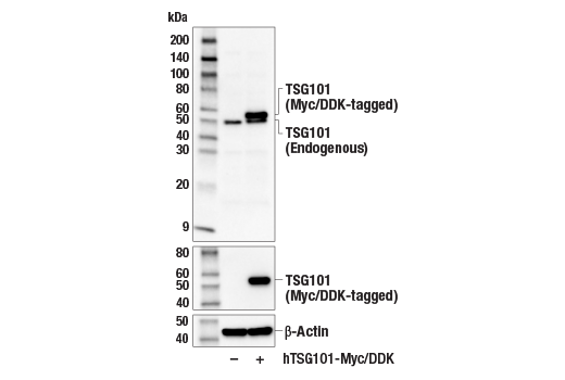
Western blot analysis of extracts from 293T cells, mock transfected (-) or transfected with a construct expressing Myc/DDK-tagged full-length human TSG101 (hTSG101-Myc/DDK; +), using TSG101 (E6V1X) Rabbit mAb (upper), DYKDDDDK Tag (D6W5B) Rabbit mAb #14793 (middle), or β-Actin (D6A8) Rabbit mAb #8457 (lower).
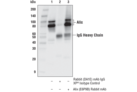
Immunoprecipitation of Alix from K-562 cell extracts. Lane 1 is 10% input, lane 2 is Rabbit (DA1E) mAb IgG XP® Isotype Control, and lane 3 is Alix (E6P9B) Rabbit mAb. Western blot was performed using Alix (E6P9B) Rabbit mAb. Anti-rabbit IgG, HRP-linked Antibody #7074 was using as a secondary antibody.
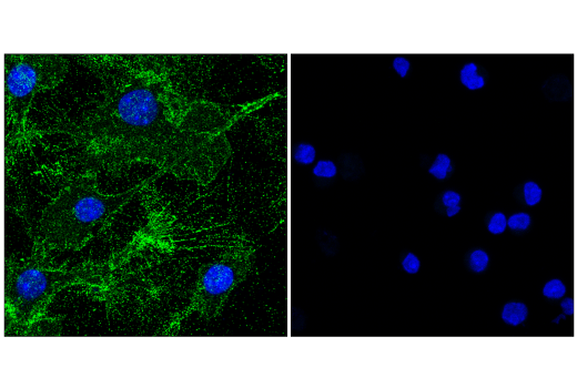
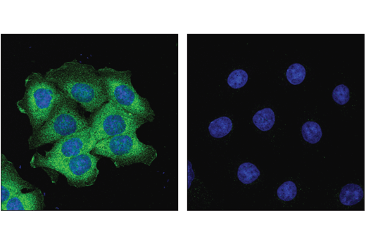
Confocal immunofluorescent analysis of RAW 264.7 cells (left, positive) or BA/F3 cells (right, negative) using CD9 (E8L5J) Rabbit mAb (green) and DAPI #4083 (blue).
Revision 1
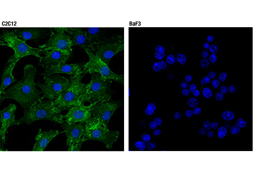
Confocal immunofluorescent analysis of C2C12 (positive, left) and BaF3 (negative, right) cells using CD81 (D5O2Q) Rabbit mAb (green). Blue pseudocolor = DRAQ5® #4084 (fluorescent DNA dye).
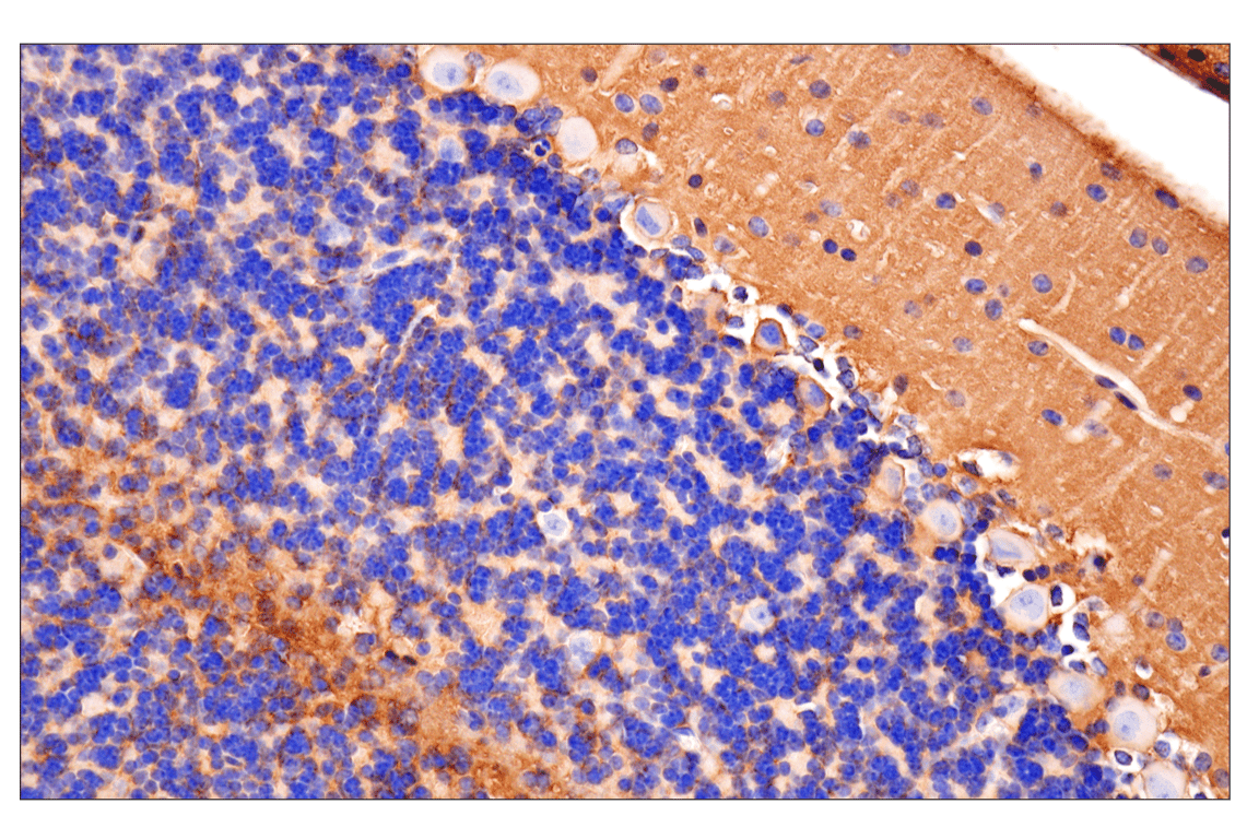
Immunohistochemical analysis of paraffin-embedded mouse cerebellum using CD81 (D5O2Q) Rabbit mAb.
Immunohistochemical analysis of paraffin-embedded human hepatocellular carcinoma using Flotillin-1 (D2V7J) XP® Rabbit mAb in the presence of control peptide (left) or antigen-specific peptide (right).
Revision 1
Immunohistochemical analysis of paraffin-embedded human lung carcinoma showing cytoplasmic localization using HSP70 Antibody.
Confocal immunofluorescent analysis of HCT 116 cells (left, positive) and HCT 116 Alix knockout (-/-) cells (right, negative) using Alix (E6P9B) Rabbit mAb (green). Blue pseudocolor = DRAQ5® #4084 (fluorescent DNA dye).
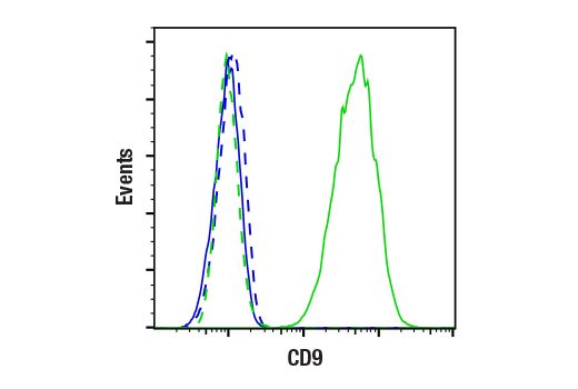
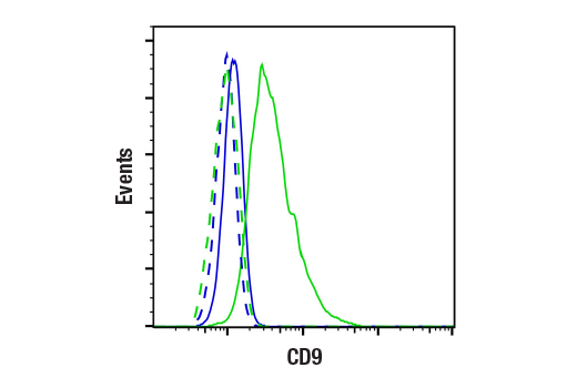
Flow cytometric analysis of live BA/F3 cells (blue, negative) and JAWSII cells (green, positive) using CD9 (E8L5J) Rabbit mAb (solid lines) or concentration-matched Rabbit (DA1E) mAb IgG XP® Isotype Control #3900 (dashed lines). Anti-rabbit IgG (H+L), F(ab')2 Fragment (Alexa Fluor® 488 Conjugate) #4412 was used as a secondary antibody.
Revision 1
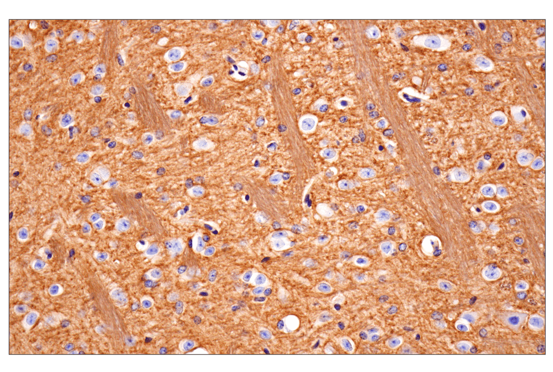
Immunohistochemical analysis of paraffin-embedded mouse striatum using CD81 (D5O2Q) Rabbit mAb.
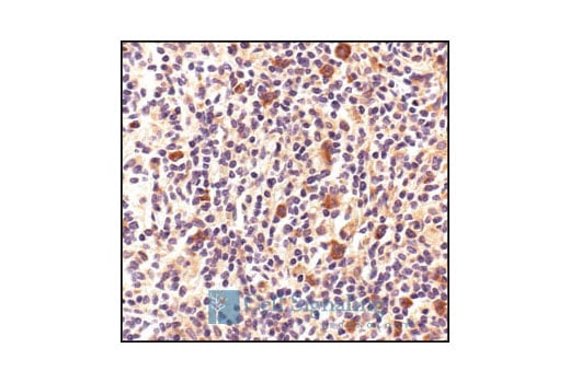
Immunohistochemical analysis of paraffin-embedded human Non-Hodgkin's lymphoma, using HSP70 Antibody.
Flow cytometric analysis of fixed and permeabilized BA/F3 cells (blue, negative) and JAWSII cells (green, positive) using CD9 (E8L5J) Rabbit mAb (solid lines) or concentration-matched Rabbit (DA1E) mAb IgG XP® Isotype Control #3900 (dashed lines). Anti-rabbit IgG (H+L), F(ab')2 Fragment (Alexa Fluor® 488 Conjugate) #4412 was used as a secondary antibody.
Revision 1
Immunohistochemical analysis of paraffin-embedded mouse brown fat using CD81 (D5O2Q) Rabbit mAb.
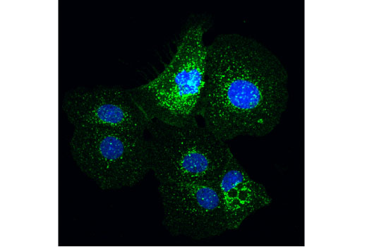
Confocal immunofluorescent analysis of BT-20 cells using Flotillin-1 (D2V7J) XP® Rabbit mAb (green). Blue pseudocolor = DRAQ5® #4084 (fluorescent DNA dye).
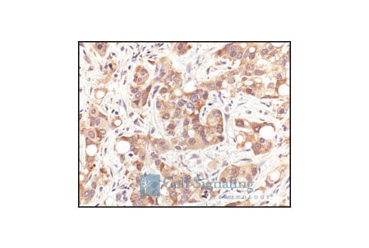
Immunohistochemical analysis of paraffin-embedded human prostate carcinoma, using HSP70 Antibody.
Revision 1
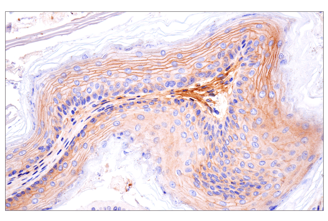
Immunohistochemical analysis of paraffin-embedded mouse colon using CD81 (D5O2Q) Rabbit mAb.
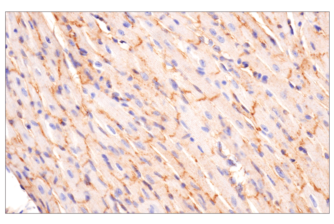
Immunohistochemical analysis of paraffin-embedded mouse heart using CD81 (D5O2Q) Rabbit mAb.
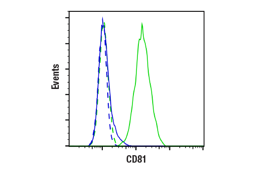
Flow cytometric analysis of live EL4 cells (blue, negative) and A20 cells (green, positive) using CD81 (D5O2Q) Rabbit mAb (solid lines) or concentration-matched Rabbit (DA1E) mAb IgG XP® Isotype Control #3900 (dashed line). Anti-rabbit IgG (H+L), F(ab')2 Fragment (Alexa Fluor® 488 Conjugate) #4412 was used as a secondary antibody.
Revision 1
Immunohistochemical analysis of paraffin-embedded mouse forestomach using CD81 (D5O2Q) Rabbit mAb.
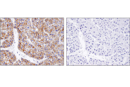
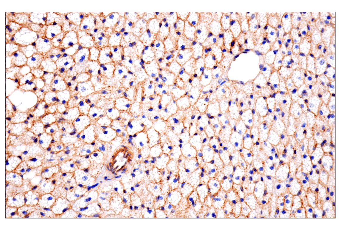
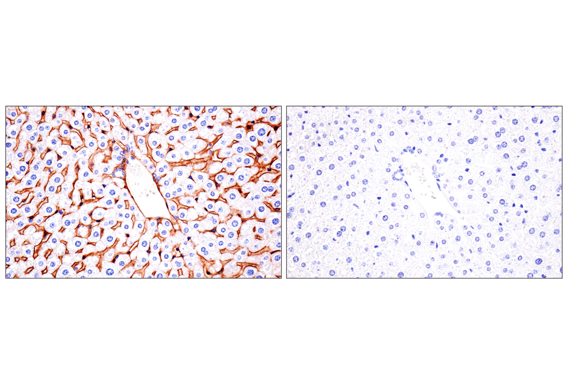
Immunohistochemical analysis of paraffin-embedded mouse liver using CD81 (D5O2Q) Rabbit mAb (left) compared to concentration-matched Rabbit (DA1E) mAb IgG XP® Isotype Control #3900 (right).
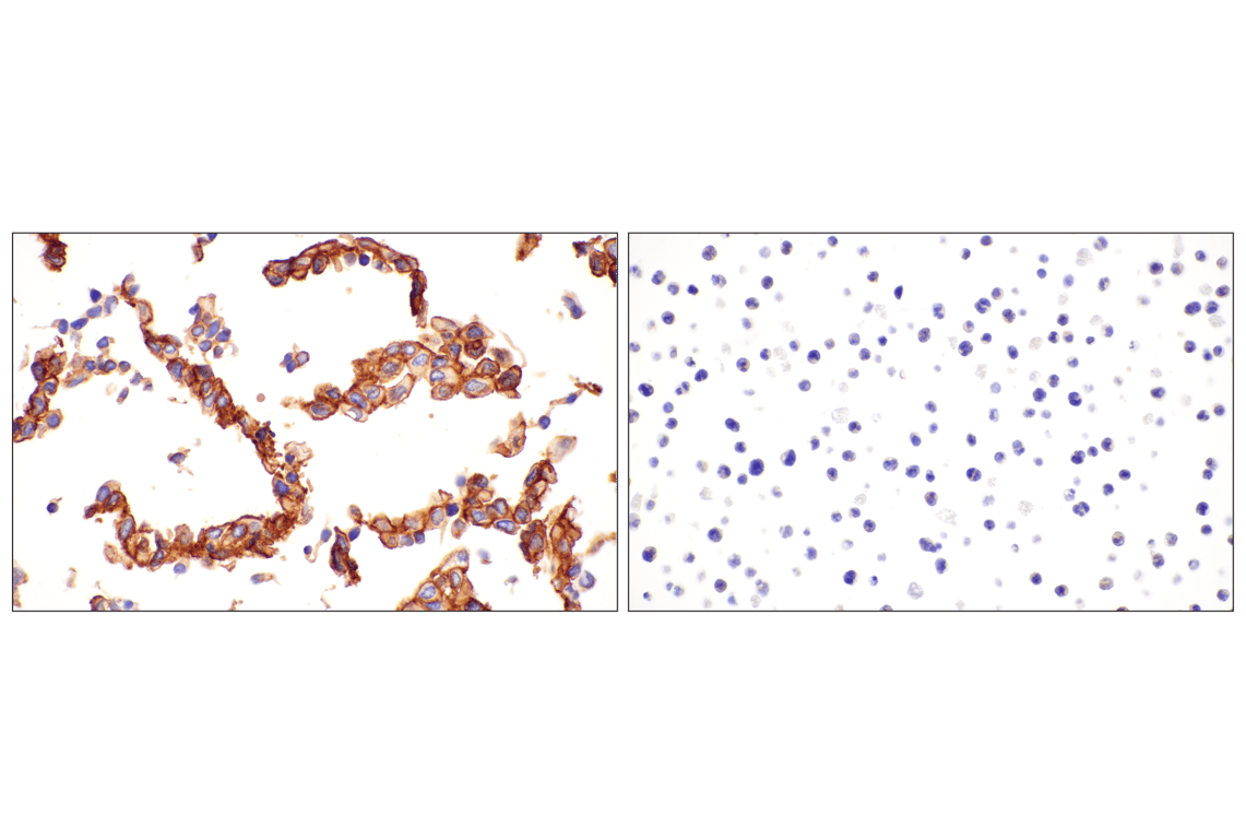
Immunohistochemical analysis of paraffin-embedded C2C12 cell pellet (left, positive) or Ba/F3 cell pellet (right, negative) using CD81 (D5O2Q) Rabbit mAb.