Revision 4
#9936
Store at -20C
NF-κB Pathway Antibody Sampler Kit
1 Kit
(7 x 20 microliters)
877-616-CELL (2355)
877-678-TECH (8324)
3 Trask Lane | Danvers | Massachusetts | 01923 | USA
For Research Use Only. Not for Use in Diagnostic Procedures.
| Product Includes | Product # | Quantity | Mol. Wt | Isotype/Source |
|---|---|---|---|---|
| IKKα (3G12) Mouse mAb | 11930 | 20 µl | 85 kDa | Mouse IgG1 |
| IKKβ (D30C6) Rabbit mAb | 8943 | 20 µl | 87 kDa | Rabbit IgG |
| Phospho-IKKα/β (Ser176/180) (16A6) Rabbit mAb | 2697 | 20 µl | 85 IKK-alpha 87 IKK-beta kDa | Rabbit IgG |
| Phospho-NF-κB p65 (Ser536) (93H1) Rabbit mAb | 3033 | 20 µl | 65 kDa | Rabbit IgG |
| IκBα (L35A5) Mouse mAb (Amino-terminal Antigen) | 4814 | 20 µl | 39 kDa | Mouse IgG1 |
| Phospho-IκBα (Ser32) (14D4) Rabbit mAb | 2859 | 20 µl | 40 kDa | Rabbit IgG |
| NF-κB p65 (D14E12) XP® Rabbit mAb | 8242 | 20 µl | 65 kDa | Rabbit IgG |
| Anti-rabbit IgG, HRP-linked Antibody | 7074 | 100 µl | Goat | |
| Anti-mouse IgG, HRP-linked Antibody | 7076 | 100 µl | Horse |
Please visit cellsignal.com for individual component applications, species cross-reactivity, dilutions, protocols, and additional product information.
Description
The NF-κB Pathway Antibody Sampler Kit contains reagents to examine the activation state and total protein levels of key proteins in the NF-κB pathway: IKKα, IKKβ, NF-κB p65/RelA, and IκBα. The kit contains enough primary and secondary antibodies to perform two Western blot experiments per primary antibody.
Storage
Background
The transcriptional nuclear factor κB (NF-κB)/Rel transcription factors are present in the cytosol in an inactive state, complexed with the inhibitory IκB proteins. Activation occurs via phosphorylation of IκBα at Ser32 and Ser36, resulting in the ubiquitin-mediated proteasome-dependent degradation of IκBα and the release and nuclear translocation of active NF-κB dimers. The regulation of IκBβ and IκBε is similar to that of IκBα, however, the phosphorylation and degradation of these proteins occurs with much slower kinetics. Phosphorylation of IκBβ occurs at Ser/Thr19 and Ser23, while IκBε can be phosphorylated at Ser18 and Ser22. The key regulatory step in this pathway involves activation of a high molecular weight IkappaB kinase (IKK) complex, consisting of three tightly associated IKK subunits. IKKα and IKKβ serve as the catalytic subunits of the kinase. Activation of IKK depends on phosphorylation at Ser177 and Ser181 in the activation loop of IKKβ (176 and 180 in IKKα). NF-κB-inducing kinase (NIK), TANK-binding kinase 1 (TBK1), and its homolog IKKε (IKKi), phosphorylate and activate IKKα and IKKβ.
The NF-κB family of transcription factors is comprised of five proteins in mammals, p65/RelA, c-Rel, RelB, NF-κB1 (p105/p50) and NF-κB2 (p100/p52). p105 and p100 are proteolytically processed to produce p50 and p52, respectively. The 50 kDa active form is produced through proteolytic processing following IKK-mediated phosphorylation of p105 at multiple sites (Ser922, 924, 928 and 933), while p100's processing to p52 is induced by phosphorylation of Ser864 and Ser868. The p50 and p52 products form dimeric complexes with Rel proteins, which are then able to bind DNA and regulate transcription. Phosphorylation of p65/RelA at Ser276 by PKA C and MSK1 enhances transcriptional activity. p65 phosphorylation at Ser536 regulates activation, nuclear localization, protein-protein interactions, and transcriptional activity. PMA-induced NF-κB transcriptional activity is dependent on the region of p65 containing the potential phosphorylation sites Ser457, Thr458, Thr464 and Ser468. Phosphorylation of Ser468 by GSK-3β inhibits basal p65 activity.
Trademarks and Patents
Cell Signaling Technology is a trademark of Cell Signaling Technology, Inc.
XP is a registered trademark of Cell Signaling Technology, Inc.
All other trademarks are the property of their respective owners. Visit cellsignal.com/trademarks for more information.
Limited Uses
Except as otherwise expressly agreed in a writing signed by a legally authorized representative of CST, the following terms apply to Products provided by CST, its affiliates or its distributors. Any Customer's terms and conditions that are in addition to, or different from, those contained herein, unless separately accepted in writing by a legally authorized representative of CST, are rejected and are of no force or effect.
Products are labeled with For Research Use Only or a similar labeling statement and have not been approved, cleared, or licensed by the FDA or other regulatory foreign or domestic entity, for any purpose. Customer shall not use any Product for any diagnostic or therapeutic purpose, or otherwise in any manner that conflicts with its labeling statement. Products sold or licensed by CST are provided for Customer as the end-user and solely for research and development uses. Any use of Product for diagnostic, prophylactic or therapeutic purposes, or any purchase of Product for resale (alone or as a component) or other commercial purpose, requires a separate license from CST. Customer shall (a) not sell, license, loan, donate or otherwise transfer or make available any Product to any third party, whether alone or in combination with other materials, or use the Products to manufacture any commercial products, (b) not copy, modify, reverse engineer, decompile, disassemble or otherwise attempt to discover the underlying structure or technology of the Products, or use the Products for the purpose of developing any products or services that would compete with CST products or services, (c) not alter or remove from the Products any trademarks, trade names, logos, patent or copyright notices or markings, (d) use the Products solely in accordance with CST Product Terms of Sale and any applicable documentation, and (e) comply with any license, terms of service or similar agreement with respect to any third party products or services used by Customer in connection with the Products.
Revision 4
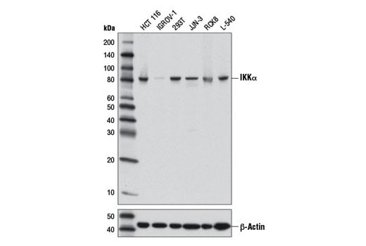
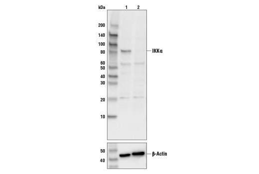
Western blot analysis of extracts from various cell lines using IKKα (3G12) Mouse mAb (upper) or β-Actin (D6A8) Rabbit mAb #8457 (lower).
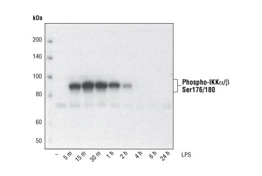
Western blot analysis of extracts from THP-1 cells, differentiated with TPA (#9905, 80 nM for 24h) and treated with 1 μg/ml LPS for the indicated times, using Phospho-IKKα/β (Ser176/180) (16A6) Rabbit mAb.
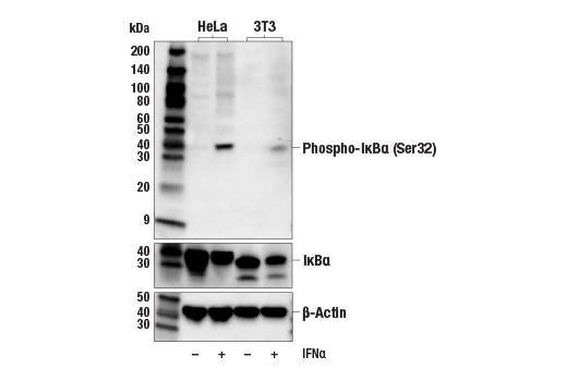
Western blot analysis of extracts from various cell lines, untreated or treated with IFNa (#36000, 20 ng/mL, 5 min); using Phospho-IκBα (Ser32) (14D4) Rabbit mAb #2859 (upper), IκBα (L35A5) Mouse mAb (Amino-terminal Antigen) #4814 (middle), and β-Actin (D6A8) Rabbit mAb #8457 (lower).
Revision 4
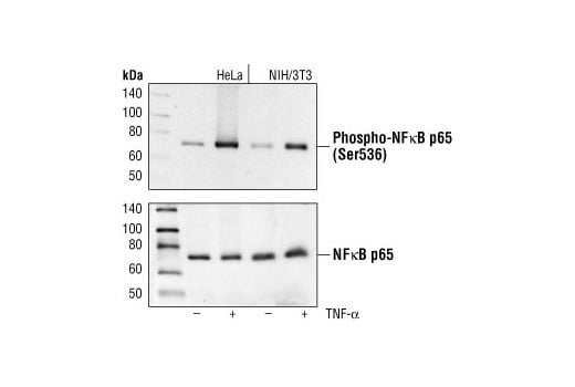
Western blot analysis of extracts from HeLa and NIH/3T3 cells, untreated or TNF-α treated (#2169, 20 ng/ml for 5 minutes), using Phospho-NF-κB p65 (Ser536) (93H1) Rabbit mAb (upper) or NF-κB p65 Antibody #3034 (lower).
Western blot analysis of HeLa cell extracts (lane 1) or IKKα knock-out HeLa cells (lane 2), using IKKα (3G12) Mouse mAb #11930 (upper), or β-actin (13E5) Rabbit mAb #4970 (lower). The absence of signal in the knockout HeLa cells confirms specificity of the antibody for IKKα.
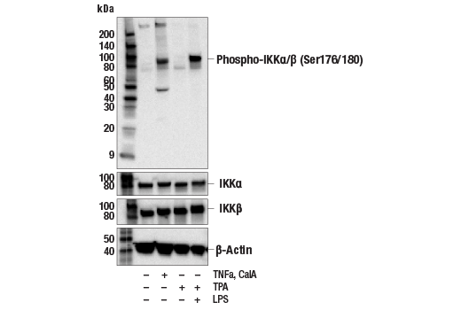
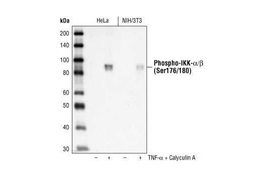
Western blot analysis of extracts from HeLa cells, untreated or treated with TNFa (#16789, 20 ng/mL, 10 min) and Calyculin A (#9902, 100 nM, 10 min); THP-1 cells differentiated with TPA (#4174, 80 nM for 16hr) untreated and treated with LPS (#14011, 1 μg/ml for 1hr) using #2697 (upper) or IKKα (D3W6N) Rabbit mAb #61294, IKKβ (D30C6) Rabbit mAb #8943 (middle), and β-Actin (D6A8) Rabbit mAb #8457 (lower).
Revision 4
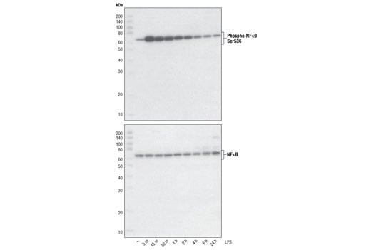
Western blot analysis of extracts from THP-1 cells, differentiated with TPA (#9905, 80 nM for 24h) and treated with 1 μg/ml LPS for the indicated times, using Phospho-NF-κB p65 (Ser536) (93H1) Rabbit mAb (upper) and NF-κB p65 (C22B4) Rabbit mAb #4764 (lower).
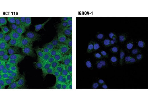
Confocal immunofluorescent analysis of HCT 116 (high expression; left) and IGROV-1 (low expression; right) cells using IKKα (3G12) Mouse mAb (green). Blue pseudocolor = DRAQ5® #4084 (fluorescent DNA dye).
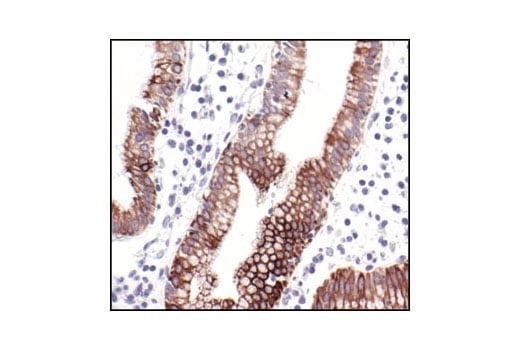
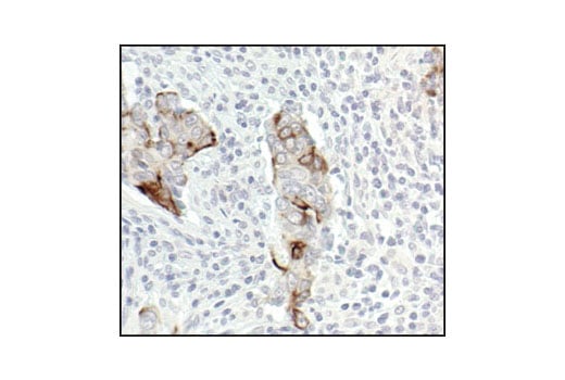
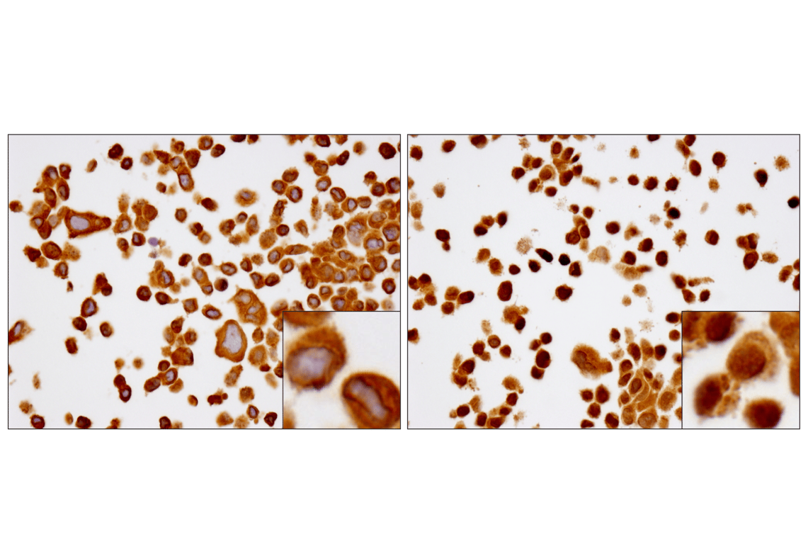
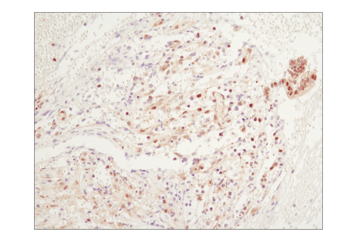
Immunohistochemical analysis of paraffin-embedded human gall bladder (chronic cholecystitis), using Phospho-IKKα/β (Ser176/180) (16A6) Rabbit mAb.
Revision 4
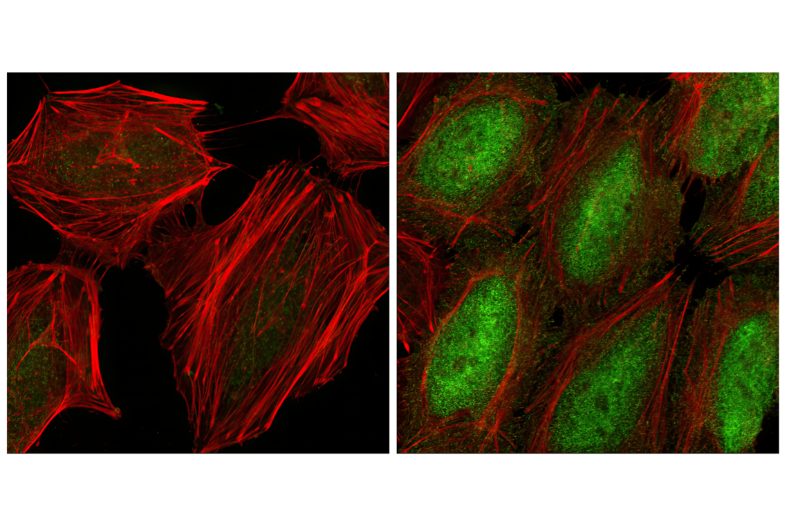
Confocal immunofluorescent analysis of HeLa cells, serum starved (left) or TNF-α treated (#8902 at 20 ng/ml for 20 min, right), using Phospho-NF-κB p65 (Ser536) (93H1) Rabbit mAb (green). Actin filaments have been labeled with Alexa Fluor® phalloidin 555 (red).
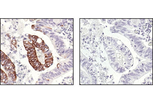
Immunohistochemical analysis of paraffin-embedded human colon carcinoma, untreated (left) or λ phosphatase-treated (right), using Phospho-IKKα/β (Ser176/180) (16A6) Rabbit mAb.
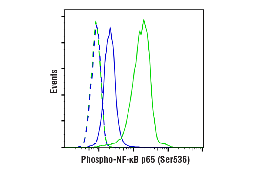
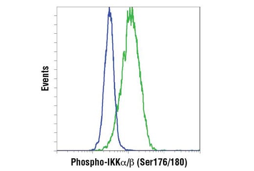
Flow cytometric analysis of HeLa cells, untreated (blue) or treated with Human Tumor Necrosis Factor-α (hTNF-α) #8902 and Calyculin A #9902 (20 ng/ml and 100 nM, 15 min; green), using Phospho-NF-κB p65 (Ser536) (93H1) Rabbit mAb (solid lines) or concentration-matched Rabbit (DA1E) mAb IgG XP® Isotype Control #3900 (dashed lines). Anti-rabbit IgG (H+L), F(ab')2 Fragment (Alexa Fluor® 488 Conjugate) #4412 was used as a secondary antibody.
Revision 4
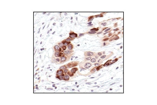
Immunohistochemical analysis of paraffin-embedded human colon carcinoma, showing cytoplasmic localization, using Phospho-IKKα/β (Ser176/180) (16A6) Rabbit mAb.
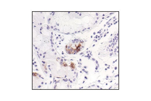
Immunohistochemical analysis of paraffin-embedded human lung (chronic bronchitis), using Phospho-IKKα/β (Ser176/180) (16A6) Rabbit mAb.
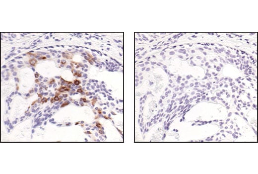
Immunohistochemical analysis of paraffin-embedded human breast carcinoma, using Phospho-IKKα/β (Ser176/180) (16A6) Rabbit mAb in the presence of control peptide (left) or Phospho-IKK-alpha/beta (Ser176/180) Blocking Peptide #1023 (right).
Revision 4
Flow cytometric analysis of THP-1 cells, untreated (blue) and with TPA and LPS (green) using IKK-α (Ser176/Ser180) phosphate Rabbit mAb. Anti-rabbit IgG (H+L), F(ab')2 Fragment (PE Conjugate) #8885 was used as a secondary antibody.
Western blot analysis of extracts from TNF-alpha and Calyculin A treated HeLa and NIH/3T3 cells, using Phospho-IKKα/β (Ser176/180) (16A6) Rabbit mAb.
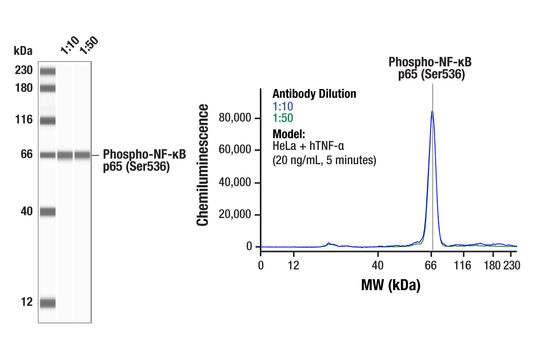
Simple Western™ analysis of lysates (1.0 mg/mL) from HeLa cells treated with hTNF-α (20 ng/mL, 5 minutes) using Phospho-NF-κB p65 (Ser536) (93H1) Rabbit mAb #3033. The virtual lane view (left) shows a single target band (as indicated) at 1:10 and 1:50 dilutions of primary antibody. The corresponding electropherogram view (right) plots chemiluminescence by molecular weight along the capillary at 1:10 (blue line) and 1:50 (green line) dilutions of primary antibody. This experiment was performed under reducing conditions on the Jess™ Simple Western instrument from ProteinSimple, a BioTechne brand, using the 12-230 kDa separation module.
Revision 4
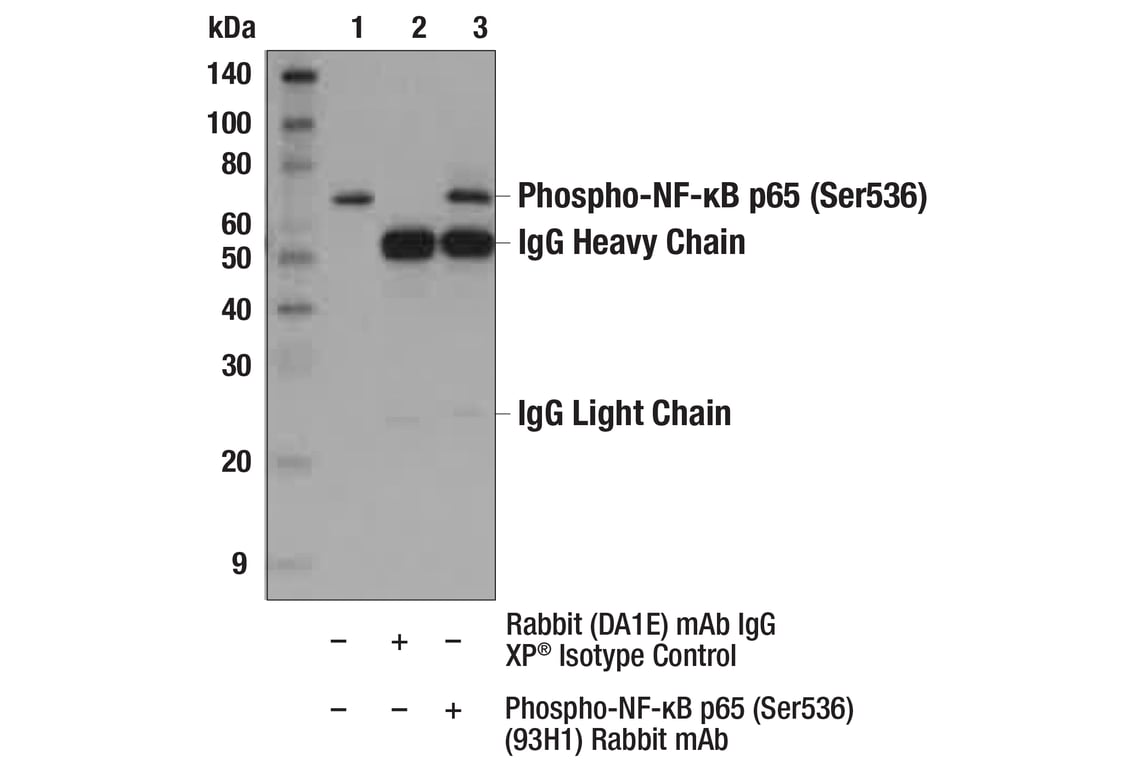
Immunoprecipitation of Phospho-NF-κB p65 (Ser536) from HeLa extracts treated with hTNF-α #8902 (20 ng/ml, 5 min). Lane 1 is 10% input, lane 2 is Rabbit (DA1E) mAb IgG XP® Isotype Control #3900, and lane 3 is Phospho-NF-κB p65 (Ser536) (93H1) Rabbit mAb. Western blot analysis was performed using Phospho-NF-κB p65 (Ser536) (93H1) Rabbit mAb. Anti-rabbit IgG, HRP-linked Antibody #7074 was used as a secondary antibody.
Western blot analysis of extracts from various cell lines using IκBα (L35A5) Mouse mAb (Amino-terminal Antigen) #4814 (upper) or β-Actin (D6A8) Rabbit mAb #8457 (lower).

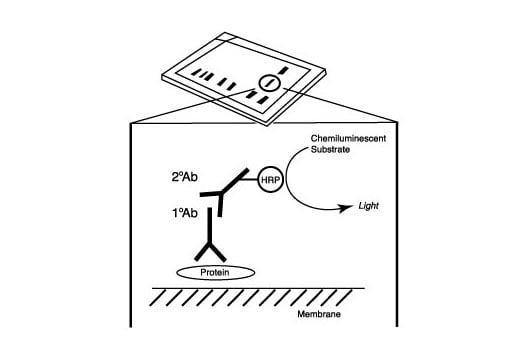
After the primary antibody is bound to the target protein, a complex with HRP-linked secondary antibody is formed. The LumiGLO® is added and emits light during enzyme catalyzed decomposition.
Revision 4
After the primary antibody is bound to the target protein, a complex with HRP-linked secondary antibody is formed. The LumiGLO* is added and emits light during enzyme catalyzed decomposition.

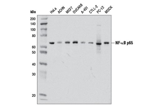
Western blot analysis of extracts from various cell lines using NF-κB p65 (D14E12) XP® Rabbit mAb.
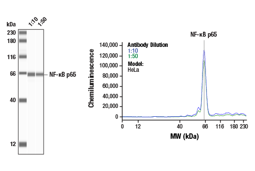
Simple Western™ analysis of lysates (1 mg/mL) from HeLa cells using NF-κB p65 (D14E12) XP® Rabbit mAb #8242. The virtual lane view (left) shows a single target band (as indicated) at 1:10 and 1:50 dilutions of primary antibody. The corresponding electropherogram view (right) plots chemiluminescence by molecular weight along the capillary at 1:10 (blue line) and 1:50 (green line) dilutions of primary antibody. This experiment was performed under reducing conditions on the Jess™ Simple Western instrument from ProteinSimple, a BioTechne brand, using the 12-230 kDa separation module.
Revision 4
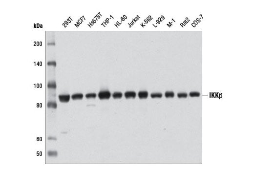
Western blot analysis of extracts from various cell lines using IKKβ (D30C6) Rabbit mAb.
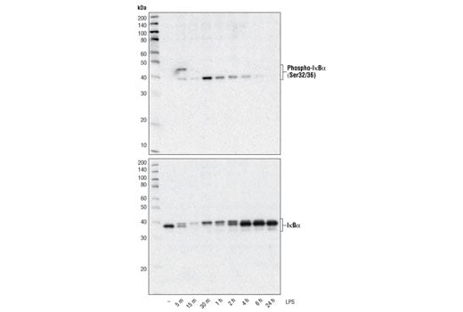
Western blot analysis of extracts from THP-1 cells, differentiated with TPA (#9905, 80 nM for 24h) and treated with 1 μg/ml LPS for the indicated times, using Phospho-IκBα (Ser32/36) (5A5) Mouse mAb #9246 (upper) and IκBα (L35A5) Mouse mAb (Amino-terminal Antigen) (lower).
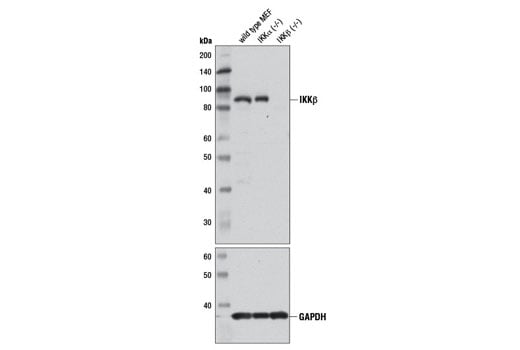
Western blot analysis of extracts from wild-type, IKKα (-/-), and IKKβ (-/-) mouse embryonic fibroblasts (MEFs) using IKKβ (D30C6) Rabbit mAb (upper) and GAPDH (14C10) Rabbit mAb #2118 (lower).
Revision 4
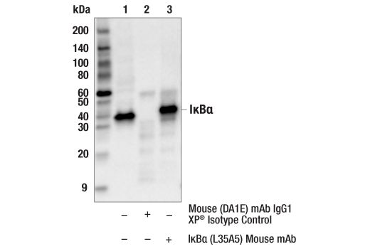
Immunoprecipitation of IkBa from HeLa cell extracts. Lane 1 is 10% input, lane 2 is precipitated with Mouse (G3A1) mAb IgG1 Isotype Control #5415, and lane 3 is IκBα (L35A5) Mouse mAb (Amino-terminal Antigen), #4814. Western blot was performed using IκBα Antibody #9242.
Immunohistochemical analysis using NF-κB p65 (D14E12) XP® Rabbit mAb on SignalSlide® NF-κB p65 IHC Controls #12873 (paraffin-embedded HCT116 cells, untreated (left) or treated with hTNF-α #8902 (right)).
Immunohistochemical analysis of paraffin-embedded human leiomyoma, using IκBα (L35A5) Mouse mAb (Amino-terminal Antigen).

Revision 4
Immunohistochemical analysis of paraffin-embedded human chronic cholecystitis using NF-κB p65 (D14E12) XP® Rabbit mAb.
Immunohistochemical analysis of paraffin-embedded human lung carcinoma, using IκBα (L35A5) Mouse mAb (Amino-terminal Antigen).
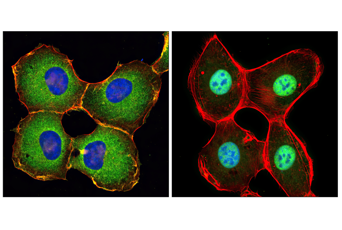
Confocal immunofluorescent analysis of HT-1080 cells, untreated (left) or treated with hTNF-α #8902 (20 ng/ml, 20 min) (right), using NF-κB p65 (D14E12) XP® Rabbit mAb (green). Actin filaments were labeled with DY-554 phalloidin (red). Blue pseudocolor = DRAQ5® #4084 (fluorescent DNA dye).
Revision 4
Immunohistochemical analysis of paraffin-embedded human renal adenocarcinoma, using IκBα (L35A5) Mouse mAb (Amino-terminal Antigen).

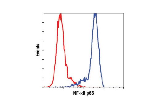
Flow cytometric analysis of HeLa cells using NF-κB p65 (D14E12) XP® Rabbit mAb (blue) compared to concentration matched Rabbit (DA1E) mAb IgG XP® Isotype Control #3900 (red).
Confocal immunofluorescent analysis of HeLa cells, untreated (left), or TNF-α-treated (#8902, 10 ng/ml for 15 min, right) using IκBα (L35A5) Mouse mAb (Amino-terminal Antigen) (red). Blue pseudocolor = DRAQ5® #4084 (fluorescent DNA dye).

Revision 4
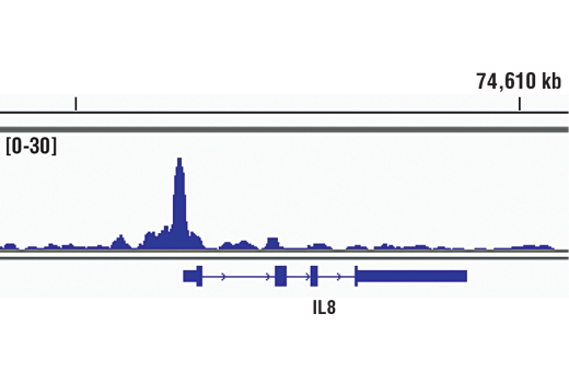
Chromatin immunoprecipitations were performed with cross-linked chromatin from HeLa cells treated with hTNF-α #8902 (30 ng/ml, 1 hr) and NF-κB p65 (D14E12) XP® Rabbit mAb, using SimpleChIP® Enzymatic Chromatin IP Kit (Magnetic Beads) #9005. DNA Libraries were prepared using DNA Library Prep Kit for Illumina® (ChIP-seq, CUT&RUN) #56795. The figure shows binding across IL-8, a known target gene of NFκB (see additional figure containing ChIP-qPCR data). For additional ChIP-seq tracks, please download the product datasheet.
Flow cytometric analysis of NIH/3T3 cells, treated with TNF-α (#8902, 10 ng/ml for 5 min; blue, negative) or untreated (green, positive) using IκBα (L35A5) Mouse mAb (Amino-terminal Antigen) (solid lines) or concentration-matched Mouse (G3A1) mAb IgG1 Isotype Control #5415 (dashed lines). Anti-mouse IgG (H+L), F(ab')2 Fragment (Alexa Fluor® 488 Conjugate) #4408 was used as a secondary antibody.

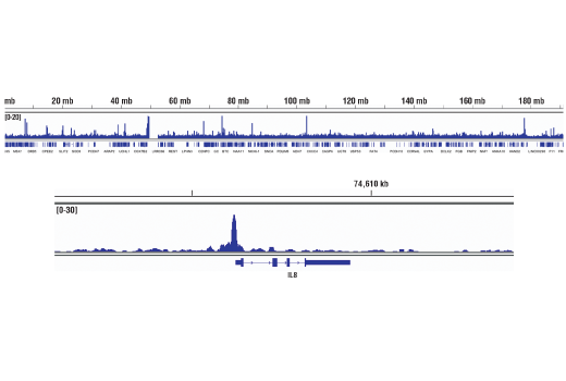
Chromatin immunoprecipitations were performed with cross-linked chromatin from HeLa cells treated with hTNF-α #8902 (30 ng/ml, 1 hr) and NF-κB p65 (D14E12) XP® Rabbit mAb, using SimpleChIP® Enzymatic Chromatin IP Kit (Magnetic Beads) #9005. DNA Libraries were prepared using DNA Library Prep Kit for Illumina® (ChIP-seq, CUT&RUN) #56795. The figure shows binding across chromosome 4 (upper), including IL-8 (lower), a known target gene of NFκB (see additional figure containing ChIP-qPCR data).
Revision 4
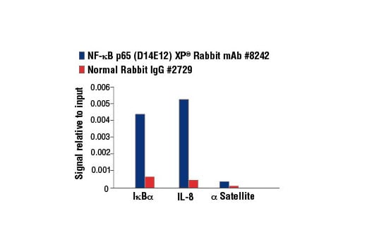
Chromatin immunoprecipitations were performed with cross-linked chromatin from HeLa cells treated with hTNF-α #8902 (30 ng/ml, 1 hr) and either NF-κB p65 (D14E12) XP® Rabbit mAb or Normal Rabbit IgG #2729 using SimpleChIP® Enzymatic Chromatin IP Kit (Magnetic Beads) #9003. The enriched DNA was quantified by Real-Time PCR using SimpleChIP® Human IκBα Promoter Primers #5552, human IL-8 promoter primers, and SimpleChIP® Human α Satellite Repeat Primers #4486. The amount of immunoprecipitated DNA in each sample is represented as signal relative to the total amount of input chromatin, which is equivalent to one.
CUT&RUN was performed with HeLa cells treated with hTNF-α #8902 (30 ng/ml, 1 hr) and NF-κB p65 (D14E12) XP® Rabbit mAb, using CUT&RUN Assay Kit #86652. DNA Libraries were prepared using DNA Library Prep Kit for Illumina® (ChIP-seq, CUT&RUN) #56795. The figure shows binding across LAMC2, a known target gene of NF-κB p65 (see additional figure containing CUT&RUN-qPCR data).
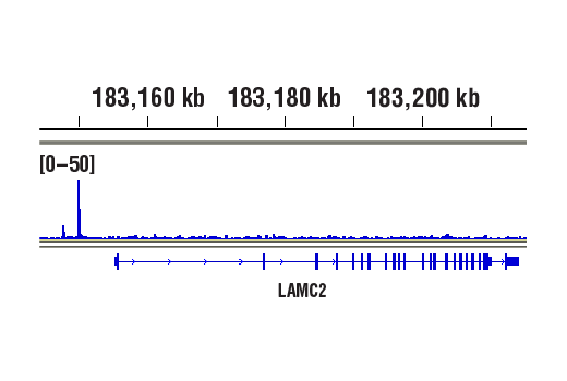
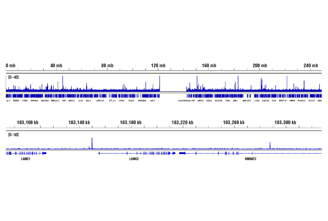
CUT&RUN was performed with HeLa cells treated with hTNF-α #8902 (30 ng/ml, 1 hr) and NF-κB p65 (D14E12) XP® Rabbit mAb, using CUT&RUN Assay Kit #86652. DNA Libraries were prepared using DNA Library Prep Kit for Illumina® (ChIP-seq, CUT&RUN) #56795. The figures show binding across chromosome 1 (upper), including LAMC2 (lower), a known target gene of NF-κB p65 (see additional figure containing CUT&RUN-qPCR data).
Revision 4
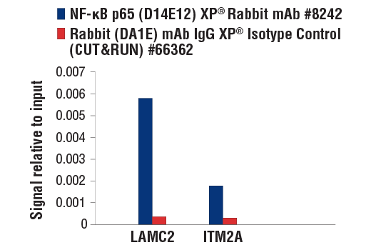
CUT&RUN was performed with HeLa cells treated with hTNF-α #8902 (30 ng/ml, 1 hr) and either NF-κB p65 (D14E12) XP® Rabbit mAb or Rabbit (DA1E) mAb IgG XP® Isotype Control (CUT&RUN) #66362, using CUT&RUN Assay Kit #86652. The enriched DNA was quantified by real-time PCR using human LAMC2 upstream primers, and human ITM2A upstream primers. The amount of immunoprecipitated DNA in each sample is represented as signal relative to the total amount of input chromatin, which is equivalent to one.
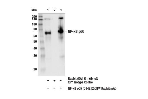
Immunoprecipitation of NF-kB p65 from CHO cell extracts. Lane 1 is 10% input, lane 2 is precipitated with Rabbit (DA1E) mAb IgG XP® Isotype Control #3900, and lane 3 is NF-κB p65 (D14E12) XP® Rabbit mAb, #8242. Western blot was performed using NF-κB p65 (L8F6) Mouse mAb, #6956.