Revision 7
#88742
Store at -20C
877-616-CELL (2355)
877-678-TECH (8324)
3 Trask Lane | Danvers | Massachusetts | 01923 | USA
For Research Use Only. Not for Use in Diagnostic Procedures.
Applications:
W, W-S, IP, IHC-P, IF-F, IF-IC, FC-FP
Reactivity:
M
Sensitivity:
Endogenous
MW (kDa):
58-75
Source/Isotype:
Rabbit IgG
UniProt ID:
#P10923
Entrez-Gene Id:
20750
Product Usage Information
| Application | Dilution |
|---|---|
| Western Blotting | 1:1000 |
| Simple Western™ | 1:10 - 1:50 |
| Immunoprecipitation | 1:200 |
| Immunohistochemistry (Paraffin) | 1:50 - 1:200 |
| Immunofluorescence (Frozen) | 1:200 - 1:800 |
| Immunofluorescence (Immunocytochemistry) | 1:200 - 1:800 |
| Flow Cytometry (Fixed/Permeabilized) | 1:200 - 1:800 |
Storage
For a carrier free (BSA and azide free) version of this product see product #17090.
Specificity/Sensitivity
Osteopontin/SPP1 (E9Z1D) Rabbit mAb recognizes endogenous levels of total osteopontin/SPP1 protein.
Source / Purification
Monoclonal antibody is produced by immunizing animals with a synthetic peptide corresponding to residues surrounding Ala149 of mouse osteopontin/SPP1 protein.
Background
Osteopontin is a highly phosphorylated secreted glycoprotein expressed in a variety of cell types. Osteopontin, encoded by the gene secreted phosphoprotein 1 (SPP1), was originally isolated and cloned from a rat osteosarcoma cell line (1). Osteopontin is a highly negatively charged protein that likely regulates extracellular matrix turnover and remodeling, both in bone and other tissue (2). Osteopontin also has three highly conserved receptor-binding domains, otherwise known as arginine-glycine-aspartic acid (RGD) domains. Binding of osteopontin to cell surface integrins likely activates cell signaling pathways that regulate cell proliferation, adhesion, invasion, migration, and fibrosis (3,4). Osteopontin may be regulated by posttranslational regulation, including phosphorylation, glycosylation, and proteolytic cleavage (5). Osteopontin is also linked to several human diseases. For example, cleaved osteopontin is higher in patients suffering with cirrhosis, a disease involving fibrosis of the liver (5). Its role in the brain is unclear, but SPP1 expression in microglia, the resident macrophages of the brain, is upregulated in mouse neurodegenerative disease models, including Alzheimer’s disease (6). Osteopontin’s role in disease likely involve its function as a key mediator of inflammatory responses (4).
Background References
- Oldberg, A. et al. (1986) Proc Natl Acad Sci U S A 83, 8819-23.
- Kazanecki, C.C. et al. (2007) J Cell Biochem 102, 912-24.
- Standal, T. et al. (2004) Exp Oncol 26, 179-84.
- Lamort, A.S. et al. (2019) Cells 8, 815. doi: 10.3390/cells8080815.
- Song, Z. et al. (2021) Hepatology 73, 1594-1608.
- Sala Frigerio, C. et al. (2019) Cell Rep 27, 1293-1306.e6.
Species Reactivity
Species reactivity is determined by testing in at least one approved application (e.g., western blot).
Western Blot Buffer
IMPORTANT: For western blots, incubate membrane with diluted primary antibody in 5% w/v BSA, 1X TBS, 0.1% Tween® 20 at 4°C with gentle shaking, overnight.
Applications Key
W: Western Blotting IP: Immunoprecipitation IHC-P: Immunohistochemistry (Paraffin) IF-F: Immunofluorescence (Frozen) FC-FP: Flow Cytometry (Fixed/Permeabilized)
Cross-Reactivity Key
H: Human M: Mouse R: Rat Hm: Hamster Mk: Monkey Vir: Virus Mi: Mink C: Chicken Dm: D. melanogaster X: Xenopus Z: Zebrafish B: Bovine Dg: Dog Pg: Pig Sc: S. cerevisiae Ce: C. elegans Hr: Horse GP: Guinea Pig Rab: Rabbit G: Goat All: All Species Expected
Trademarks and Patents
Cell Signaling Technology is a trademark of Cell Signaling Technology, Inc.
Alexa Fluor is a registered trademark of Life Technologies Corporation.
All other trademarks are the property of their respective owners. Visit cellsignal.com/trademarks for more information.
Limited Uses
Except as otherwise expressly agreed in a writing signed by a legally authorized representative of CST, the following terms apply to Products provided by CST, its affiliates or its distributors. Any Customer's terms and conditions that are in addition to, or different from, those contained herein, unless separately accepted in writing by a legally authorized representative of CST, are rejected and are of no force or effect.
Products are labeled with For Research Use Only or a similar labeling statement and have not been approved, cleared, or licensed by the FDA or other regulatory foreign or domestic entity, for any purpose. Customer shall not use any Product for any diagnostic or therapeutic purpose, or otherwise in any manner that conflicts with its labeling statement. Products sold or licensed by CST are provided for Customer as the end-user and solely for research and development uses. Any use of Product for diagnostic, prophylactic or therapeutic purposes, or any purchase of Product for resale (alone or as a component) or other commercial purpose, requires a separate license from CST. Customer shall (a) not sell, license, loan, donate or otherwise transfer or make available any Product to any third party, whether alone or in combination with other materials, or use the Products to manufacture any commercial products, (b) not copy, modify, reverse engineer, decompile, disassemble or otherwise attempt to discover the underlying structure or technology of the Products, or use the Products for the purpose of developing any products or services that would compete with CST products or services, (c) not alter or remove from the Products any trademarks, trade names, logos, patent or copyright notices or markings, (d) use the Products solely in accordance with CST Product Terms of Sale and any applicable documentation, and (e) comply with any license, terms of service or similar agreement with respect to any third party products or services used by Customer in connection with the Products.
Revision 7
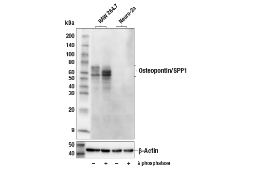
Western blot analysis of extracts from RAW 264.7 and Neuro-2a cells, untreated (-) or treated with λ phosphatase (+), using Osteopontin/SPP1 (E9Z1D) Rabbit mAb (upper) or β-Actin (D6A8) Rabbit mAb #8457 (lower).
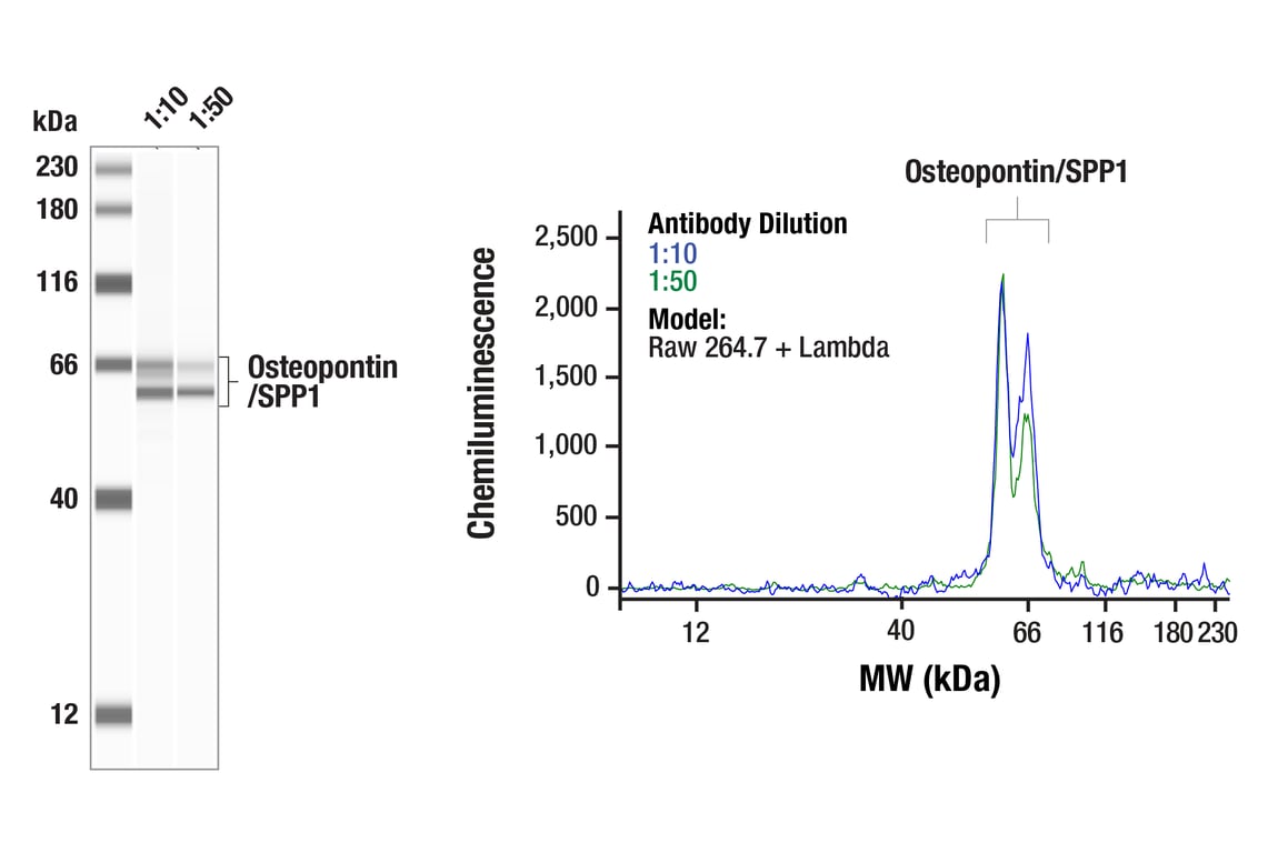
Simple Western™ analysis of lysates (0.1 mg/mL) from Raw 264.7 cells treated with Lambda using Osteopontin/SPP1 (E9Z1D) Rabbit mAb #88742. The virtual lane view (left) shows the target band (as indicated) at 1:10 and 1:50 dilutions of primary antibody. The corresponding electropherogram view (right) plots chemiluminescence by molecular weight along the capillary at 1:10 (blue line) and 1:50 (green line) dilutions of primary antibody. This experiment was performed under reducing conditions on the Jess™ Simple Western instrument from ProteinSimple, a BioTechne brand, using the 12-230 kDa separation module.
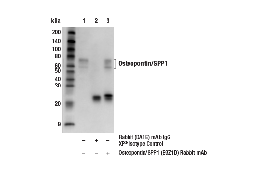
Immunoprecipitation of osteopontin/SPP1 protein from RAW 264.7 cell extracts. Lane 1 is 10% input, lane 2 is Rabbit (DA1E) mAb IgG XP® Isotype Control #3900, and lane 3 is Osteopontin/SPP1 (E9Z1D) Rabbit mAb. Western blot analysis was performed using Osteopontin/SPP1 (E4O2F) Rabbit mAb #27927. Mouse Anti-Rabbit IgG (Light-Chain Specific) (D4W3E) mAb (HRP Conjugate) #93702 was used as a secondary antibody.
Revision 7
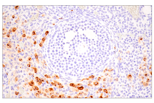
Immunohistochemical analysis of paraffin-embedded mouse ovary using Osteopontin/SPP1 (E9Z1D) Rabbit mAb.
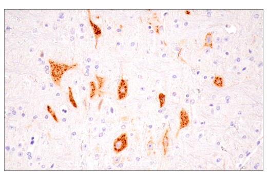
Immunohistochemical analysis of paraffin-embedded mouse pancreas using Osteopontin/SPP1 (E9Z1D) Rabbit mAb.
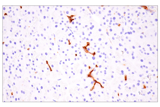
Immunohistochemical analysis of paraffin-embedded pons region of mouse brain using Osteopontin/SPP1 (E9Z1D) Rabbit mAb.
Revision 7
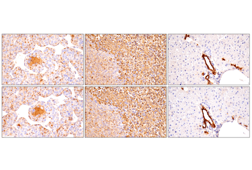
Immunohistochemical analysis of paraffin-embedded renca syngeneic tumor (left), 4T1 syngeneic tumor (middle), or mouse liver (right) using Osteopontin/SPP1 (E9Z1D) Rabbit mAb (top) or an Osteopontin/SPP1 Rabbit mAb (bottom). These two antibodies detect unique, non-overlapping epitopes on mouse Osteopontin/SPP1. The similar staining patterns obtained with both antibodies help to confirm the specificity of the staining.
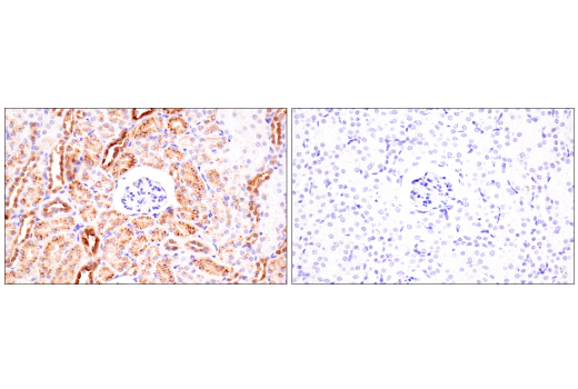
Immunohistochemical analysis of paraffin-embedded mouse kidney using Osteopontin/SPP1 (E9Z1D) Rabbit mAb (left) compared to concentration-matched Rabbit (DA1E) mAb IgG XP® Isotype Control #3900 (right).
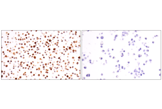
Immunohistochemical analysis of paraffin-embedded RAW 264.7 cell pellet (left, positive) or Neuro-2a cell pellet (right, negative) using Osteopontin/SPP1 (E9Z1D) Rabbit mAb.
Revision 7
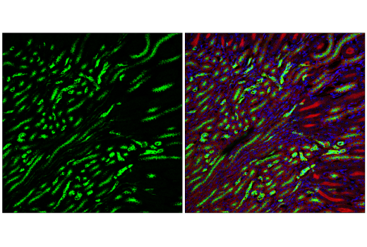
Confocal immunofluorescent analysis of fixed frozen mouse kidney labeled with Osteopontin/SPP1 (E9Z1D) Rabbit mAb (left, green). Free secondary binding sites were then blocked with Rabbit (DA1E) mAb IgG XP® Isotype Control #3900 prior to co-labeling with Ras (E4K9L) Rabbit mAb (Alexa Fluor® 488 Conjugate) #51813 (right, red pseudocolor) and DAPI #4083 (right, blue).
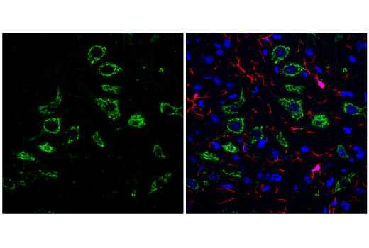
Confocal immunofluorescent analysis of fixed frozen mouse thalamus labeled with Osteopontin/SPP1 (E9Z1D) Rabbit mAb (left, green). Free secondary binding sites were then blocked with Rabbit (DA1E) mAb IgG XP® Isotype Control #3900 prior to co-labeling with Iba1/AIF-1 (E4O4W) XP® Rabbit mAb (Alexa Fluor® 647 Conjugate) #78060 (right, red) and DAPI #4083 (right, blue).
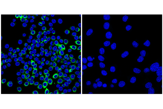
Confocal immunofluorescent analysis of RAW 264.7 cells (left, positive) and Neuro-2a cells (right, negative) using Osteopontin/SPP1 (E9Z1D) Rabbit mAb (green) and DAPI #4083 (blue). Cells were treated with Brefeldin A #9972 (100 ng/mL, 90 min) to inhibit protein secretion.