Revision 6
#39692
Store at -20C
877-616-CELL (2355)
877-678-TECH (8324)
3 Trask Lane | Danvers | Massachusetts | 01923 | USA
For Research Use Only. Not for Use in Diagnostic Procedures.
Applications:
W, IP, IHC-P, IF-IC
Reactivity:
H
Sensitivity:
Endogenous
MW (kDa):
75
Source/Isotype:
Rabbit IgG
UniProt ID:
#Q9H3D4
Entrez-Gene Id:
8626
Product Usage Information
| Application | Dilution |
|---|---|
| Western Blotting | 1:1000 |
| Immunoprecipitation | 1:50 |
| Immunohistochemistry (Paraffin) | 1:450 - 1:1800 |
| Immunofluorescence (Immunocytochemistry) | 1:400 - 1:1600 |
Storage
For a carrier-free (BSA and azide free) version of this product see product #64471.
Specificity/Sensitivity
p63 (D9L7L) XP® Rabbit mAb recognizes endogenous levels of total p63. Based on the sequence of the immunogenic peptide, this antibody is expected to recognize both full length (TA) p63 as well as DeltaN p63 isoforms that contain exon 4, such as alpha, beta, and gamma. This antibody will not detect DeltaNp73L (Q9H3D4-10/NM_001329146.1).
Source / Purification
Monoclonal antibody is produced by immunizing animals with a synthetic peptide corresponding to residues surrounding Asn118 of human p63 protein.
Background
The p53 tumor suppressor protein plays a major role in cellular response to DNA damage and other genomic aberrations. Activation of p53 can lead to either cell cycle arrest and DNA repair or apoptosis (1). In addition to p53, mammalian cells contain two p53 family members, p63 and p73, which are similar to p53 in both structure and function (2). While p63 can induce p53-responsive genes and apoptosis, mutation of p63 rarely results in tumors (2). Research investigators frequently observe amplification of the p63 gene in squamous cell carcinomas of the lung, head, and neck (2,3). The p63 gene contains an alternative transcription initiation site that yields a truncated ΔNp63 lacking the transactivation domain, and alternative splicing at the carboxy terminus yields the α, β, and γ isoforms (3,4).
Species Reactivity
Species reactivity is determined by testing in at least one approved application (e.g., western blot).
Western Blot Buffer
IMPORTANT: For western blots, incubate membrane with diluted primary antibody in 5% w/v BSA, 1X TBS, 0.1% Tween® 20 at 4°C with gentle shaking, overnight.
Applications Key
W: Western Blotting IP: Immunoprecipitation IHC-P: Immunohistochemistry (Paraffin) IF-IC: Immunofluorescence (Immunocytochemistry)
Cross-Reactivity Key
H: Human M: Mouse R: Rat Hm: Hamster Mk: Monkey Vir: Virus Mi: Mink C: Chicken Dm: D. melanogaster X: Xenopus Z: Zebrafish B: Bovine Dg: Dog Pg: Pig Sc: S. cerevisiae Ce: C. elegans Hr: Horse GP: Guinea Pig Rab: Rabbit G: Goat All: All Species Expected
Trademarks and Patents
Cell Signaling Technology is a trademark of Cell Signaling Technology, Inc.
XP is a registered trademark of Cell Signaling Technology, Inc.
All other trademarks are the property of their respective owners. Visit cellsignal.com/trademarks for more information.
Limited Uses
Except as otherwise expressly agreed in a writing signed by a legally authorized representative of CST, the following terms apply to Products provided by CST, its affiliates or its distributors. Any Customer's terms and conditions that are in addition to, or different from, those contained herein, unless separately accepted in writing by a legally authorized representative of CST, are rejected and are of no force or effect.
Products are labeled with For Research Use Only or a similar labeling statement and have not been approved, cleared, or licensed by the FDA or other regulatory foreign or domestic entity, for any purpose. Customer shall not use any Product for any diagnostic or therapeutic purpose, or otherwise in any manner that conflicts with its labeling statement. Products sold or licensed by CST are provided for Customer as the end-user and solely for research and development uses. Any use of Product for diagnostic, prophylactic or therapeutic purposes, or any purchase of Product for resale (alone or as a component) or other commercial purpose, requires a separate license from CST. Customer shall (a) not sell, license, loan, donate or otherwise transfer or make available any Product to any third party, whether alone or in combination with other materials, or use the Products to manufacture any commercial products, (b) not copy, modify, reverse engineer, decompile, disassemble or otherwise attempt to discover the underlying structure or technology of the Products, or use the Products for the purpose of developing any products or services that would compete with CST products or services, (c) not alter or remove from the Products any trademarks, trade names, logos, patent or copyright notices or markings, (d) use the Products solely in accordance with CST Product Terms of Sale and any applicable documentation, and (e) comply with any license, terms of service or similar agreement with respect to any third party products or services used by Customer in connection with the Products.
Revision 6
#39692
p63 (D9L7L) XP® Rabbit mAb
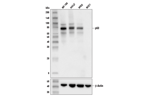
Western blot analysis of extracts from various cell lines cells using p63 (D9L7L) XP® Rabbit mAb (upper) or β-Actin (D6A8) Rabbit mAb #8457 (lower).
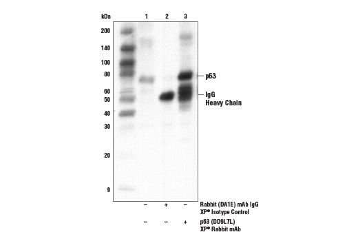
Immunoprecipitation of ME-180 cells. Lane 1 is 10% input, lane 2 is Rabbit (DA1E) mAb IgG XP® Isotype Control #3900, and lane 3 is p63 (D9L7L) XP® Rabbit mAb. Western blot analysis was performed using p63 (D9L7L) XP® Rabbit mAb.

Immunoprecipitation of ME-180 cells. Lane 1 is 10% input, lane 2 is Rabbit (DA1E) mAb IgG XP® Isotype Control #3900, and lane 3 is p63 (D9L7L) XP® Rabbit mAb. Western blot analysis was performed using p63 (D9L7L) XP® Rabbit mAb.
Revision 6
#39692
p63 (D9L7L) XP® Rabbit mAb
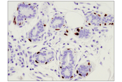
Immunohistochemical analysis of paraffin-embedded human intraductal and infiltrating carcinoma of the breast using p63 (D9L7L) XP® Rabbit mAb.
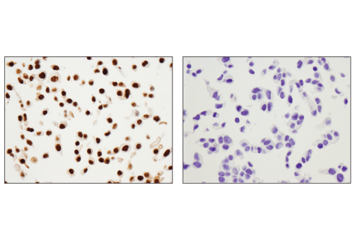
Immunohistochemical analysis of paraffin-embedded ME-180 cell pellet (left, positive) and MCF7 cell pellet (right, negative) using p63 (D9L7L) XP® Rabbit mAb.
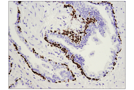
Immunohistochemical analysis of paraffin-embedded human prostate carcinoma using p63 (D9L7L) XP® Rabbit mAb.
Revision 6
#39692
p63 (D9L7L) XP® Rabbit mAb
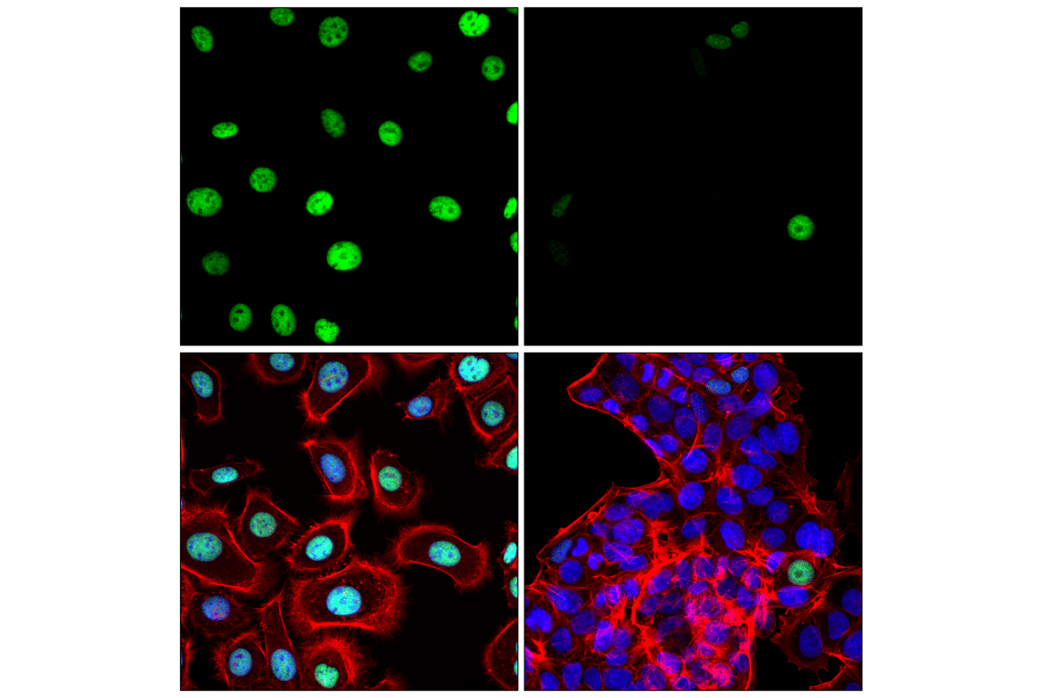
Confocal immunofluorescent analysis of ME-180 cells (left, positive) and MCF7 cells (right, negative excluding presumed cancer stem cells) using p63 (D9L7L) XP® Rabbit mAb (green), DyLight™ 554 Phalloidin #13054 (red), and DAPI #4083 (blue).