Revision 6
#5536
Store at -20C
877-616-CELL (2355)
877-678-TECH (8324)
3 Trask Lane | Danvers | Massachusetts | 01923 | USA
For Research Use Only. Not for Use in Diagnostic Procedures.
Applications:
W, W-S, IP, IF-IC
Reactivity:
H M R Mk
Sensitivity:
Endogenous
MW (kDa):
289
Source/Isotype:
Rabbit IgG
UniProt ID:
#P42345
Entrez-Gene Id:
2475
Product Usage Information
| Application | Dilution |
|---|---|
| Western Blotting | 1:1000 |
| Simple Western™ | 1:10 - 1:50 |
| Immunoprecipitation | 1:50 |
| Immunofluorescence (Immunocytochemistry) | 1:50 - 1:100 |
Storage
Specificity/Sensitivity
Phospho-mTOR (Ser2448) (D9C2) XP® Rabbit mAb detects endogenous levels of mTOR protein only when phosphorylated at Ser2448.
Species predicted to react based on 100% sequence homology
Rat, Chicken, Pig, Horse
Source / Purification
Monoclonal antibody is produced by immunizing animals with a synthetic phosphopeptide corresponding to residues surrounding Ser2448 of human mTOR protein.
Background
The mammalian target of rapamycin (mTOR, FRAP, RAFT) is a Ser/Thr protein kinase (1-3) that functions as an ATP and amino acid sensor to balance nutrient availability and cell growth (4,5). When sufficient nutrients are available, mTOR responds to a phosphatidic acid-mediated signal to transmit a positive signal to p70 S6 kinase and participate in the inactivation of the eIF4E inhibitor, 4E-BP1 (6). These events result in the translation of specific mRNA subpopulations. mTOR is phosphorylated at Ser2448 via the PI3 kinase/Akt signaling pathway and autophosphorylated at Ser2481 (7,8). mTOR plays a key role in cell growth and homeostasis and may be abnormally regulated in tumors. For these reasons, mTOR is currently under investigation as a potential target for anti-cancer therapy (9).
Background References
- Sabers, C.J. et al. (1995) J Biol Chem 270, 815-22.
- Brown, E.J. et al. (1994) Nature 369, 756-8.
- Sabatini, D.M. et al. (1994) Cell 78, 35-43.
- Gingras, A.C. et al. (2001) Genes Dev 15, 807-26.
- Dennis, P.B. et al. (2001) Science 294, 1102-5.
- Fang, Y. et al. (2001) Science 294, 1942-5.
- Navé, B.T. et al. (1999) Biochem J 344 Pt 2, 427-31.
- Peterson, R.T. et al. (2000) J Biol Chem 275, 7416-23.
- Huang, S. and Houghton, P.J. (2003) Curr Opin Pharmacol 3, 371-7.
Species Reactivity
Species reactivity is determined by testing in at least one approved application (e.g., western blot).
Western Blot Buffer
IMPORTANT: For western blots, incubate membrane with diluted primary antibody in 5% w/v BSA, 1X TBS, 0.1% Tween® 20 at 4°C with gentle shaking, overnight.
Applications Key
W: Western Blotting IP: Immunoprecipitation IF-IC: Immunofluorescence (Immunocytochemistry)
Cross-Reactivity Key
H: Human M: Mouse R: Rat Hm: Hamster Mk: Monkey Vir: Virus Mi: Mink C: Chicken Dm: D. melanogaster X: Xenopus Z: Zebrafish B: Bovine Dg: Dog Pg: Pig Sc: S. cerevisiae Ce: C. elegans Hr: Horse GP: Guinea Pig Rab: Rabbit G: Goat All: All Species Expected
Trademarks and Patents
Cell Signaling Technology is a trademark of Cell Signaling Technology, Inc.
XP is a registered trademark of Cell Signaling Technology, Inc.
All other trademarks are the property of their respective owners. Visit cellsignal.com/trademarks for more information.
Limited Uses
Except as otherwise expressly agreed in a writing signed by a legally authorized representative of CST, the following terms apply to Products provided by CST, its affiliates or its distributors. Any Customer's terms and conditions that are in addition to, or different from, those contained herein, unless separately accepted in writing by a legally authorized representative of CST, are rejected and are of no force or effect.
Products are labeled with For Research Use Only or a similar labeling statement and have not been approved, cleared, or licensed by the FDA or other regulatory foreign or domestic entity, for any purpose. Customer shall not use any Product for any diagnostic or therapeutic purpose, or otherwise in any manner that conflicts with its labeling statement. Products sold or licensed by CST are provided for Customer as the end-user and solely for research and development uses. Any use of Product for diagnostic, prophylactic or therapeutic purposes, or any purchase of Product for resale (alone or as a component) or other commercial purpose, requires a separate license from CST. Customer shall (a) not sell, license, loan, donate or otherwise transfer or make available any Product to any third party, whether alone or in combination with other materials, or use the Products to manufacture any commercial products, (b) not copy, modify, reverse engineer, decompile, disassemble or otherwise attempt to discover the underlying structure or technology of the Products, or use the Products for the purpose of developing any products or services that would compete with CST products or services, (c) not alter or remove from the Products any trademarks, trade names, logos, patent or copyright notices or markings, (d) use the Products solely in accordance with CST Product Terms of Sale and any applicable documentation, and (e) comply with any license, terms of service or similar agreement with respect to any third party products or services used by Customer in connection with the Products.
Revision 6
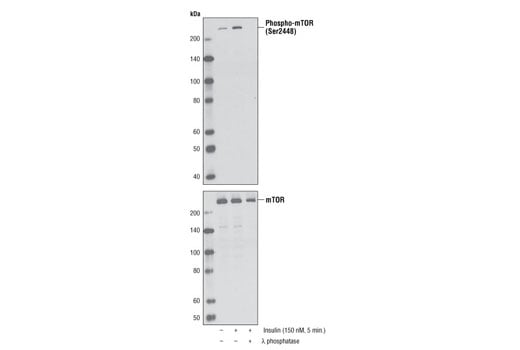
Western blot analysis of extracts from serum-starved NIH/3T3 cells, untreated or insulin-treated (150 nM, 5 minutes), alone or in combination with λ-phosphatase, using Phospho-mTOR (Ser2448) (D9C2) XP® Rabbit mAb (upper) or mTOR (7C10) Rabbit mAb #2983.
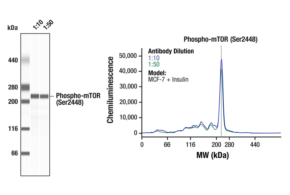
Simple WesternTM analysis of lysates (0.1 mg/mL) from MCF-7 cells treated with insulin (100nM, 10 minutes) using Phospho-mTOR (Ser2448) (D9C2) XP® Rabbit mAb #5536. The virtual lane view (left) shows a single target band (as indicated) at 1:10 and 1:50 dilutions of primary antibody. The corresponding electropherogram view (right) plots chemiluminescence by molecular weight along the capillary at 1:10 (blue line) and 1:50 (green line) dilutions of primary antibody. This experiment was performed under reducing conditions on the JessTM Simple Western instrument from ProteinSimple, a BioTechne brand, using the 66-440 kDa separation module.
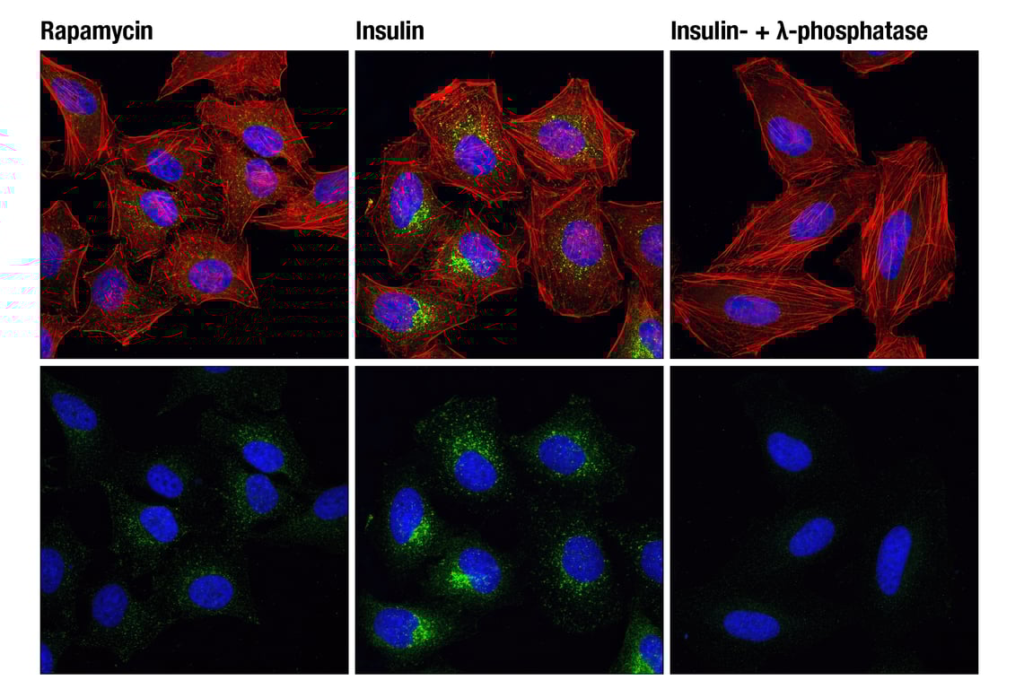
Confocal immunofluorescent analysis of HeLa cells, rapamycin-treated (#9904, 10 nM for 2 hours, left), insulin-treated (150 nM for 6 minutes, middle) or insulin- and λ-phosphatase-treated (right), using Phospho-mTOR (Ser2448) (D9C2) XP® Rabbit mAb (green). Actin filaments were labeled with DY-554 phalloidin. Blue pseudocolor = DRAQ5® #4084 (fluorescent DNA dye).