Revision 5
#37115
Store at -20C
877-616-CELL (2355)
877-678-TECH (8324)
3 Trask Lane | Danvers | Massachusetts | 01923 | USA
For Research Use Only. Not for Use in Diagnostic Procedures.
Applications:
W, IP, IF-IC, FC-FP
Reactivity:
H M R Mk
Sensitivity:
Endogenous
MW (kDa):
43
Source/Isotype:
Rabbit IgG
UniProt ID:
#P08559, #P29803
Entrez-Gene Id:
5160, 5161
Product Usage Information
| Application | Dilution |
|---|---|
| Western Blotting | 1:1000 |
| Immunoprecipitation | 1:50 |
| Immunofluorescence (Immunocytochemistry) | 1:400 - 1:1600 |
| Flow Cytometry (Fixed/Permeabilized) | 1:400 - 1:1600 |
Storage
For a carrier free (BSA and azide free) version of this product see product #48447.
Specificity/Sensitivity
Phospho-Pyruvate Dehydrogenase α1 (Ser293) (E4V9L) Rabbit mAb recognizes endogenous levels of pyruvate dehydrogenase α1 protein only when phosphorylated at Ser293. Based on amino acid sequence comparisons, this antibody is predicted to detect endogenous levels of pyruvate dehydrogenase α2 protein only when phosphorylated at Ser291 residue.
Source / Purification
Monoclonal antibody is produced by immunizing animals with a synthetic phosphopeptide corresponding to residues surrounding Ser293 of human pyruvate dehydrogenase α1 protein.
Background
The pyruvate dehydrogenase complex catalyzes the conversion of pyruvate and CoA into acetyl-CoA and CO2 in the presence of NAD+. Acetyl-CoA then goes into the citric acid cycle where it reacts with oxaloacetate to form citrate. The reaction of oxidative decarboxylation of pyruvate serves as a critical link between glycolysis and the citric acid cycle. In mammalian cells, the pyruvate dehydrogenase complex is located in the mitochondrial matrix (1). This complex is composed of three enzymes: pyruvate dehydrogenase (E1), dihydrolipoamide acetyltransferase (E2), and dihydrolipoamide dehydrogenase (E3). Pyruvate dehydrogenase (E1) consists of two subunits: α and β. This enzyme catalyzes the removal of CO2 from pyruvate. Mutations in the α subunits of pyruvate dehydrogenase (E1) lead to congenital defects that are usually associated with lactic acidosis, neurodegeneration, and early death (2).
Pyruvate dehydrogenase kinase 1 phosphorylates pyruvate dehydrogenase (E1) α1 subunit at Ser293 to inactivate its activity (3,4). This phosphorylation contributes to the tumor metabolic reprogramming toward glycolysis in hypoxia by inhibiting the citric acid cycle (4).
Species Reactivity
Species reactivity is determined by testing in at least one approved application (e.g., western blot).
Western Blot Buffer
IMPORTANT: For western blots, incubate membrane with diluted primary antibody in 5% w/v BSA, 1X TBS, 0.1% Tween® 20 at 4°C with gentle shaking, overnight.
Applications Key
W: Western Blotting IP: Immunoprecipitation IF-IC: Immunofluorescence (Immunocytochemistry) FC-FP: Flow Cytometry (Fixed/Permeabilized)
Cross-Reactivity Key
H: Human M: Mouse R: Rat Hm: Hamster Mk: Monkey Vir: Virus Mi: Mink C: Chicken Dm: D. melanogaster X: Xenopus Z: Zebrafish B: Bovine Dg: Dog Pg: Pig Sc: S. cerevisiae Ce: C. elegans Hr: Horse GP: Guinea Pig Rab: Rabbit G: Goat All: All Species Expected
Trademarks and Patents
Cell Signaling Technology is a trademark of Cell Signaling Technology, Inc.
XP is a registered trademark of Cell Signaling Technology, Inc.
All other trademarks are the property of their respective owners. Visit cellsignal.com/trademarks for more information.
Limited Uses
Except as otherwise expressly agreed in a writing signed by a legally authorized representative of CST, the following terms apply to Products provided by CST, its affiliates or its distributors. Any Customer's terms and conditions that are in addition to, or different from, those contained herein, unless separately accepted in writing by a legally authorized representative of CST, are rejected and are of no force or effect.
Products are labeled with For Research Use Only or a similar labeling statement and have not been approved, cleared, or licensed by the FDA or other regulatory foreign or domestic entity, for any purpose. Customer shall not use any Product for any diagnostic or therapeutic purpose, or otherwise in any manner that conflicts with its labeling statement. Products sold or licensed by CST are provided for Customer as the end-user and solely for research and development uses. Any use of Product for diagnostic, prophylactic or therapeutic purposes, or any purchase of Product for resale (alone or as a component) or other commercial purpose, requires a separate license from CST. Customer shall (a) not sell, license, loan, donate or otherwise transfer or make available any Product to any third party, whether alone or in combination with other materials, or use the Products to manufacture any commercial products, (b) not copy, modify, reverse engineer, decompile, disassemble or otherwise attempt to discover the underlying structure or technology of the Products, or use the Products for the purpose of developing any products or services that would compete with CST products or services, (c) not alter or remove from the Products any trademarks, trade names, logos, patent or copyright notices or markings, (d) use the Products solely in accordance with CST Product Terms of Sale and any applicable documentation, and (e) comply with any license, terms of service or similar agreement with respect to any third party products or services used by Customer in connection with the Products.
Revision 5
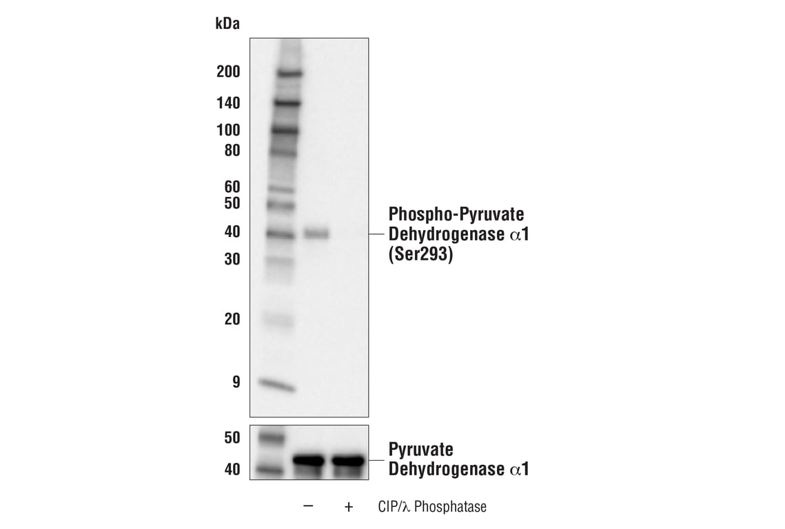
Western blot analysis of extracts from HeLa cells, vehicle-treated (-) or treated (+) with calf intestinal alkaline phosphatase (CIP)/λ phosphatase, using Phospho-Pyruvate Dehydrogenase α1 (Ser293) (E4V9L) Rabbit mAb (upper) or Pyruvate Dehydrogenase (C54G1) Rabbit mAb #3205 (lower).
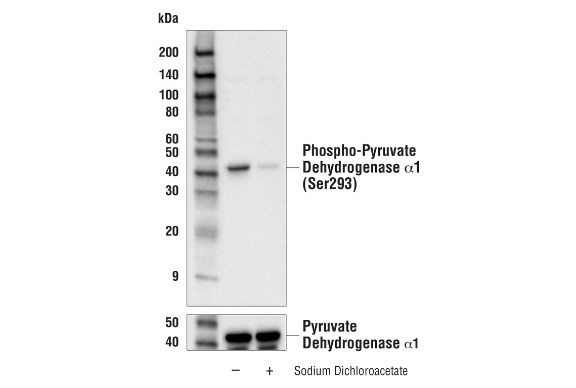
Western blot analysis of extracts from HeLa cells, vehicle-treated (-) or treated (+) with sodium dichloroacetate to reduce phosphorylation of Ser293, using Phospho-Pyruvate Dehydrogenase α1 (Ser293) (E4V9L) Rabbit mAb (upper) or Pyruvate Dehydrogenase (C54G1) Rabbit mAb #3205 (lower).
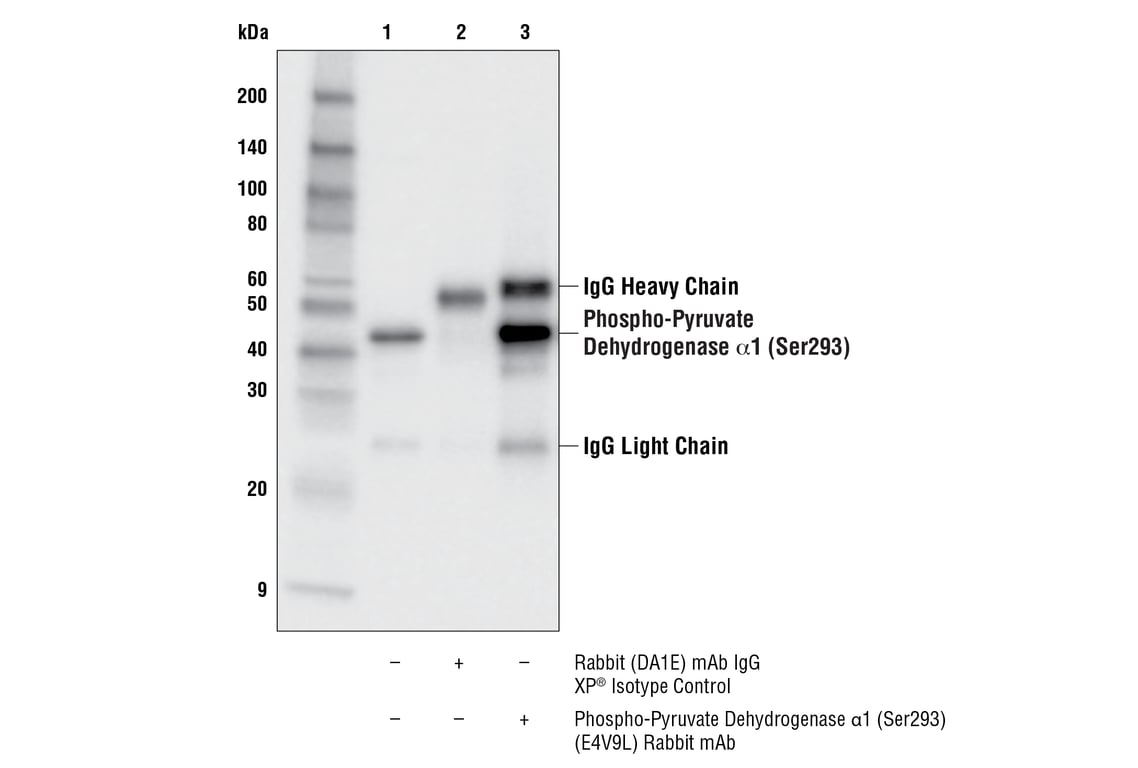
Immunoprecipitation of phospho-pyruvate dehydrogenase α1 protein from HeLa cell extracts. Lane 1 is 10% input, lane 2 is Rabbit (DA1E) mAb IgG XP® Isotype Control #3900, and lane 3 is Phospho-Pyruvate Dehydrogenase α1 (Ser293) (E4V9L) Rabbit mAb. Western blot analysis was performed using Phospho-Pyruvate Dehydrogenase α1 (Ser293) (E4V9L) Rabbit mAb. Anti-rabbit IgG, HRP-linked Antibody #7074 was used as the secondary antibody.
Revision 5
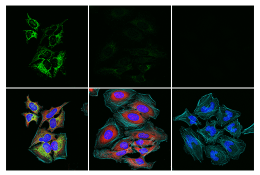
Confocal immunofluorescent analysis of HeLa cells (left, high-expressing), treated with 5 mM sodium dichloroacetate for 16 hours to reduce phosphorylation of Ser293 (middle, low-expressing), or post-processed with λ-phosphatase (right, negative) using Phospho-Pyruvate Dehydrogenase α1 (Ser293) (E4V9L) Rabbit mAb (green) and Phospho-S6 Ribosomal Protein (Ser235/236) (E2R1O) Mouse mAb #62016 (red). Actin filaments were labeled with DyLight™ 554 Phalloidin #13054 (cyan pseudocolor). Samples were mounted in ProLong® Gold Antifade Reagent with DAPI #8961 (blue).
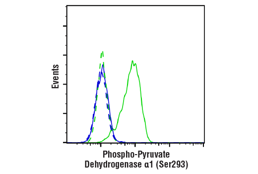
Flow cytometric analysis of Hela cells, vehicle-treated (green) or λ phosphatase-treated (blue) using Phospho-Pyruvate Dehydrogenase α1 (Ser293) (E4V9L) Rabbit mAb (solid lines) or concentration-matched Rabbit (DA1E) mAb IgG XP® Isotype Control #3900 (dashed lines). Anti-rabbit IgG (H+L), F(ab')2 Fragment (Alexa Fluor® 488 Conjugate) #4412 was used as a secondary antibody.