Revision 1
#99215
Store at -20C
877-616-CELL (2355)
877-678-TECH (8324)
3 Trask Lane | Danvers | Massachusetts | 01923 | USA
For Research Use Only. Not for Use in Diagnostic Procedures.
Applications:
W, IP, FC-FP
Reactivity:
H R
Sensitivity:
Endogenous
MW (kDa):
153
Source/Isotype:
Rabbit IgG
UniProt ID:
#Q92878
Entrez-Gene Id:
10111
Product Usage Information
| Application | Dilution |
|---|---|
| Western Blotting | 1:1000 |
| Immunoprecipitation | 1:200 |
| Flow Cytometry (Fixed/Permeabilized) | 1:200 - 1:800 |
Storage
Specificity/Sensitivity
Phospho-Rad50 (Ser635) (F5H8B) Rabbit mAb recognizes endogenous levels of Rad50 protein only when phosphorylated at Ser635.
Source / Purification
Monoclonal antibody is produced by immunizing animals with a synthetic peptide corresponding to residues surrounding Ser635 of human Rad50 protein.
Background
The DNA repair protein Rad50 is a member of the structural maintenance of chromosomes (SMC) family and plays an important role in cell cycle checkpoint signaling and double-strand break repair in response to DNA damage (1-4). Rad50 forms a complex with Mre11 and NBS1 that becomes activated in response to DNA damage (3). In normal human cells, the MRN complex acts to tether linear DNA molecules, providing a flexible link between DNA ends (1). Genomic instability and cancer have been shown to develop in cells with genetic mutations affecting the proteins in the MRN complex (2). ATM-dependent phosphorylation of Rad50 at Ser635 in response to DNA damage is important in regulating downstream signaling, DNA repair, and checkpoint control (5).
Species Reactivity
Species reactivity is determined by testing in at least one approved application (e.g., western blot).
Western Blot Buffer
IMPORTANT: For western blots, incubate membrane with diluted primary antibody in 5% w/v BSA, 1X TBS, 0.1% Tween® 20 at 4°C with gentle shaking, overnight.
Applications Key
W: Western Blotting IP: Immunoprecipitation FC-FP: Flow Cytometry (Fixed/Permeabilized)
Cross-Reactivity Key
H: Human M: Mouse R: Rat Hm: Hamster Mk: Monkey Vir: Virus Mi: Mink C: Chicken Dm: D. melanogaster X: Xenopus Z: Zebrafish B: Bovine Dg: Dog Pg: Pig Sc: S. cerevisiae Ce: C. elegans Hr: Horse GP: Guinea Pig Rab: Rabbit G: Goat All: All Species Expected
Trademarks and Patents
Cell Signaling Technology is a trademark of Cell Signaling Technology, Inc.
All other trademarks are the property of their respective owners. Visit cellsignal.com/trademarks for more information.
Limited Uses
Except as otherwise expressly agreed in a writing signed by a legally authorized representative of CST, the following terms apply to Products provided by CST, its affiliates or its distributors. Any Customer's terms and conditions that are in addition to, or different from, those contained herein, unless separately accepted in writing by a legally authorized representative of CST, are rejected and are of no force or effect.
Products are labeled with For Research Use Only or a similar labeling statement and have not been approved, cleared, or licensed by the FDA or other regulatory foreign or domestic entity, for any purpose. Customer shall not use any Product for any diagnostic or therapeutic purpose, or otherwise in any manner that conflicts with its labeling statement. Products sold or licensed by CST are provided for Customer as the end-user and solely for research and development uses. Any use of Product for diagnostic, prophylactic or therapeutic purposes, or any purchase of Product for resale (alone or as a component) or other commercial purpose, requires a separate license from CST. Customer shall (a) not sell, license, loan, donate or otherwise transfer or make available any Product to any third party, whether alone or in combination with other materials, or use the Products to manufacture any commercial products, (b) not copy, modify, reverse engineer, decompile, disassemble or otherwise attempt to discover the underlying structure or technology of the Products, or use the Products for the purpose of developing any products or services that would compete with CST products or services, (c) not alter or remove from the Products any trademarks, trade names, logos, patent or copyright notices or markings, (d) use the Products solely in accordance with CST Product Terms of Sale and any applicable documentation, and (e) comply with any license, terms of service or similar agreement with respect to any third party products or services used by Customer in connection with the Products.
Revision 1
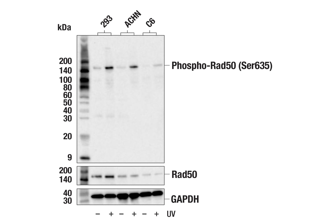
Western blot analysis of extracts from 293, ACHN, and C6 cells, untreated (-) or treated with UV (100 mJ, 4 hr, 4 hr, and 2 hr respectively; +), using Phospho-Rad50 (Ser635) (F5H8B) Rabbit mAb (upper), Rad50 Antibody #3427 (middle), or GAPDH (D16H11) XP® Rabbit mAb #5174 (lower). Phosphorylation of Rad50 protein at Ser635 is induced by UV treatment as expected.
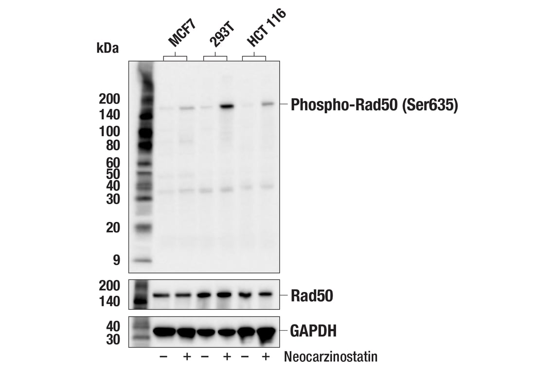
Western blot analysis of extracts from MCF7, 293T, and HCT 116 cells, untreated (-) or treated with Neocarzinostatin (0.5 μg/mL, 1 hr; +), using Phospho-Rad50 (Ser635) (F5H8B) Rabbit mAb (upper), Rad50 (E3I8K) Rabbit mAb #86225 (middle), or GAPDH (D16H11) XP® Rabbit mAb #5174 (lower). Phosphorylation of Rad50 protein at Ser635 is induced by neocarzinostatin treatment as expected.
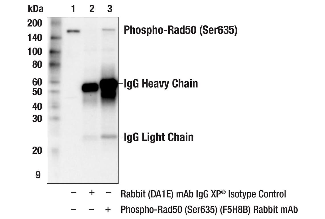
Immunoprecipitation of phospho-Rad50 (Ser635) protein from 293 cell extracts. Cells were treated with UV (100 mJ, 4 hr). Lane 1 is 10% input, lane 2 is Rabbit (DA1E) mAb IgG XP® Isotype Control #3900, and lane 3 is Phospho-Rad50 (Ser635) (F5H8B) Rabbit mAb. Western blot analysis was performed using Phospho-Rad50 (Ser635) (F5H8B) Rabbit mAb. Anti-rabbit IgG, HRP-linked Antibody #7074 was used as a secondary antibody.
Revision 1
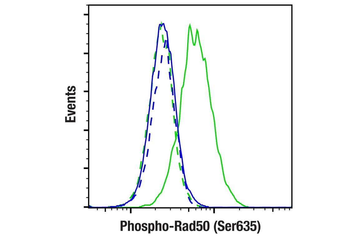
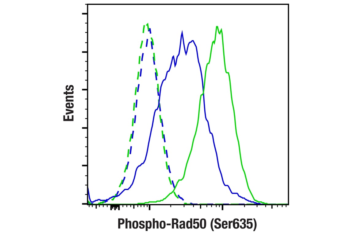
Flow cytometric analysis of untreated MCF7 cells (blue) and MCF7 cells treated with neocarzinostatin (0.5 µg/mL, 1 hr; green) using Phospho-Rad50 (Ser635) (F5H8B) Rabbit mAb (solid lines) or concentration-matched Rabbit (DA1E) mAb IgG XP® Isotype Control #3900 (dashed lines). Anti-rabbit IgG (H+L), F(ab')2 Fragment (Alexa Fluor® 488 Conjugate) #4412 was used as a secondary antibody.
Flow cytometric analysis of MCF7 cells treated with neocarzinostatin (0.5 µg/mL, 1 hr), and then treated post-fixation with λ phosphatase (blue) or untreated (green), using Phospho-Rad50 (Ser635) (F5H8B) Rabbit mAb (solid lines) or concentration-matched Rabbit (DA1E) mAb IgG XP® Isotype Control #3900 (dashed lines). Anti-rabbit IgG (H+L), F(ab')2 Fragment (Alexa Fluor® 488 Conjugate) #4412 was used as a secondary antibody.