Revision 1
#12599
Store at -20C
Phospho-VEGF Receptor 2 Antibody Sampler Kit
1 Kit
(5 x 20 microliters)
877-616-CELL (2355)
877-678-TECH (8324)
3 Trask Lane | Danvers | Massachusetts | 01923 | USA
For Research Use Only. Not for Use in Diagnostic Procedures.
| Product Includes | Product # | Quantity | Mol. Wt | Isotype/Source |
|---|---|---|---|---|
| Phospho-VEGF Receptor 2 (Tyr951) (15D2) Rabbit mAb | 4991 | 20 µl | 230 kDa | Rabbit IgG |
| Phospho-VEGF Receptor 2 (Tyr996) Antibody | 2474 | 20 µl | 230 kDa | Rabbit |
| Phospho-VEGF Receptor 2 (Tyr1059) (D5A6) Rabbit mAb | 3817 | 20 µl | 230 kDa | Rabbit IgG |
| Phospho-VEGF Receptor 2 (Tyr1175) (D5B11) Rabbit mAb | 3770 | 20 µl | 230 kDa | Rabbit IgG |
| VEGF Receptor 2 (D5B1) Rabbit mAb | 9698 | 20 µl | 210, 230 kDa | Rabbit IgG |
| Anti-rabbit IgG, HRP-linked Antibody | 7074 | 100 µl | Goat |
Please visit cellsignal.com for individual component applications, species cross-reactivity, dilutions, protocols, and additional product information.
Description
The Phospho-VEGF Receptor 2 Antibody Sampler Kit provides an economical means of evaluating the VEGFR2 tyrosine kinase and several phosphorylation sites that are involved in its activation. The kit includes enough antibody to perform two western blot experiments with each primary antibody.
Storage
Background
Vascular endothelial growth factor receptor 2 (VEGFR2, KDR, Flk-1) is a major receptor for VEGF-induced signaling in endothelial cells. Upon ligand binding, VEGFR2 undergoes autophosphorylation and becomes activated (1). Major autophosphorylation sites of VEGFR2 are located in the kinase insert domain (Tyr951/996) and in the tyrosine kinase catalytic domain (Tyr1054/1059) (2). Activation of the receptor leads to rapid recruitment of adaptor proteins, including Shc, GRB2, PI3 kinase, NCK, and the protein tyrosine phosphatases SHP-1 and SHP-2 (3). Phosphorylation at Tyr1212 provides a docking site for GRB2 binding and phospho-Tyr1175 binds the p85 subunit of PI3 kinase and PLCγ, as well as Shb (1,4,5). Signaling from VEGFR2 is necessary for the execution of VEGF-stimulated proliferation, chemotaxis and sprouting, as well as survival of cultured endothelial cells in vitro and angiogenesis in vivo (6-8).
Background References
- Meyer, M. et al. (1999) EMBO J 18, 363-74.
- Dougher-Vermazen, M. et al. (1994) Biochem Biophys Res Commun 205, 728-38.
- Kroll, J. and Waltenberger, J. (1997) J Biol Chem 272, 32521-7.
- Takahashi, T. et al. (2001) EMBO J 20, 2768-78.
- Holmqvist, K. et al. (2004) J Biol Chem 279, 22267-75.
- Karkkainen, M.J. and Petrova, T.V. (2000) Oncogene 19, 5598-605.
- Rahimi, N. et al. (2000) J Biol Chem 275, 16986-92.
- Claesson-Welsh, L. (2003) Biochem Soc Trans 31, 20-4.
Trademarks and Patents
Cell Signaling Technology is a trademark of Cell Signaling Technology, Inc.
All other trademarks are the property of their respective owners. Visit cellsignal.com/trademarks for more information.
Limited Uses
Except as otherwise expressly agreed in a writing signed by a legally authorized representative of CST, the following terms apply to Products provided by CST, its affiliates or its distributors. Any Customer's terms and conditions that are in addition to, or different from, those contained herein, unless separately accepted in writing by a legally authorized representative of CST, are rejected and are of no force or effect.
Products are labeled with For Research Use Only or a similar labeling statement and have not been approved, cleared, or licensed by the FDA or other regulatory foreign or domestic entity, for any purpose. Customer shall not use any Product for any diagnostic or therapeutic purpose, or otherwise in any manner that conflicts with its labeling statement. Products sold or licensed by CST are provided for Customer as the end-user and solely for research and development uses. Any use of Product for diagnostic, prophylactic or therapeutic purposes, or any purchase of Product for resale (alone or as a component) or other commercial purpose, requires a separate license from CST. Customer shall (a) not sell, license, loan, donate or otherwise transfer or make available any Product to any third party, whether alone or in combination with other materials, or use the Products to manufacture any commercial products, (b) not copy, modify, reverse engineer, decompile, disassemble or otherwise attempt to discover the underlying structure or technology of the Products, or use the Products for the purpose of developing any products or services that would compete with CST products or services, (c) not alter or remove from the Products any trademarks, trade names, logos, patent or copyright notices or markings, (d) use the Products solely in accordance with CST Product Terms of Sale and any applicable documentation, and (e) comply with any license, terms of service or similar agreement with respect to any third party products or services used by Customer in connection with the Products.
Revision 1
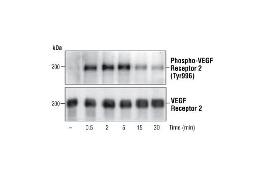
Western blot analysis of extracts from CKR/PAE cells expressing chimeric receptors containing human CSF-1 extracellular binding domain/mouse VEGF receptor 2 intracellular domains (Rahimi, N. et al. (2000) J. Biol. Chem. 275, 16986-16992), using Phospho-VEGF Receptor 2 (Tyr996) Antibody (upper) or VEGF receptor 2 antibody (lower).
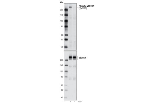
Western blot analysis of extracts from HUVEC, untreated or stimulated with VEGF #9943, using Phospho-VEGF Receptor 2 (Tyr1175) (D5B11) Rabbit mAb (upper) and VEGF Receptor 2 (55B11) Rabbit mAb (lower) #2479.
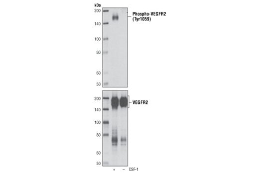
Western blot analysis of PAE/CKR cells, untreated or stimulated with CSF-1, using Phospho-VEGF Receptor 2 (Tyr1059) (D5A6) Rabbit mAb (upper) and VEGF Receptor 2 (55B11) Rabbit mAb #2479 (lower). PAE/CKR cells express a chimeric receptor made up of human CSF-1 receptor extracellular domain and mouse VEGF receptor 2 transmembrane and intracellular domains.
Revision 1
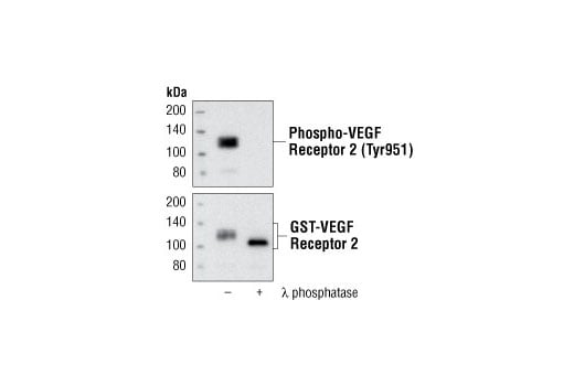
Western blot analysis of recombinant human GST-VEGF Receptor 2 (Val789-Val1356), untreated or λ phosphatase-treated, using Phospho-VEGF Receptor 2 (Tyr951) (15D2) Rabbit mAb (upper) and VEGF Receptor 2 Antibody # 2472 (lower).
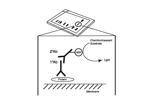
After the primary antibody is bound to the target protein, a complex with HRP-linked secondary antibody is formed. The LumiGLO® is added and emits light during enzyme catalyzed decomposition.
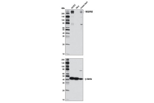
Western blot analysis of extracts from HUVE cells, HeLa cells and mouse heart using VEGF Receptor 2 (D5B1) Rabbit mAb for VEGFR2 expression (upper) and β-Actin (D6A8) Rabbit mAb (#8457) for loading control. (The 80 kDa bands represent partial degradation product).
Revision 1
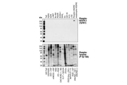
Phospho-VEGF Receptor 2 (Tyr951) (15D2) Rabbit mAb specifically binds to phosphorylated VEGFR2, but not other phosphorylated tyrosine kinases. Western blot analysis of extracts from cells expressing different activated tyrosine kinase proteins, using Phospho-VEGF Receptor-2 (Tyr951) (15D2) Rabbit mAb (upper) or Phospho-Tyrosine Mouse mAb (P-Tyr-100) #9411 (lower). CKR/PAE cells (lanes 13 and 14) express chimeric receptors containing human CSF-1 extracellular binding domain/mouse VEGF receptor-2 intracellular domain (5). CSF-1 stimulates phosphorylation of Tyr951 of intracellular VEGF receptor-2 domain (lane 13) , which was specifically detected by Phospho-VEGF Receptor-2 (Tyr951) (15D2) Rabbit mAb.
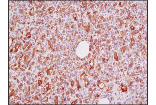
Immunohistochemical analysis of paraffin-embedded human angiosarcoma using VEGF Receptor 2 (D5B1) Rabbit mAb.
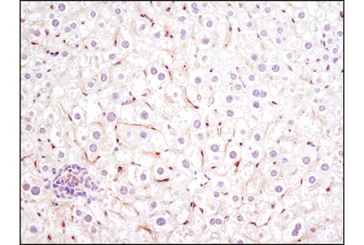
Immunohistochemical analysis of paraffin-embedded mouse liver using VEGF Receptor 2 (D5B1) Rabbit mAb.
Revision 1
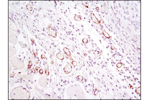
Immunohistochemical analysis of paraffin-embedded HCC827 xenograft using VEGFR2 (D5B1) Rabbit mAb.
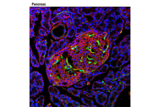
Confocal immunofluorescent analysis of rat pancreas using VEGF Receptor 2 (D5B1) Rabbit mAb (green) and β-Catenin (L54E2) Mouse mAb (IF Preferred) #2677 (red). Blue pseudocolor = DRAQ5® #4084 (fluorescent DNA dye).
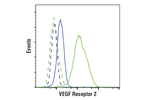
Flow cytometric analysis of fixed and permeabilized HeLa cells (blue, negative) and HUVEC cells (green, positive) using VEGF Receptor 2 (D5B1) Rabbit mAb (solid lines) or a concentration-matched Rabbit (DA1E) mAb IgG XP® Isotype Control #3900 (dashed lines). Anti-rabbit IgG (H+L), F(ab')2 Fragment (Alexa Fluor® 488 Conjugate) #4412 was used as a secondary antibody.