Revision 1
#61381
Store at -20C
PICALM Signaling Antibody Sampler Kit
1 Kit
(5 x 20 microliters)
877-616-CELL (2355)
877-678-TECH (8324)
3 Trask Lane | Danvers | Massachusetts | 01923 | USA
For Research Use Only. Not for Use in Diagnostic Procedures.
| Product Includes | Product # | Quantity | Mol. Wt | Isotype/Source |
|---|---|---|---|---|
| PICALM (E3J9R) Rabbit mAb | 26765 | 20 µl | 68, 70 kDa | Rabbit IgG |
| Clathrin Heavy Chain (D3C6) XP® Rabbit mAb | 4796 | 20 µl | 190 kDa | Rabbit IgG |
| LAMP1 (D2D11) XP® Rabbit mAb | 9091 | 20 µl | 42 (non-glycosylated), 90-120 (glycosylated) kDa | Rabbit IgG |
| EEA1 (C45B10) Rabbit mAb | 3288 | 20 µl | 170 kDa | Rabbit IgG |
| Cathepsin D (E7Z4L) XP® Rabbit mAb | 88239 | 20 µl | 46, 43, 28 kDa | Rabbit IgG |
| Anti-rabbit IgG, HRP-linked Antibody | 7074 | 100 µl | Goat |
Please visit cellsignal.com for individual component applications, species cross-reactivity, dilutions, protocols, and additional product information.
Description
The PICALM Signaling Antibody Sampler Kit provides an economical means of investigating PICALM signaling by western blot and labeling endo-lysosomal components by immunofluorescence (IF). This kit includes enough primary antibodies to perform two western blot experiments or at least 40 IF tests per primary antibody.
Storage
Background
The antibodies in this kit serve to characterize phosphatidylinositol-binding clathrin assembly protein (PICALM)-mediated lysosomal maturation, as endo-lysosomal systems are important for normal physiology and prevention of common late-onset neurodegenerative diseases such as Alzheimer's disease (AD).
PICALM is a clathrin-binding protein involved in the endo-lysosomal pathway, where it has been genetically associated with AD (1,2). Clathrin is a triskelion-shaped protein that plays a major role in the formation of coated vesicles (1). Formation of these vesicles is critical for shaping the cell membrane to promote intracellular trafficking for multiple membrane trafficking pathways in the cell, including the trans-Golgi network as well as transport to and from the cell membrane and endosomal compartments. PICALM disruption increases the number of early endosomes, which is linked to exacerbated tau aggregation (2). Early endosome antigen 1 (EEA1) is an early endosomal marker and a Rab5 effector protein essential for early endosomal membrane fusion and trafficking (3,4). Lysosome-associated membrane protein 1 (LAMP1) is an abundant lysosomal membrane protein involved in regulating lysosomal motility during lysosome-phagosome fusion (5,6). Cathepsin D (CSTD) is a ubiquitously expressed lysosomal aspartyl protease involved in the normal degradation of proteins (7). Mutations in PICALM were shown to cause lysosomal enzymes and membrane proteins to be mis-trafficked and accumulated; for example, immature forms of CTSD accumulate abnormally within endosomes. These changes correlate with reduced turnover of lysosomal cargoes generated by autophagy and endocytosis (2).
Background References
- Kaksonen, M. and Roux, A. (2018) Nat Rev Mol Cell Biol 19, 313-326.
- Hattersley, K.J. et al. (2021) Biochem Biophys Res Commun 570, 103-109.
- Mu, F.T. et al. (1995) J Biol Chem 270, 13503-11.
- Christoforidis, S. et al. (1999) Nature 397, 621-5.
- Eskelinen, E.L. et al. (2003) Trends Cell Biol 13, 137-45.
- Huynh, K.K. et al. (2007) EMBO J 26, 313-24.
- Faust, P.L. et al. (1985) Proc Natl Acad Sci USA 82, 4910-4.
Trademarks and Patents
Cell Signaling Technology is a trademark of Cell Signaling Technology, Inc.
XP is a registered trademark of Cell Signaling Technology, Inc.
U.S. Patent No. 7,429,487, foreign equivalents, and child patents deriving therefrom.
All other trademarks are the property of their respective owners. Visit cellsignal.com/trademarks for more information.
Limited Uses
Except as otherwise expressly agreed in a writing signed by a legally authorized representative of CST, the following terms apply to Products provided by CST, its affiliates or its distributors. Any Customer's terms and conditions that are in addition to, or different from, those contained herein, unless separately accepted in writing by a legally authorized representative of CST, are rejected and are of no force or effect.
Products are labeled with For Research Use Only or a similar labeling statement and have not been approved, cleared, or licensed by the FDA or other regulatory foreign or domestic entity, for any purpose. Customer shall not use any Product for any diagnostic or therapeutic purpose, or otherwise in any manner that conflicts with its labeling statement. Products sold or licensed by CST are provided for Customer as the end-user and solely for research and development uses. Any use of Product for diagnostic, prophylactic or therapeutic purposes, or any purchase of Product for resale (alone or as a component) or other commercial purpose, requires a separate license from CST. Customer shall (a) not sell, license, loan, donate or otherwise transfer or make available any Product to any third party, whether alone or in combination with other materials, or use the Products to manufacture any commercial products, (b) not copy, modify, reverse engineer, decompile, disassemble or otherwise attempt to discover the underlying structure or technology of the Products, or use the Products for the purpose of developing any products or services that would compete with CST products or services, (c) not alter or remove from the Products any trademarks, trade names, logos, patent or copyright notices or markings, (d) use the Products solely in accordance with CST Product Terms of Sale and any applicable documentation, and (e) comply with any license, terms of service or similar agreement with respect to any third party products or services used by Customer in connection with the Products.
Revision 1
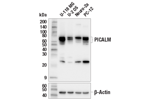
Western blot analysis of extracts from various cell lines using PICALM (E3J9R) Rabbit mAb (upper) or β-Actin (D6A8) Rabbit mAb #8457 (lower).
Western blot analysis of extracts from various cell lines using EEA1 (C45B10) Rabbit mAb.

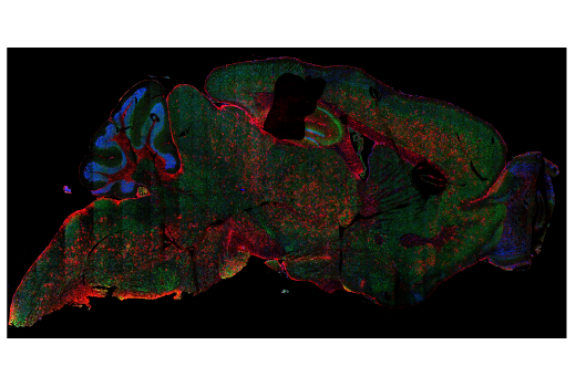
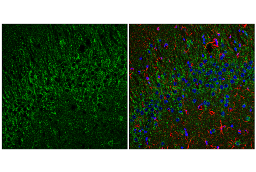
Confocal immunofluorescent tile scan of a fixed frozen brain from an amyloid mouse model of Alzheimer's disease using EEA1 (C45B10) Rabbit mAb #3288 (green), GFAP (GA5) Mouse mAb #3670 (red), and DAPI #4083 (blue).
Revision 1
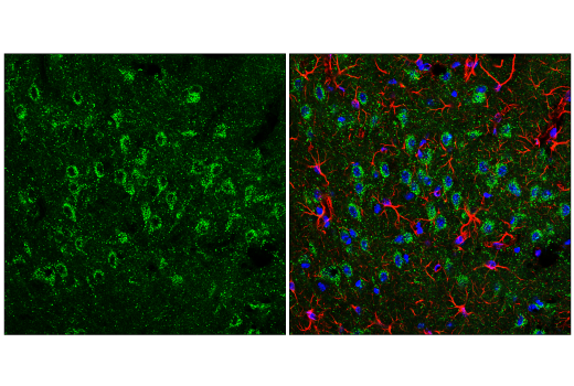
Confocal immunofluorescent analysis of fixed frozen mouse thalamus, labeled with EEA1 (C45B10) Rabbit mAb #3288 (left, green) and co-labeled with GFAP (GA5) Mouse mAb #3670 (right, red) and DAPI #4083 (right, blue).
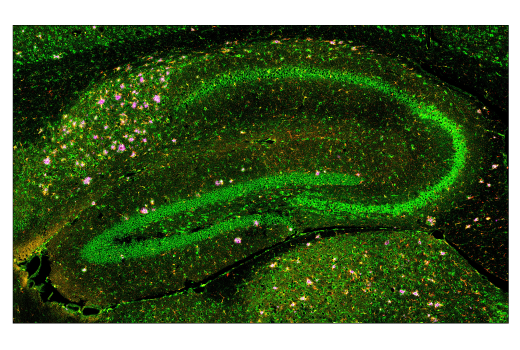
Confocal immunofluorescent analysis of fixed frozen mouse hippocampus, labeled with EEA1 (C45B10) Rabbit mAb #3288 (left, green) and co-labeled with GFAP (GA5) Mouse mAb #3670 (right, red) and DAPI #4093 (right, blue).
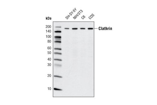
Western blot analysis of extracts from various cell lines using Clathrin Heavy Chain (D3C6) XP® Rabbit mAb.
Revision 1
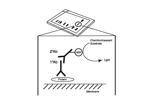
After the primary antibody is bound to the target protein, a complex with HRP-linked secondary antibody is formed. The LumiGLO® is added and emits light during enzyme catalyzed decomposition.
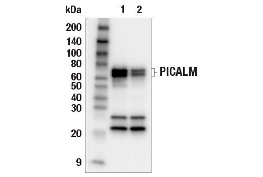
Western blot analysis of extracts from control HCT 116 cells (lane 1) or PICALM knockdown (KD) HCT 116 cells (lane 2) using PICALM (E3J9R) Rabbit mAb (upper) or β-Actin (D6A8) Rabbit mAb #8457 (lower).
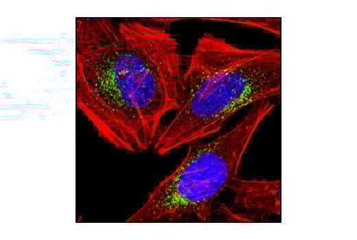
Confocal immunofluorescent analysis of HeLa cells using EEA1 (C45B10) Rabbit mAb (green). Actin filaments have been labeled with DY-554 phalloidin (red). Blue pseudocolor = DRAQ5™ (fluorescent DNA dye).
Revision 1
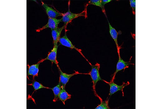
Confocal immunofluorescent analysis of SH-SY5Y cells using Clathrin Heavy Chain (D3C6) XP® Rabbit mAb (green). Actin filaments were labeled with DY-554 phalloidin (red). Blue pseudocolor = DRAQ5® #4084 (fluorescent DNA dye).
Immunoprecipitation of PICALM protein from U-118 MG extracts. Lane 1 is 10% input, lane 2 is Rabbit (DA1E) mAb IgG XP® Isotype Control #3900 and lane 3 is PICALM (E3J9R) Rabbit mAb. Western blot analysis was performed using PICALM (E3J9R) Rabbit mAb. Mouse Anti-Rabbit IgG (Light-Chain Specific) (D4W3E) mAb #93702 was used as the secondary antibody.

Confocal immunofluorescent analysis of fixed frozen mouse cerebellum, labeled with PICALM (E3J9R) Rabbit mAb (green) and DAPI #4083 (blue).

Revision 1
Confocal immunofluorescent analysis of fixed frozen brain from an amyloid mouse model of Alzheimer’s disease labeled with PICALM (E3J9R) Rabbit mAb (left, red). Free secondary binding sites were then blocked with Rabbit (DA1E) mAb IgG XP® Isotype Control #3900 prior to co-labeling with HS1 (D5A9) XP® Rabbit mAb (Rodent Specific) (Alexa Fluor® 488 Conjugate) #68206 (right, green).
Confocal immunofluorescent analysis of fixed frozen mouse kidney, labeled with PICALM (E3J9R) Rabbit mAb (green) and DAPI #4083 (blue).
Immunohistochemical analysis of paraffin-embedded mouse cerebellum using Cathepsin D (E7Z4L) XP® Rabbit mAb.
Revision 1
Confocal immunofluorescent analysis of fixed frozen mouse liver, labeled with PICALM (E3J9R) Rabbit mAb (green) and DAPI #4083 (blue).

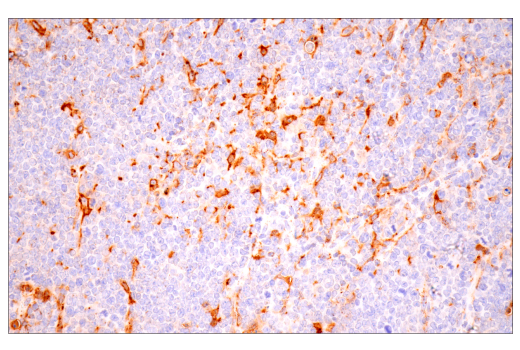
Immunohistochemical analysis of paraffin-embedded A20 syngeneic tumor using Cathepsin D (E7Z4L) XP® Rabbit mAb.
Confocal immunofluorescent analysis of HCT 116 cells, either mock transfected (left, high-expressing) or transfected with siRNA directed against PICALM (right, low-expressing), using PICALM (E3J9R) Rabbit mAb (green), DyLight™ 554 Phalloidin #13054 (red), and DAPI #4083 (blue).

Revision 1
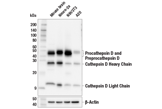
Western blot analysis of extracts from various cell lines and mouse brain tissue using Cathepsin D (E7Z4L) XP® Rabbit mAb (upper) or β-Actin (D6A8) Rabbit mAb #8457 (lower).
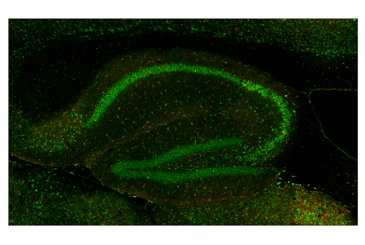
Confocal immunofluorescent analysis of fixed frozen wild-type mouse brain using Cathepsin D (E7Z4L) XP® Rabbit mAb (green). After blocking free secondary antibody binding sites with Rabbit (DA1E) mAb IgG XP® Isotype Control #3900, the tissue was then labeled using Iba1/AIF-1 (E4O4W) XP® Rabbit mAb (Alexa Fluor® 555 Conjugate) #36618 (red) and β-Amyloid (D54D2) XP® Rabbit mAb (Alexa Fluor® 594 Conjugate) #35363 (magenta pseudocolor).
Confocal immunofluorescent analysis of fixed frozen brain from an amyloid mouse model of Alzheimer's disease using Cathepsin D (E7Z4L) XP® Rabbit mAb (green). After blocking free secondary antibody binding sites with Rabbit (DA1E) mAb IgG XP® Isotype Control #3900, the tissue was then labeled using Iba1/AIF-1 (E4O4W) XP® Rabbit mAb (Alexa Fluor® 555 Conjugate) #36618 (red) and β-Amyloid (D54D2) XP® Rabbit mAb (Alexa Fluor® 594 Conjugate) #35363 (magenta pseudocolor).
Revision 1
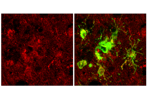
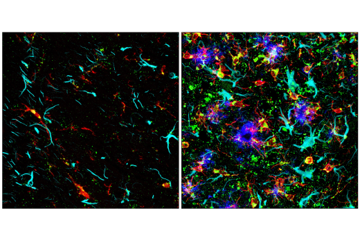
Confocal immunofluorescent analysis of fixed frozen wild-type mouse brain (left) or brain from an amyloid mouse model of Alzheimer's disease (right) using Cathepsin D (E7Z4L) XP® Rabbit mAb (green). After blocking free secondary antibody binding sites with Rabbit (DA1E) mAb IgG XP® Isotype Control #3900, the tissue was then labeled using Iba1/AIF-1 (E4O4W) XP® Rabbit mAb (Alexa Fluor® 555 Conjugate) #36618 (red), GFAP (GA5) Mouse mAb (Alexa Fluor® 647 Conjugate) #3657 (cyan pseudocolor), and β-Amyloid (D54D2) XP® Rabbit mAb (Alexa Fluor® 594 Conjugate) #35363 (blue pseudocolor).
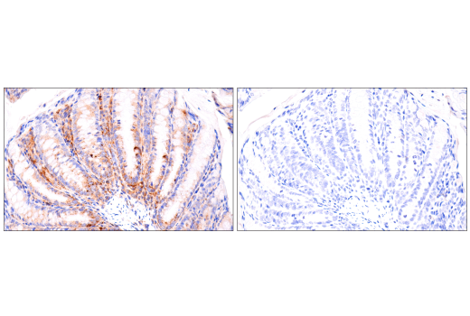
Immunohistochemical analysis of paraffin-embedded mouse colon using Cathepsin D (E7Z4L) XP® Rabbit mAb (left) compared to concentration-matched Rabbit (DA1E) mAb IgG XP® Isotype Control #3900 (right).
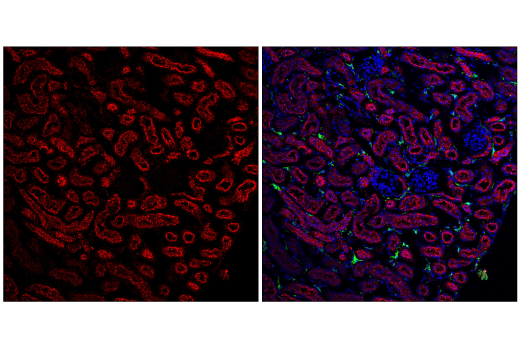
Projected confocal z-stack of fixed frozen mouse kidney using Cathepsin D (E7Z4L) XP® Rabbit mAb (red). After blocking free secondary antibody binding sites with Rabbit (DA1E) mAb IgG XP® Isotype Control #3900, the tissue was then labeled using using Iba1/AIF-1 (E4O4W) XP® Rabbit mAb (Alexa Fluor® 488 Conjugate) #20825 (green) and DAPI #4083 (blue).
Revision 1
Confocal immunofluorescent analysis of NIH/3T3 cells using Cathepsin D (E7Z4L) XP® Rabbit mAb (green) and DAPI #4083 (blue).
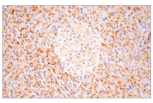
Immunohistochemical analysis of paraffin-embedded mouse liver using Cathepsin D (E7Z4L) XP® Rabbit mAb.
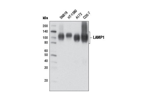
Western blot analysis of extracts from various cell lines using LAMP1 (D2D11) XP® Rabbit mAb.
Revision 1
Immunohistochemical analysis of paraffin-embedded mouse kidney using Cathepsin D (E7Z4L) XP® Rabbit mAb.
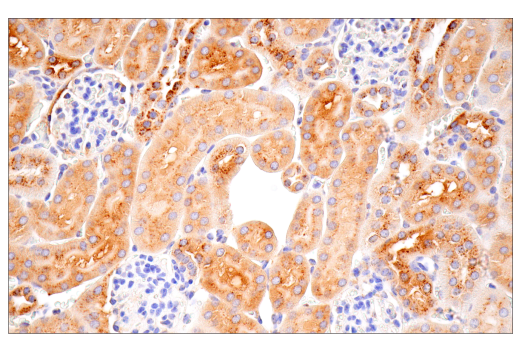
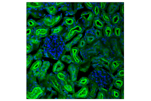
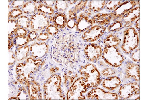
Immunohistochemical analysis of normal human kidney using LAMP1 (D2D11) XP® Rabbit mAb.
Immunohistochemical analysis of paraffin-embedded mouse lung using Cathepsin D (E7Z4L) XP® Rabbit mAb.
Revision 1
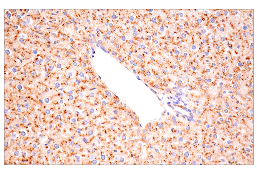
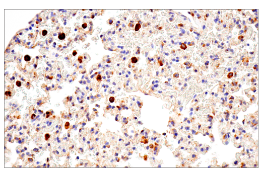
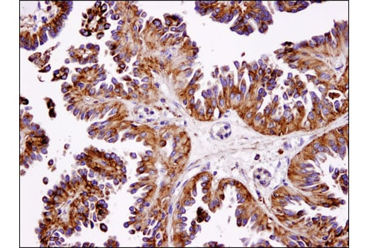
Immunohistochemical analysis of human lung carcinoma using LAMP1 (D2D11) XP® Rabbit mAb.
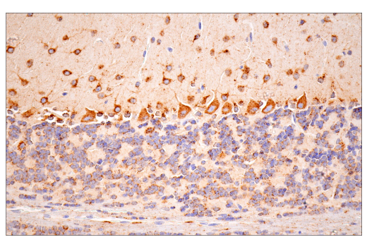
Immunohistochemical analysis of paraffin-embedded mouse pancreas using Cathepsin D (E7Z4L) XP® Rabbit mAb.
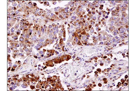
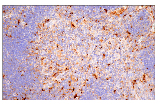
Immunohistochemical analysis of human ovarian serous cystadenoma using LAMP1 (D2D11) XP® Rabbit mAb.
Revision 1
Immunohistochemical analysis of paraffin-embedded mouse thymus using Cathepsin D (E7Z4L) XP® Rabbit mAb.
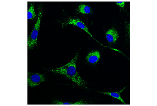
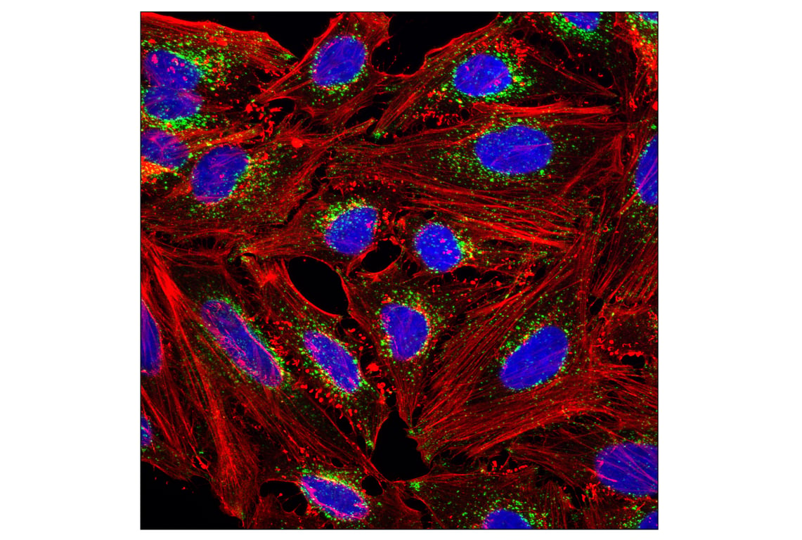
Confocal immunofluorescent analysis of HeLa cells using LAMP1 (D2D11) XP® Rabbit mAb (green). Actin filaments were labeled with DY-554 phalloidin (red). Blue pseudocolor = DRAQ5® #4084 (fluorescent DNA dye).
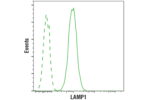
Flow cytometric analysis of Jurkat cells using LAMP1 (D2D11) XP® Rabbit mAb (solid line) compared to concentration-matched Rabbit (DA1E) mAb IgG XP® Isotype Control #3900 (dashed line). Anti-rabbit IgG (H+L), F(ab')2 Fragment (Alexa Fluor® 488 Conjugate) #4412 was used as a secondary antibody.
Revision 1
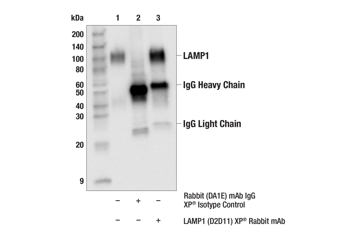
Immunoprecipitation of LAMP1 from SNB-19 cell extracts. Lane 1 is 10% input, lane 2 is Rabbit (DA1E) mAb IgG XP® Isotype Control #3900, and lane 3 is LAMP1 (D2D11) XP® Rabbit mAb. Western blot was performed using LAMP1 (D2D11) XP® Rabbit mAb. Anti-rabbit IgG, HRP-linked Antibody #7074 was used as a secondary antibody.