Revision 1
#97211
Store at -20C
877-616-CELL (2355)
877-678-TECH (8324)
3 Trask Lane | Danvers | Massachusetts | 01923 | USA
For Research Use Only. Not for Use in Diagnostic Procedures.
Applications:
W, IP, IF-F
Reactivity:
H M
Sensitivity:
Endogenous
MW (kDa):
150
Source/Isotype:
Rabbit IgG
UniProt ID:
#Q8CIH5
Entrez-Gene Id:
234779
Product Usage Information
| Application | Dilution |
|---|---|
| Western Blotting | 1:1000 |
| Immunoprecipitation | 1:50 |
| Immunofluorescence (Frozen) | 1:200 - 1:400 |
Storage
Specificity/Sensitivity
PLCγ2 (F9L8Z) Rabbit mAb recognizes endogenous levels of total PLCγ2 protein. PLCγ2 (F9L8Z) Rabbit mAb lacks sensitivity by immunofluorescence in human samples. This antibody has nonspecific nuclear background in cultured mouse and human cells. Nonspecific puncta are detected by immunofluorescence in fixed frozen mouse brain.
Source / Purification
Monoclonal antibody is produced by immunizing animals with a synthetic peptide corresponding to residues specific to the carboxy terminus of mouse PLCγ2 protein.
Background
Phosphoinositide-specific phospholipase C (PLC) plays a significant role in transmembrane signaling. In response to extracellular stimuli, such as hormones, growth factors, and neurotransmitters, PLC hydrolyzes phosphatidylinositol 4,5-bisphosphate (PIP2) to generate two secondary messengers: inositol 1,4,5-triphosphate (IP3) and diacylglycerol (DAG) (1). At least four families of PLCs have been identified: PLCβ, PLCγ, PLCδ, and PLCε. Phosphorylation is one of the key mechanisms that regulate the activity of PLC. PLCγ is activated by both receptor and non-receptor tyrosine kinases (2). PLCγ forms a complex with EGF and PDGF receptors, which leads to the phosphorylation of PLCγ at Tyr771, 783, and 1248 (3). Phosphorylation by Syk at Tyr783 activates the enzymatic activity of PLCγ1 (4). PLCγ2 is engaged in antigen-dependent signaling in B cells and collagen-dependent signaling in platelets. Phosphorylation by Btk or Lck at Tyr753, 759, 1197, and 1217 is correlated with PLCγ2 activity (5,6).
Background References
Species Reactivity
Species reactivity is determined by testing in at least one approved application (e.g., western blot).
Western Blot Buffer
IMPORTANT: For western blots, incubate membrane with diluted primary antibody in 5% w/v nonfat dry milk, 1X TBS, 0.1% Tween® 20 at 4°C with gentle shaking, overnight.
Applications Key
W: Western Blotting IP: Immunoprecipitation IF-F: Immunofluorescence (Frozen)
Cross-Reactivity Key
H: Human M: Mouse R: Rat Hm: Hamster Mk: Monkey Vir: Virus Mi: Mink C: Chicken Dm: D. melanogaster X: Xenopus Z: Zebrafish B: Bovine Dg: Dog Pg: Pig Sc: S. cerevisiae Ce: C. elegans Hr: Horse GP: Guinea Pig Rab: Rabbit G: Goat All: All Species Expected
Trademarks and Patents
Cell Signaling Technology is a trademark of Cell Signaling Technology, Inc.
All other trademarks are the property of their respective owners. Visit cellsignal.com/trademarks for more information.
Limited Uses
Except as otherwise expressly agreed in a writing signed by a legally authorized representative of CST, the following terms apply to Products provided by CST, its affiliates or its distributors. Any Customer's terms and conditions that are in addition to, or different from, those contained herein, unless separately accepted in writing by a legally authorized representative of CST, are rejected and are of no force or effect.
Products are labeled with For Research Use Only or a similar labeling statement and have not been approved, cleared, or licensed by the FDA or other regulatory foreign or domestic entity, for any purpose. Customer shall not use any Product for any diagnostic or therapeutic purpose, or otherwise in any manner that conflicts with its labeling statement. Products sold or licensed by CST are provided for Customer as the end-user and solely for research and development uses. Any use of Product for diagnostic, prophylactic or therapeutic purposes, or any purchase of Product for resale (alone or as a component) or other commercial purpose, requires a separate license from CST. Customer shall (a) not sell, license, loan, donate or otherwise transfer or make available any Product to any third party, whether alone or in combination with other materials, or use the Products to manufacture any commercial products, (b) not copy, modify, reverse engineer, decompile, disassemble or otherwise attempt to discover the underlying structure or technology of the Products, or use the Products for the purpose of developing any products or services that would compete with CST products or services, (c) not alter or remove from the Products any trademarks, trade names, logos, patent or copyright notices or markings, (d) use the Products solely in accordance with CST Product Terms of Sale and any applicable documentation, and (e) comply with any license, terms of service or similar agreement with respect to any third party products or services used by Customer in connection with the Products.
Revision 1
#97211
PLCγ2 (F9L8Z) Rabbit mAb
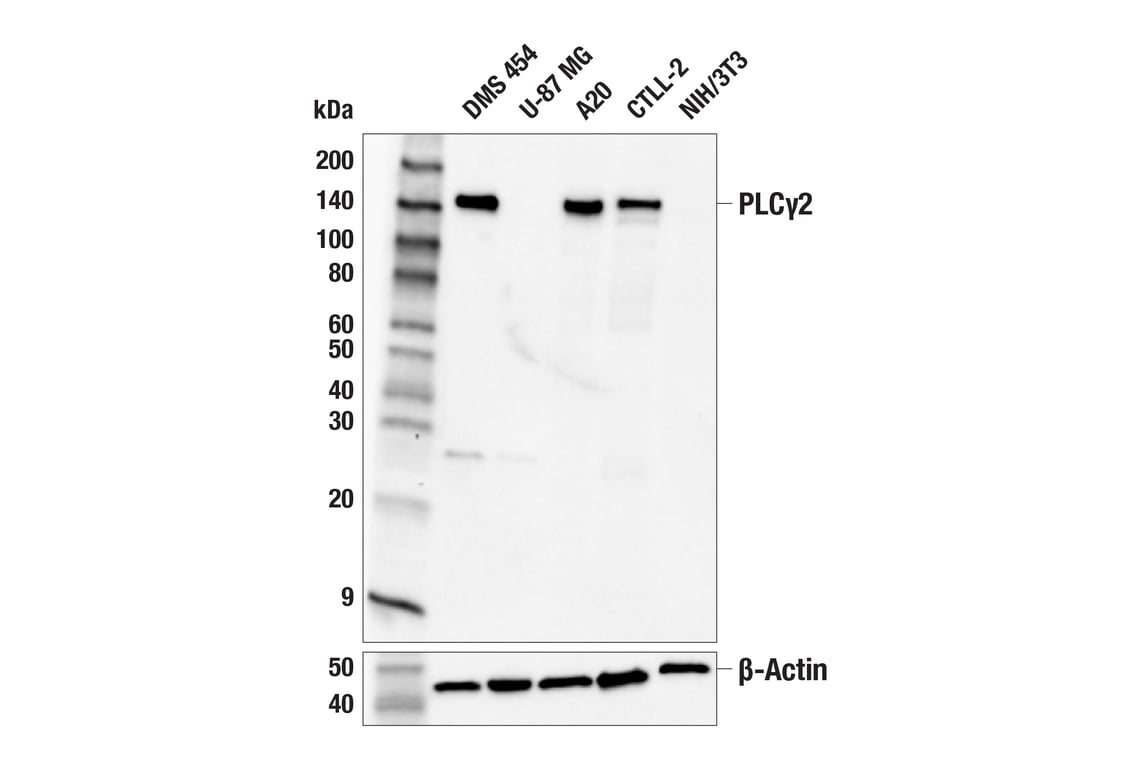
Western blot analysis of extracts from various cell lines using PLCγ2 (F9L8Z) Rabbit mAb (upper) or β-Actin (D6A8) Rabbit mAb #8457 (lower). Negative expression of PLCγ2 protein in U-87 MG and NIH/3T3 cells is consistent with the predicted expression pattern.
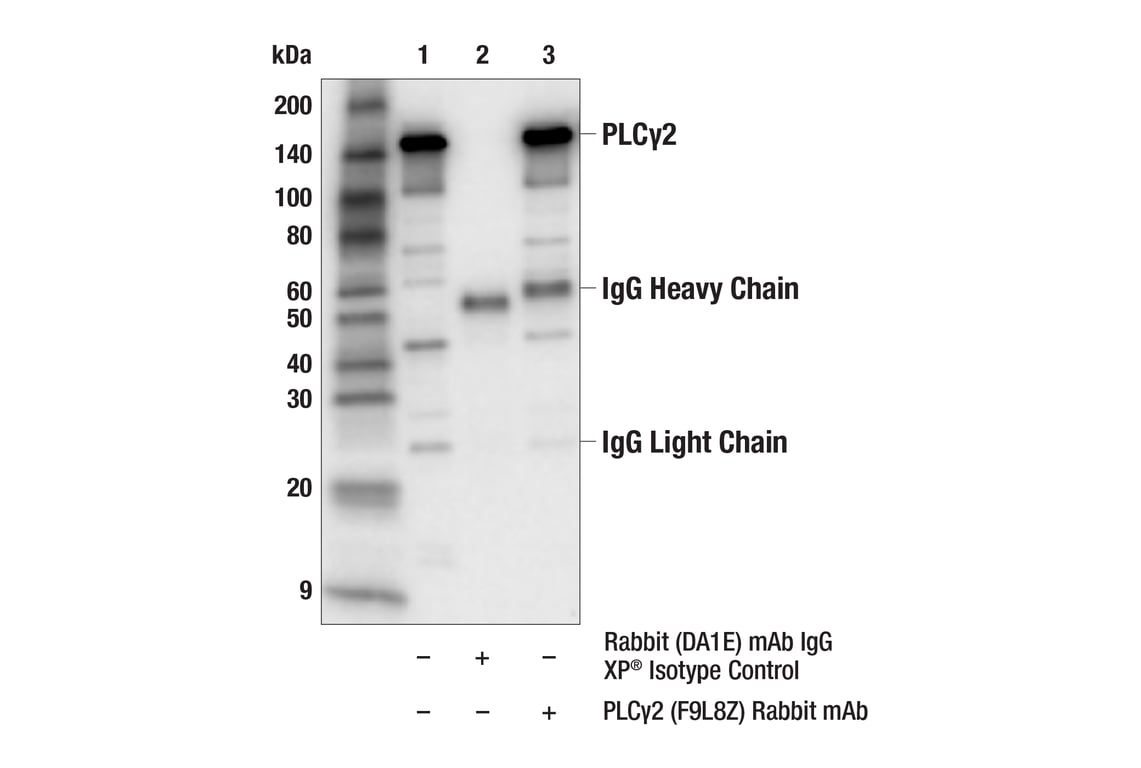
Immunoprecipitation of PLCγ2 protein from A20 cell extracts. Lane 1 is 10% input, lane 2 is Rabbit (DA1E) mAb IgG XP® Isotype Control #3900, and lane 3 is PLCγ2 (F9L8Z) Rabbit mAb. Western blot analysis was performed using PLCγ2 (F9L8Z) Rabbit mAb. Anti-rabbit IgG, HRP-linked Antibody #7074 was used as a secondary antibody.
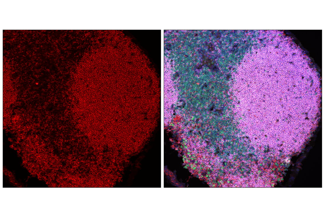
Confocal immunofluorescent analysis of fixed frozen mouse spleen labeled with PLCγ2 (F9L8Z) Rabbit mAb (red), CD3 (17A2) Rat mAb (FITC Conjugate) #86603 (green), CD45R/B220 (RA3-6B2) Rat mAb (redFluor 710 Conjugate) #82984 (gray pseudocolor), and ProLong Gold Antifade Reagent with DAPI #8961 (blue).
Revision 1
#97211
PLCγ2 (F9L8Z) Rabbit mAb
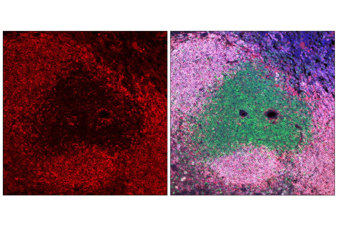
Confocal immunofluorescent analysis of fixed frozen mouse lymph node labeled with PLCγ2 (F9L8Z) Rabbit mAb (red), CD3 (17A2) Rat mAb (FITC Conjugate) #86603 (green), CD45R/B220 (RA3-6B2) Rat mAb (redFluor 710 Conjugate) #82984 (gray pseudocolor), and ProLong Gold Antifade Reagent with DAPI #8961 (blue).