| Product Includes | Product # | Quantity | Mol. Wt | Isotype/Source |
|---|---|---|---|---|
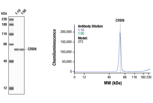
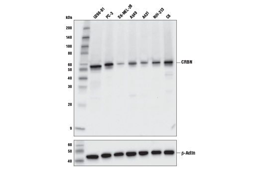
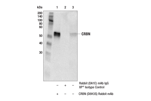
| CRBN (D8H3S) Rabbit mAb | 71810 | 20 µl | 55 kDa | Rabbit IgG |
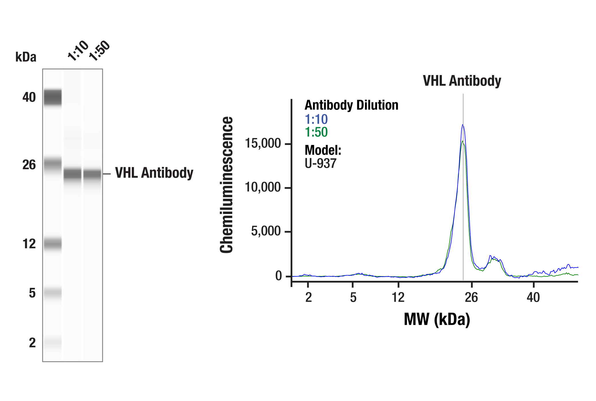
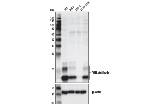
| VHL Antibody | 68547 | 20 µl | 18-22 kDa | Rabbit |
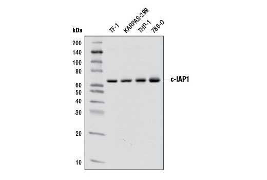
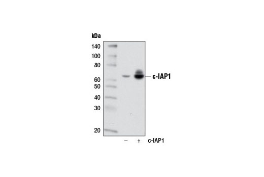
| c-IAP1 (D5G9) Rabbit mAb | 7065 | 20 µl | 62 kDa | Rabbit IgG |
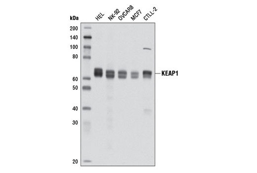
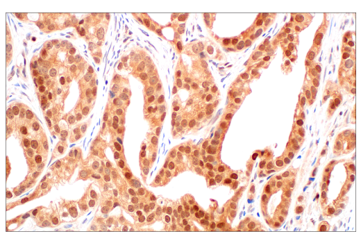
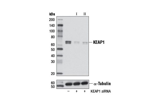
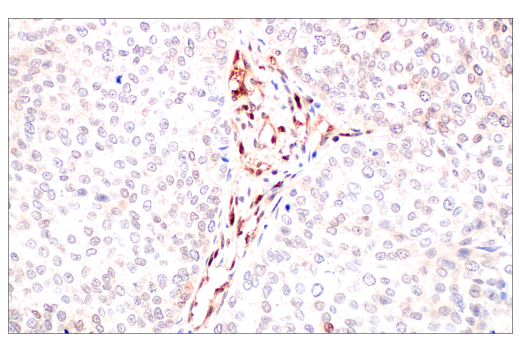
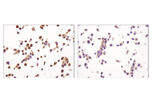
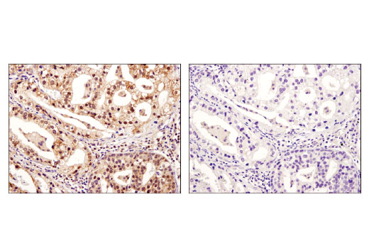
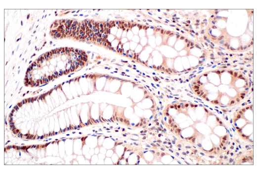
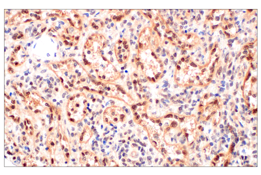
| KEAP1 (D6B12) Rabbit mAb | 8047 | 20 µl | 60-64 kDa | Rabbit IgG |
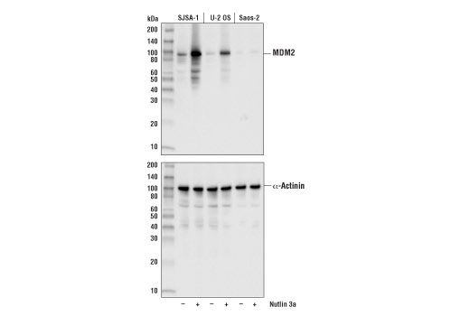
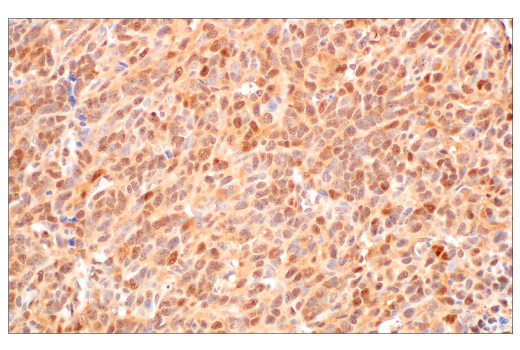
| MDM2 (D1V2Z) Rabbit mAb | 86934 | 20 µl | 90 kDa | Rabbit IgG |
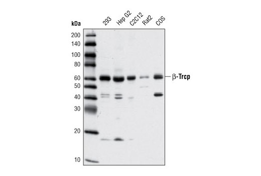
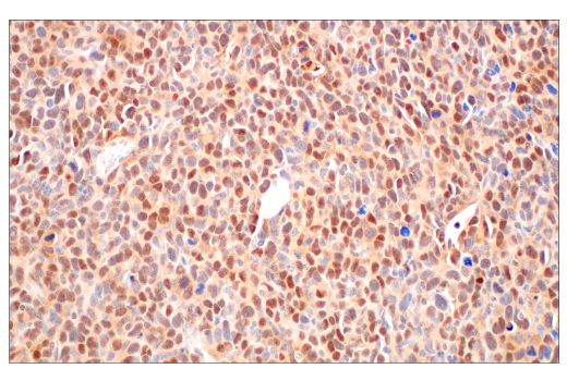
| β-TrCP (D13F10) Rabbit mAb | 4394 | 20 µl | 62 kDa | Rabbit IgG |
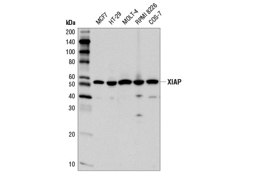
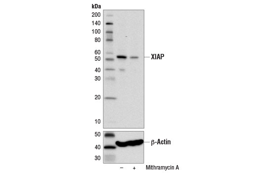
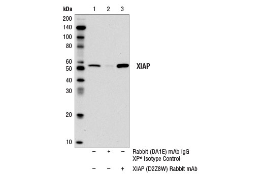
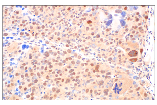
| XIAP (D2Z8W) Rabbit mAb | 14334 | 20 µl | 53 kDa | Rabbit IgG |
| Anti-rabbit IgG, HRP-linked Antibody | 7074 | 100 µl | Goat |
Please visit cellsignal.com for individual component applications, species cross-reactivity, dilutions, protocols, and additional product information.
Description
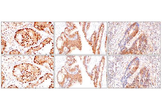
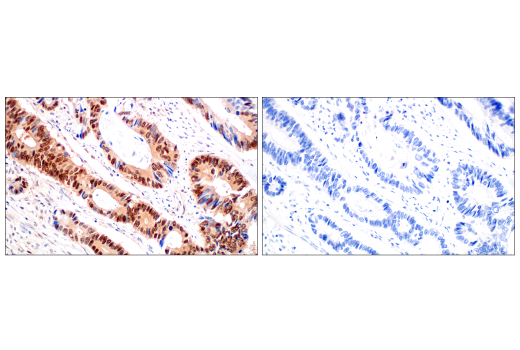
The PROTAC E3 Ligase Profiling Antibody Sampler Kit provides an economical means of detecting selected PROTAC E3 ligases. The kit includes enough antibodies to perform two western blot experiments with each primary antibody.
Storage
Background
PROTAC (proteolysis-targeting chimera) is a technique that uses a class of small molecules to target specific proteins for degradation (1). The small molecules are heterobifunctional, consisting of two protein binding parts joined by a linker. One part of the molecule binds a protein of interest (POI), while the other part binds to an E3 ubiquitin ligase, bringing the E3 ligase close to the POI for ubiquitination coupled protein degradation (2). The first successful PROTAC study used the chimeric PROTAC-1 molecule to recruit the β-TrCP E3 ligase to dictate the ubiquitination and degradation of MetAP2 (3). There are over 600 E3 ligases, and only a few have been successfully developed by PROTAC to degrade target proteins (4). Among them, CRBN- and VHL-based PROTAC have been extensively explored and successfully applied for multiple disease treatments (5,6). The E3 ligased MDM2, c-IAP, and XIAP are fruitful in using PROTAC strategy for cancer treatment (7-9). PROTAC design using the KEAP1 E3 ligase has been developed for degradation of BRD3, BRD4, and Tau. KEAP1 is another promising member of the PROTAC team (10,11).
- Békés, M. et al. (2022) Nat Rev Drug Discov 21, 181-200.
- Li, X. and Song, Y. (2020) J Hematol Oncol 13, 50.
- Sakamoto, K.M. et al. (2001) Proc Natl Acad Sci USA 98, 8554-9.
- Zou, Y. et al. (2019) Cell Biochem Funct 37, 21-30.
- Wang, C. et al. (2021) Eur J Med Chem 225, 113749.
- Wang, C. et al. (2022) Eur J Med Chem 227, 113906.
- Naito, M. et al. (2019) Drug Discov Today Technol 31, 35-42.
- Vicente, A.T.S. and Salvador, J.A.R. (2022) Int J Mol Sci 23, 11068. doi: 10.3390/ijms231911068.
- Park, S. et al. (2023) Eur J Med Chem 245, 114910.
- Wei, J. et al. (2021) J Am Chem Soc 143, 15073-15083.
- Lu, M. et al. (2018) Eur J Med Chem 146, 251-259.
Background References
Trademarks and Patents
Limited Uses
Except as otherwise expressly agreed in a writing signed by a legally authorized representative of CST, the following terms apply to Products provided by CST, its affiliates or its distributors. Any Customer's terms and conditions that are in addition to, or different from, those contained herein, unless separately accepted in writing by a legally authorized representative of CST, are rejected and are of no force or effect.
Products are labeled with For Research Use Only or a similar labeling statement and have not been approved, cleared, or licensed by the FDA or other regulatory foreign or domestic entity, for any purpose. Customer shall not use any Product for any diagnostic or therapeutic purpose, or otherwise in any manner that conflicts with its labeling statement. Products sold or licensed by CST are provided for Customer as the end-user and solely for research and development uses. Any use of Product for diagnostic, prophylactic or therapeutic purposes, or any purchase of Product for resale (alone or as a component) or other commercial purpose, requires a separate license from CST. Customer shall (a) not sell, license, loan, donate or otherwise transfer or make available any Product to any third party, whether alone or in combination with other materials, or use the Products to manufacture any commercial products, (b) not copy, modify, reverse engineer, decompile, disassemble or otherwise attempt to discover the underlying structure or technology of the Products, or use the Products for the purpose of developing any products or services that would compete with CST products or services, (c) not alter or remove from the Products any trademarks, trade names, logos, patent or copyright notices or markings, (d) use the Products solely in accordance with CST Product Terms of Sale and any applicable documentation, and (e) comply with any license, terms of service or similar agreement with respect to any third party products or services used by Customer in connection with the Products.