| Product Includes | Product # | Quantity | Mol. Wt | Isotype/Source |
|---|---|---|---|---|
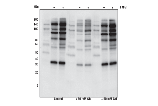
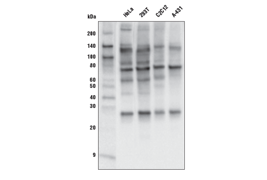
| O-GlcNAc MultiMab® Rabbit mAb mix | 82332 | 20 µl | Rabbit IgG | |
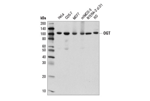
| OGT (D1D8Q) Rabbit mAb | 24083 | 20 µl | 110 kDa | Rabbit IgG |
| MGEA5/OGA (E9C5U) Rabbit mAb | 60406 | 20 µl | 138 kDa | Rabbit IgG |
| Anti-rabbit IgG, HRP-linked Antibody | 7074 | 100 µl | Goat |
Please visit cellsignal.com for individual component applications, species cross-reactivity, dilutions, protocols, and additional product information.
Description
The Protein Glycosylation Antibody Sampler Kit provides an economical means of detecting select components involved in protein glycosylation. The kit includes enough antibodies to perform two western blot experiments with each primary antibody.
Storage
Background
A distinct form of protein glycosylation, β-D-N-acetylglucosamine (GlcNAc) moieties can be added to serine or threonine residues of proteins (1,2). This differs from other glycosylation forms, as it typically is a single moiety rather than the complex branched sugars that are more commonly studied. It is thought that these modifications happen in a much more dynamic cycle reminiscent of phosphorylation modifications. GlcNAc modified proteins are found in the cytoplasm and nucleus. They are modulated by means of specific O-GlcNAc transferases (OGT) as well as GlcNAcase activity that can be inhibited using the Thiamet-G (TMG) inhibitor. Mass spectrometry analysis of this modification has been complicated due to the loss of the GlcNAc group during ionization and fragmentation, but methods and technologies such as electron transfer dissociation (ETD) are opening up new avenues to study these modifications. O-GlcNAc could play an important role in many cellular processes, including metabolism, growth, morphogenesis, apoptosis, and transcription, and it may play a critical role in cancer (3).
O-linked N-acetylglucosaminylation (O-GlcNAcylation) is a post-translational modification where GlcNAc is covalently linked to cytoplasmic and nuclear proteins at serine or threonine residues (1,2). This modification is important in many cellular processes, including metabolism, cell growth and morphogenesis, apoptosis, and transcription (2,3). The reversible protein modification by O-GlcNAc, which has been suggested to be a nutrient and stress sensor, is catalyzed by two highly conserved enzymes, O-GlcNAc transferase (OGT) and human meningioma-expressed antigen 5 (MGEA5)/O-GlcNAcase (OGA) (4). Research studies have implicated O-GlcNAcylation in cancer (1). This modification has also been proposed to have protective effects against the production and aggregation of toxic protein species associated with neurodegenerative diseases, including amyloid β (Aβ) and tau (Alzheimer's disease), and α-synuclein (Parkinson's disease) (5-7).
- Comer, F.I. et al. (2001) Anal Biochem 293, 169-77.
- Slawson, C. and Hart, G.W. (2011) Nat Rev Cancer 11, 678-84.
- Capotosti, F. et al. (2011) Cell 144, 376-88.
- Hart, G.W. et al. (2007) Nature 446, 1017-22.
- Wani, W.Y. et al. (2017) Brain Res Bull 133, 80-87.
- Yang, X. and Qian, K. (2017) Nat Rev Mol Cell Biol 18, 452-465.
- Saha, A. et al. (2021) Chem Soc Rev 50, 10451-10485.
Background References
Trademarks and Patents
Limited Uses
Except as otherwise expressly agreed in a writing signed by a legally authorized representative of CST, the following terms apply to Products provided by CST, its affiliates or its distributors. Any Customer's terms and conditions that are in addition to, or different from, those contained herein, unless separately accepted in writing by a legally authorized representative of CST, are rejected and are of no force or effect.
Products are labeled with For Research Use Only or a similar labeling statement and have not been approved, cleared, or licensed by the FDA or other regulatory foreign or domestic entity, for any purpose. Customer shall not use any Product for any diagnostic or therapeutic purpose, or otherwise in any manner that conflicts with its labeling statement. Products sold or licensed by CST are provided for Customer as the end-user and solely for research and development uses. Any use of Product for diagnostic, prophylactic or therapeutic purposes, or any purchase of Product for resale (alone or as a component) or other commercial purpose, requires a separate license from CST. Customer shall (a) not sell, license, loan, donate or otherwise transfer or make available any Product to any third party, whether alone or in combination with other materials, or use the Products to manufacture any commercial products, (b) not copy, modify, reverse engineer, decompile, disassemble or otherwise attempt to discover the underlying structure or technology of the Products, or use the Products for the purpose of developing any products or services that would compete with CST products or services, (c) not alter or remove from the Products any trademarks, trade names, logos, patent or copyright notices or markings, (d) use the Products solely in accordance with CST Product Terms of Sale and any applicable documentation, and (e) comply with any license, terms of service or similar agreement with respect to any third party products or services used by Customer in connection with the Products.