Revision 3
#57906
Store at -20C
877-616-CELL (2355)
877-678-TECH (8324)
3 Trask Lane | Danvers | Massachusetts | 01923 | USA
For Research Use Only. Not for Use in Diagnostic Procedures.
Applications:
W, IP, IF-F, IF-IC, FC-FP
Reactivity:
M
Sensitivity:
Endogenous
MW (kDa):
42
Source/Isotype:
Mouse IgG1 kappa
UniProt ID:
#P17433
Entrez-Gene Id:
20375
Product Usage Information
| Application | Dilution |
|---|---|
| Western Blotting | 1:1000 |
| Immunoprecipitation | 1:50 |
| Immunofluorescence (Frozen) | 1:200 - 1:800 |
| Immunofluorescence (Immunocytochemistry) | 1:200 - 1:800 |
| Flow Cytometry (Fixed/Permeabilized) | 1:200 - 1:800 |
Storage
Specificity/Sensitivity
PU.1 (F2D5A) Mouse mAb recognizes endogenous levels of total PU.1 protein.
Source / Purification
Monoclonal antibody is produced by immunizing animals with a synthetic peptide corresponding to residues surrounding Leu27 of mouse PU.1 protein.
Background
PU.1 is a member of the Ets family of transcription factors and activates target genes through the purine-rich PU-box (1). PU.1 plays a pivotal role in the differentiation of myeloid cells and lymphocytes and is expressed in several hematopoietic cells, including B lymphocytes, macrophages, neutrophils, mast cells, early erythroid cells, and megakaryocytes (1,2). The concentration of PU.1 is critical for both the determination of hematopoietic cell lineage and the regulation of differentiation versus stem cell proliferation (3,4). In addition, PU.1 activity is influenced by phosphorylation and interactions with other hematopoietic transcription factors. Phosphorylation of PU.1 at Ser146 by CK2 promotes binding to IRF-4 and synergistic activation through the immunoglobulin κ 3' enhancer (5). Treatment of pro-B cells with IL-3 leads to phosphorylation of PU.1 at Ser140, resulting in increased PU.1 activity and activation of the anti-apoptotic gene MCL-1 (6). GATA1 binding blocks PU.1 activity during erythroid cell development (7). Overexpression of PU.1 resulting from proviral insertion during Friend virus infection can induce erythroleukemia, while reduced expression has been associated with acute myeloid leukemia (8).
Background References
- Lloberas, J. et al. (1999) Immunol Today 20, 184-9.
- Klemsz, M.J. et al. (1990) Cell 61, 113-24.
- Dahl, R. and Simon, M.C. (2003) Blood Cells Mol Dis 31, 229-33.
- DeKoter, R.P. and Singh, H. (2000) Science 288, 1439-41.
- Pongubala, J.M. et al. (1993) Science 259, 1622-5.
- Wang, J.M. et al. (2003) Mol Cell Biol 23, 1896-909.
- Zhang, P. et al. (1999) Proc Natl Acad Sci U S A 96, 8705-10.
- Moreau-Gachelin, F. et al. (1988) Nature 331, 277-80.
Species Reactivity
Species reactivity is determined by testing in at least one approved application (e.g., western blot).
Western Blot Buffer
IMPORTANT: For western blots, incubate membrane with diluted primary antibody in 5% w/v nonfat dry milk, 1X TBS, 0.1% Tween® 20 at 4°C with gentle shaking, overnight.
Applications Key
W: Western Blotting IP: Immunoprecipitation IF-F: Immunofluorescence (Frozen) FC-FP: Flow Cytometry (Fixed/Permeabilized)
Cross-Reactivity Key
H: Human M: Mouse R: Rat Hm: Hamster Mk: Monkey Vir: Virus Mi: Mink C: Chicken Dm: D. melanogaster X: Xenopus Z: Zebrafish B: Bovine Dg: Dog Pg: Pig Sc: S. cerevisiae Ce: C. elegans Hr: Horse GP: Guinea Pig Rab: Rabbit G: Goat All: All Species Expected
Trademarks and Patents
Cell Signaling Technology is a trademark of Cell Signaling Technology, Inc.
All other trademarks are the property of their respective owners. Visit cellsignal.com/trademarks for more information.
Limited Uses
Except as otherwise expressly agreed in a writing signed by a legally authorized representative of CST, the following terms apply to Products provided by CST, its affiliates or its distributors. Any Customer's terms and conditions that are in addition to, or different from, those contained herein, unless separately accepted in writing by a legally authorized representative of CST, are rejected and are of no force or effect.
Products are labeled with For Research Use Only or a similar labeling statement and have not been approved, cleared, or licensed by the FDA or other regulatory foreign or domestic entity, for any purpose. Customer shall not use any Product for any diagnostic or therapeutic purpose, or otherwise in any manner that conflicts with its labeling statement. Products sold or licensed by CST are provided for Customer as the end-user and solely for research and development uses. Any use of Product for diagnostic, prophylactic or therapeutic purposes, or any purchase of Product for resale (alone or as a component) or other commercial purpose, requires a separate license from CST. Customer shall (a) not sell, license, loan, donate or otherwise transfer or make available any Product to any third party, whether alone or in combination with other materials, or use the Products to manufacture any commercial products, (b) not copy, modify, reverse engineer, decompile, disassemble or otherwise attempt to discover the underlying structure or technology of the Products, or use the Products for the purpose of developing any products or services that would compete with CST products or services, (c) not alter or remove from the Products any trademarks, trade names, logos, patent or copyright notices or markings, (d) use the Products solely in accordance with CST Product Terms of Sale and any applicable documentation, and (e) comply with any license, terms of service or similar agreement with respect to any third party products or services used by Customer in connection with the Products.
Revision 3
#57906
PU.1 (F2D5A) Mouse mAb
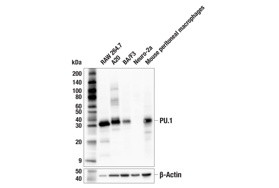
Western blot analysis of extracts from various cell lines using PU.1 (F2D5A) Mouse mAb (upper) or β-Actin (D6A8) Rabbit mAb #8457 (lower). Negative expression of PU.1 protein in Neuro-2a cells is consistent with the predicted expression pattern.
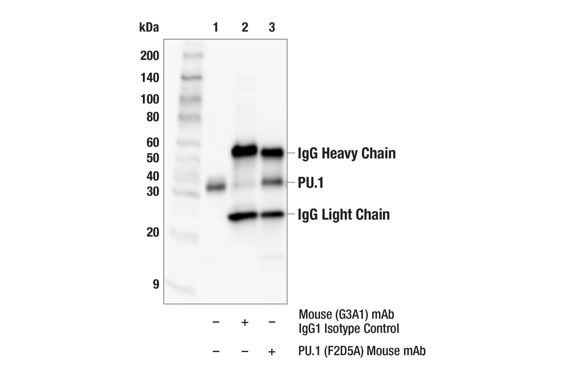
Immunoprecipitation of PU.1 protein from RAW 264.7 cell extracts. Lane 1 is 10% input, lane 2 is Mouse (G3A1) mAb IgG1 Isotype Control #5415, and lane 3 is PU.1 (F2D5A) Mouse mAb. Western blot analysis was performed using PU.1 (F2D5A) Mouse mAb. Anti-mouse IgG, HRP-linked Antibody #7076 was used as a secondary antibody.
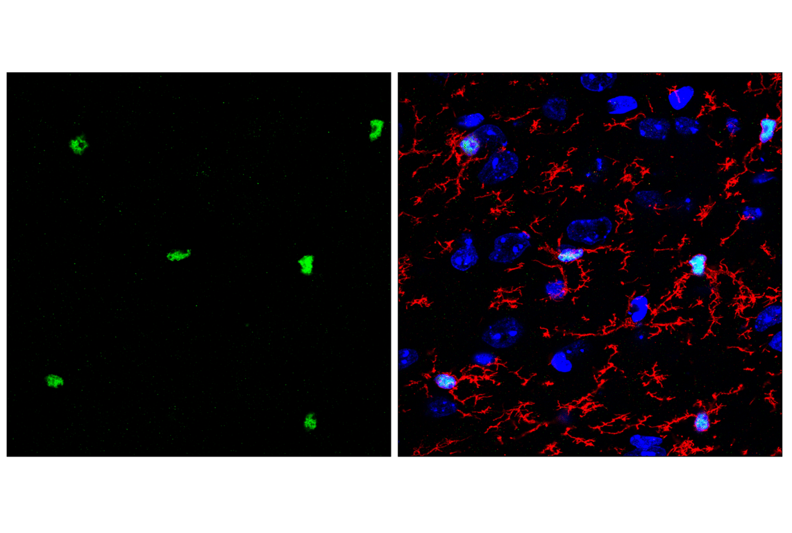
Confocal immunofluorescent analysis of fixed frozen mouse cortex, labeled with PU.1 (F2D5A) Mouse mAb (left, green) and co-labeled with TMEM119 (E3E1O) Rabbit mAb #90840 (right, red) and ProLong Gold Antifade Reagent with DAPI #8961 (right, blue).
Revision 3
#57906
PU.1 (F2D5A) Mouse mAb
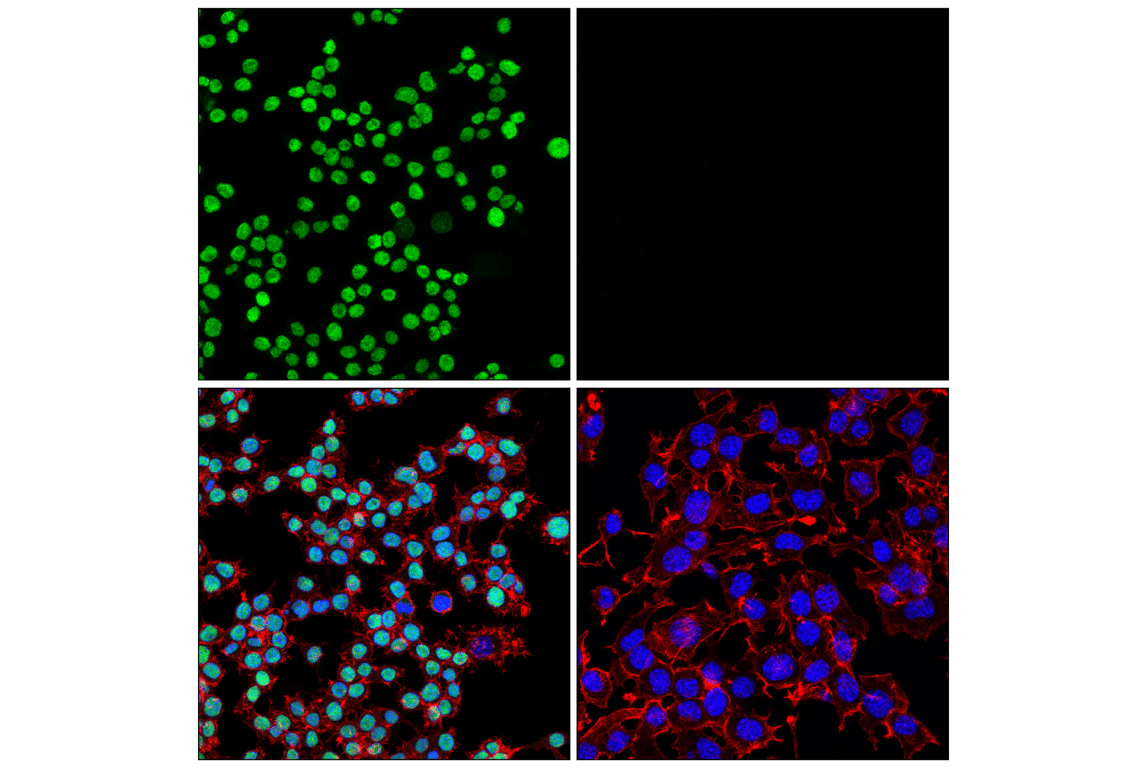
Confocal immunofluorescent analysis of RAW 264.7 cells (left, positive) and Neuro-2a cells (right, negative) using PU.1 (F2D5A) Mouse mAb (green), DyLight 554 Phalloidin #13054 (red), and DAPI #4083 (blue).
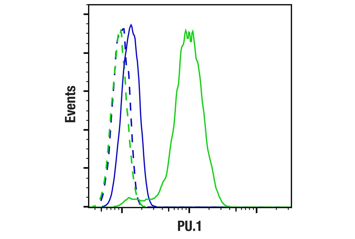
Flow cytometric analysis of Neuro-2a cells (blue, negative) and RAW 264.7 cells (green, positive) using PU.1 (F2D5A) Mouse mAb (solid lines) or a concentration-matched Mouse (G3A1) mAb IgG1 Isotype Control #5415 (dashed lines). Anti-mouse IgG (H+L), F(ab')2 Fragment (Alexa Fluor® 488 Conjugate) #4408 was used as a secondary antibody.