Revision 2
#8207
Store at -20C
877-616-CELL (2355)
877-678-TECH (8324)
3 Trask Lane | Danvers | Massachusetts | 01923 | USA
For Research Use Only. Not for Use in Diagnostic Procedures.
| Product Includes | Product # | Quantity | Mol. Wt | Isotype/Source |
|---|---|---|---|---|
| Phospho-S6 Ribosomal Protein (Ser235/236) (D57.2.2E) XP® Rabbit mAb | 4858 | 100 µl | 32 kDa | Rabbit IgG |
| S6 Ribosomal Protein (5G10) Rabbit mAb | 2217 | 100 µl | 32 kDa | Rabbit IgG |
Please visit cellsignal.com for individual component applications, species cross-reactivity, dilutions, protocols, and additional product information.
UniProt ID:
#P62753
Entrez-Gene Id:
6194
Description
PhosphoPlus® Duets from Cell Signaling Technology (CST) provide a means to assess protein activation status. Each Duet contains an activation-state and total protein antibody to your target of interest. These antibodies have been selected from CST's product offering based upon superior performance in specified applications.
Storage
Background
One way that growth factors and mitogens effectively promote sustained cell growth and proliferation is by upregulating mRNA translation (1,2). Growth factors and mitogens induce the activation of p70 S6 kinase and the subsequent phosphorylation of S6 ribosomal protein. Phosphorylation of S6 ribosomal protein correlates with an increase in translation of mRNA transcripts that contain an oligopyrimidine tract in their 5' untranslated regions (2). These particular mRNA transcripts (5'TOP) encode proteins involved in cell cycle progression, as well as ribosomal proteins and elongation factors necessary for translation (2,3). Important S6 ribosomal protein phosphorylation sites include several residues (Ser235, Ser236, Ser240, and Ser244) located within a small, carboxy-terminal region of S6 protein (4,5).
Background References
Trademarks and Patents
Cell Signaling Technology is a trademark of Cell Signaling Technology, Inc.
PhosphoPlus is a registered trademark of Cell Signaling Technology, Inc.
All other trademarks are the property of their respective owners. Visit cellsignal.com/trademarks for more information.
Limited Uses
Except as otherwise expressly agreed in a writing signed by a legally authorized representative of CST, the following terms apply to Products provided by CST, its affiliates or its distributors. Any Customer's terms and conditions that are in addition to, or different from, those contained herein, unless separately accepted in writing by a legally authorized representative of CST, are rejected and are of no force or effect.
Products are labeled with For Research Use Only or a similar labeling statement and have not been approved, cleared, or licensed by the FDA or other regulatory foreign or domestic entity, for any purpose. Customer shall not use any Product for any diagnostic or therapeutic purpose, or otherwise in any manner that conflicts with its labeling statement. Products sold or licensed by CST are provided for Customer as the end-user and solely for research and development uses. Any use of Product for diagnostic, prophylactic or therapeutic purposes, or any purchase of Product for resale (alone or as a component) or other commercial purpose, requires a separate license from CST. Customer shall (a) not sell, license, loan, donate or otherwise transfer or make available any Product to any third party, whether alone or in combination with other materials, or use the Products to manufacture any commercial products, (b) not copy, modify, reverse engineer, decompile, disassemble or otherwise attempt to discover the underlying structure or technology of the Products, or use the Products for the purpose of developing any products or services that would compete with CST products or services, (c) not alter or remove from the Products any trademarks, trade names, logos, patent or copyright notices or markings, (d) use the Products solely in accordance with CST Product Terms of Sale and any applicable documentation, and (e) comply with any license, terms of service or similar agreement with respect to any third party products or services used by Customer in connection with the Products.
Revision 2
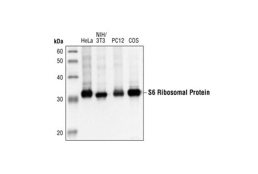
Western blot analysis of extracts from HeLa, NIH/3T3, PC12 and COS cells using S6 Ribosomal Protein (5G10) Rabbit mAb.
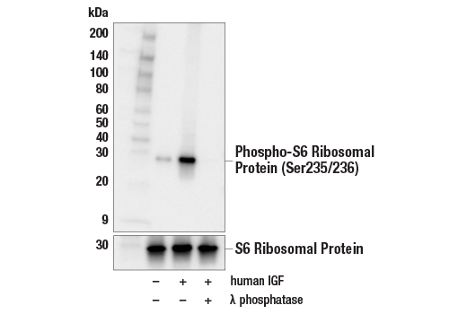
Western blot analysis of extracts from serum-starved MCF7 cells, untreated (-) or treated (+) with combinations of the following treatments as indicated: human IGF-1 (100 ng/mL, 10 min) and λ phosphatase, using Phospho-S6 Ribosomal Protein (Ser235/236) (D57.2.2E) XP® Rabbit mAb (upper) or S6 Ribosomal Protein (5G10) Rabbit mAb #2217 (lower).
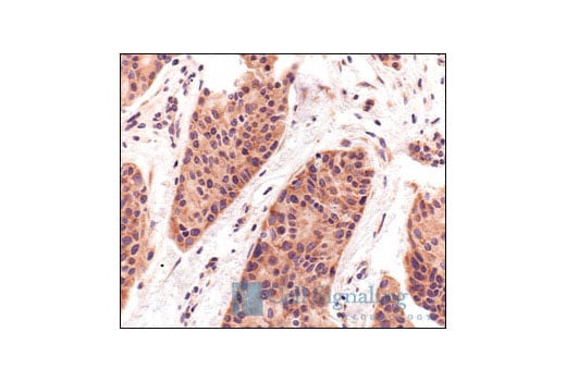
Immunohistochemical analysis of paraffin-embedded human breast carcinoma using S6 Ribosomal Protein (5G10) Rabbit mAb.
Revision 2
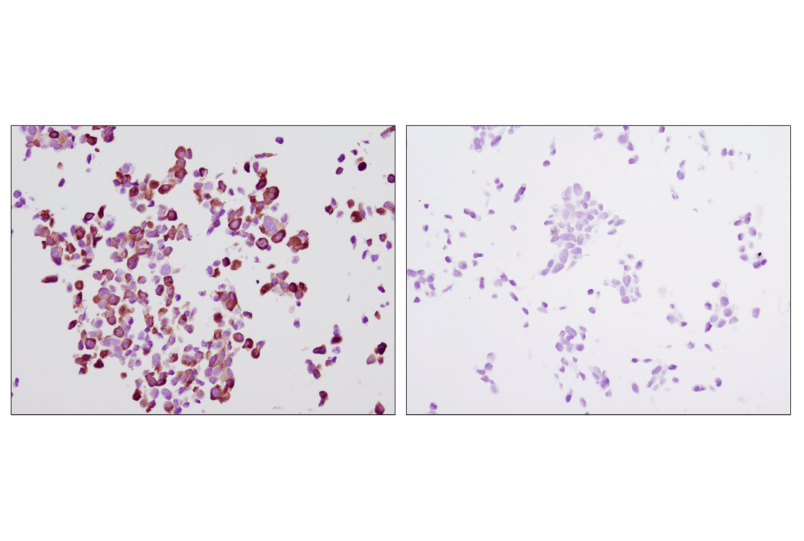
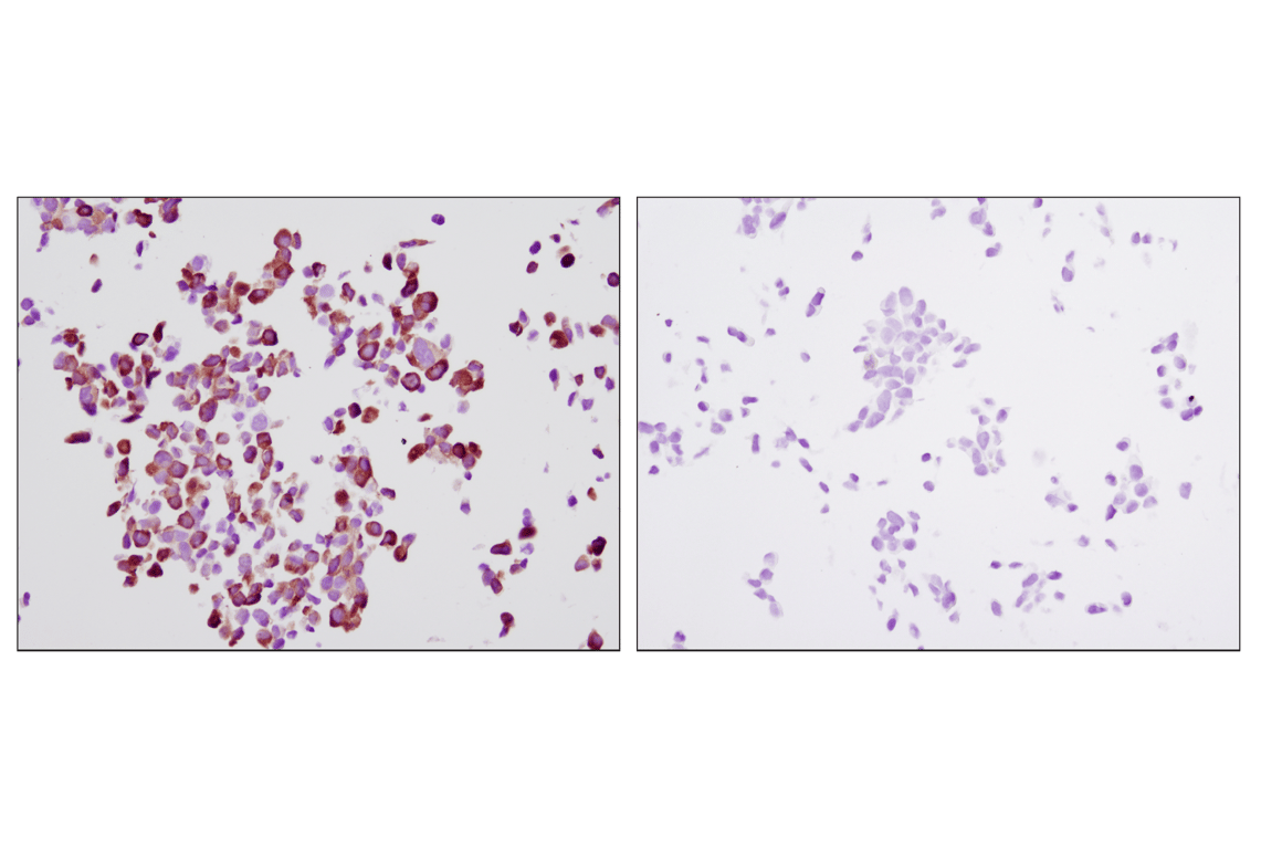
Immunohistochemical analysis of paraffin-embedded LNCaP cells, untreated (left) or LY294002-treated (right), using Phospho-S6 Ribosomal Protein (S235/236) (D57.2.2E) XP® Rabbit mAb on SignalSlide® Phospho-Akt (Ser473) IHC Controls #8101.
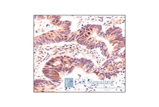
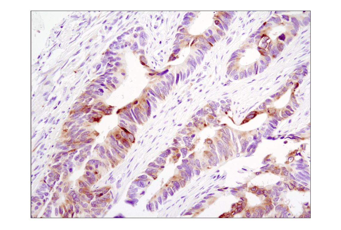
Immunohistochemical analysis of paraffin-embedded human colon carcinoma using S6 Ribosomal Protein (5G10) Rabbit mAb.
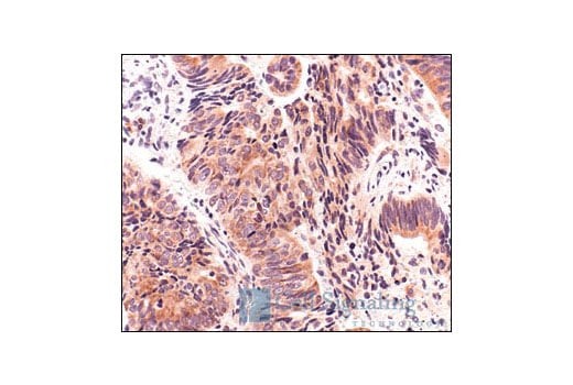
Immunohistochemical analysis of paraffin-embedded human lung carcinoma showing cytoplasmic localization using S6 Ribosomal Protein (5G10) Rabbit mAb.
Revision 2
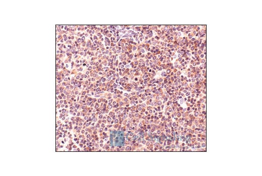
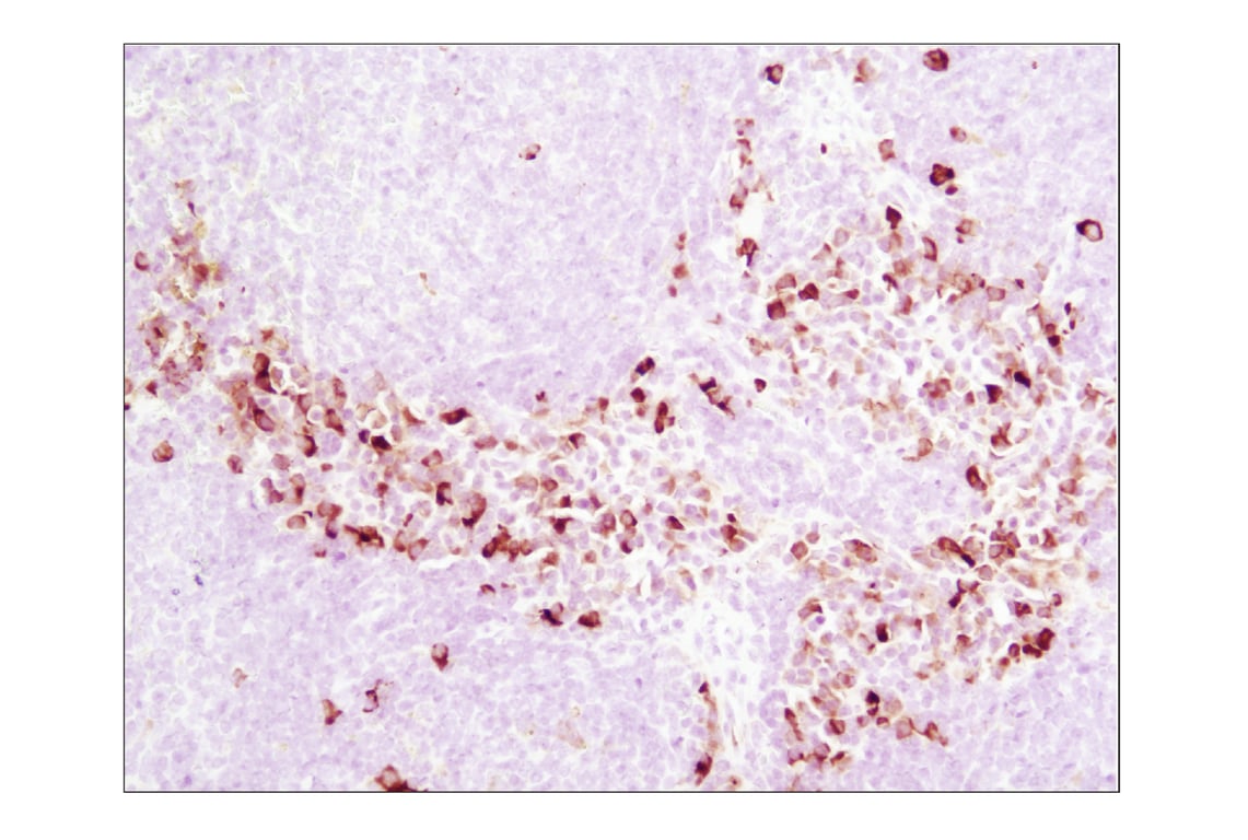
Immunohistochemical analysis of paraffin-embedded human Non-Hodgkin lymphoma, using S6 Ribosomal Protein (5G10) Rabbit mAb.
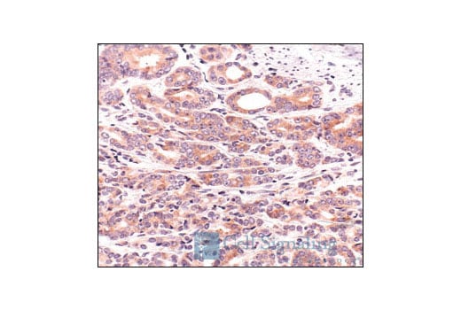
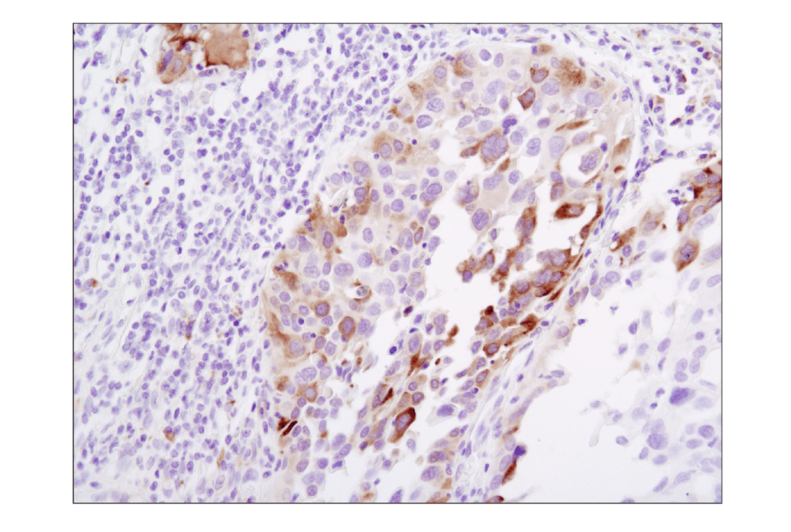
Immunohistochemical analysis of paraffin-embedded human prostate carcinoma using S6 Ribosomal Protein (5G10) Rabbit mAb.
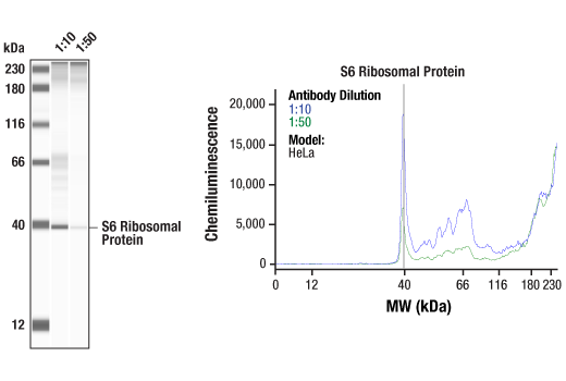
Simple Western™ analysis of lysates (1.0 mg/mL) from Hela cells using S6 Ribosomal Protein (5G10) Rabbit mAb #2217. The virtual lane view (left) shows a single target band (as indicated) at 1:10 and 1:50 dilutions of primary antibody. The corresponding electropherogram view (right) plots chemiluminescence by molecular weight along the capillary at 1:10 (blue line) and 1:50 (green line) dilutions of primary antibody. This experiment was performed under reducing conditions on the Jess™ Simple Western instrument from ProteinSimple, a BioTechne brand, using the 12-230 kDa separation module.
Revision 2
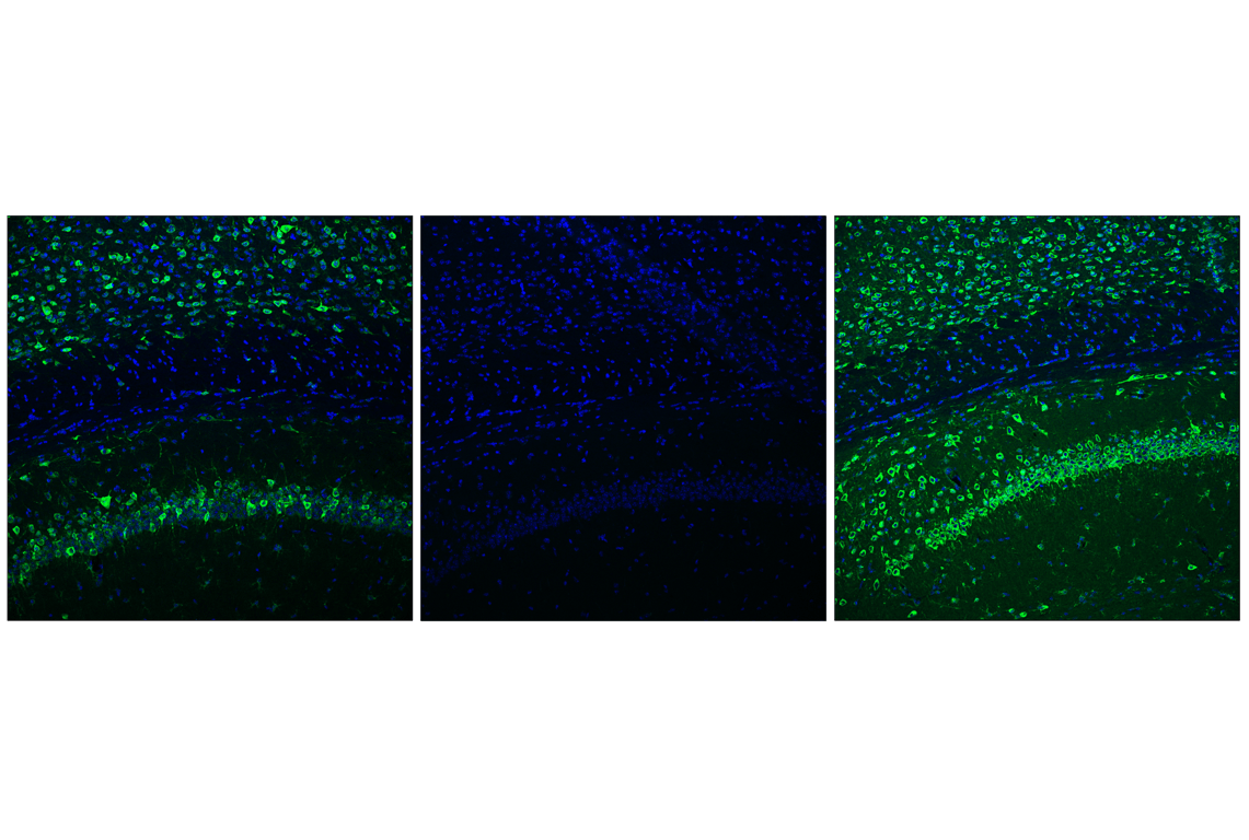
Confocal immunofluorescent analysis of fixed frozen mouse hippocampus, untreated (left) or post-processed with λ phosphatase (middle), using Phospho-S6 Ribosomal Protein (Ser235/236) (D57.2.2E) XP® Rabbit mAb (green) and, untreated (right), using S6 Ribosomal Protein (5G10) Rabbit mAb #2217 (green) and ProLong® Gold Antifade Reagent with DAPI #8961 (blue).
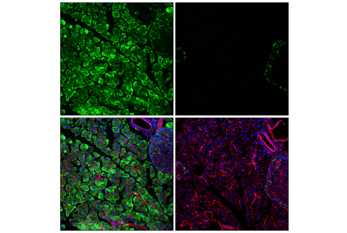
Confocal immunofluorescent analysis of fixed frozen mouse pancreas, untreated (left) or post-processed with λ phosphatase (right), using Phospho-S6 Ribosomal Protein (Ser235/236) (D57.2.2E) XP® Rabbit mAb (green), DyLight™ 554 Phalloidin #13054 (red), and ProLong® Gold Antifade Reagent with DAPI #8961 (blue).
Immunohistochemical analysis of paraffin-embedded human colon carcinoma using Phospho-S6 Ribosomal Protein (Ser235/236) (D57.2.2E) XP® Rabbit mAb.
Revision 2
Immunohistochemical analysis of paraffin-embedded human lung carcinoma using Phospho-S6 Ribosomal Protein (Ser235/236) (D57.2.2E) XP® Rabbit mAb.
Immunohistochemical analysis of paraffin-embedded mouse spleen using Phospho-S6 Ribosomal Protein (Ser235/236) (D57.2.2E) XP® Rabbit mAb.
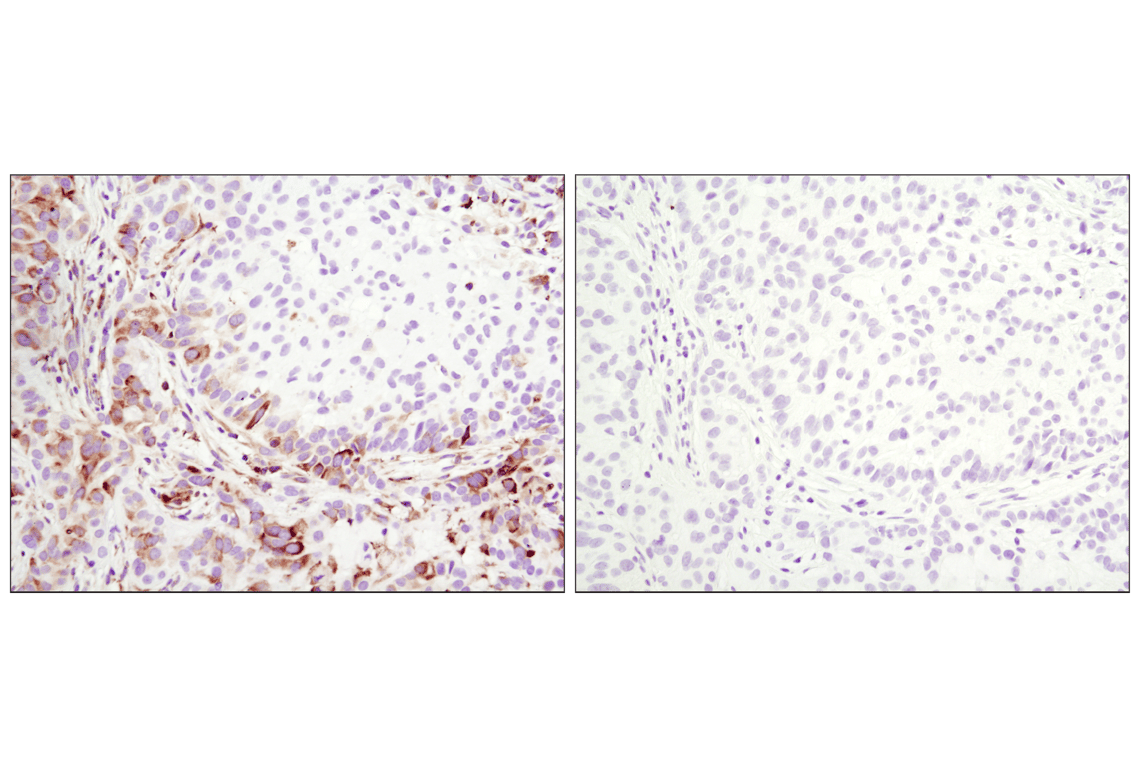
Immunohistochemical analysis of paraffin-embedded A549 xenograft, untreated (left) or λ phosphatase-treated (right), using Phospho-S6 Ribosomal Protein (Ser235/236) (D57.2.2E) XP® Rabbit mAb.
Revision 2
Immunohistochemical analysis of paraffin-embedded human breast carcinoma using Phospho-S6 Ribosomal Protein (Ser235/236) (D57.2.2E) XP® Rabbit mAb in the presence of control peptide (left) or Phospho-S6 Ribosomal Protein (Ser235/236) Blocking Peptide #1220 (right).
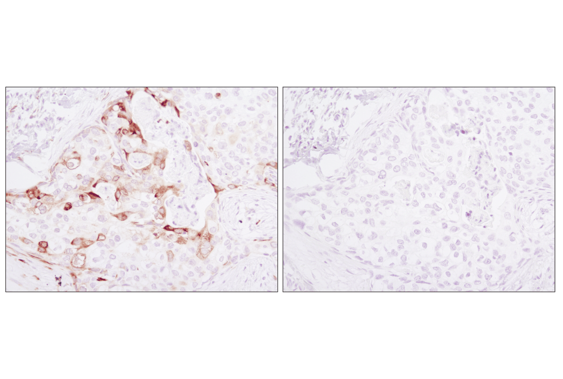
Immunohistochemical analysis of paraffin-embedded LNCaP cells, untreated (left) or rapamycin-treated (right), using Phospho-S6 Ribosomal Protein (Ser235/236) (D57.2.2E) XP® Rabbit mAb.
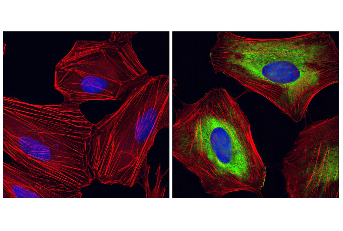
Confocal immunofluorescent analysis of HeLa cells, rapamycin-treated (left) or 20% serum-treated (right), using Phospho-S6 Ribosomal protein (Ser235/Ser236) (D57.2.2E) XP® Rabbit mAb (green). Actin filaments have been labeled with Alexa Fluor® 555 phalloidin (red). Blue pseudocolor = DRAQ5® #4084 (fluorescent DNA dye).
Revision 2
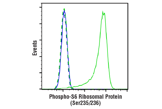
Flow cytometric analysis of Jurkat cells, untreated (green) or treated with LY294002 #9901, Wortmannin #9951, and U0126 #9903 (50 μM, 1 μM, and 10 μM, 2 hr; blue) using Phospho-S6 Ribosomal Protein (Ser235/236) (D57.2.2E) XP® Rabbit mAb (solid lines) or concentration-matched Rabbit (DA1E) mAb IgG XP® Isotype Control #3900 (dashed lines). Anti-rabbit IgG (H+L), F(ab')2 Fragment (Alexa Fluor® 488 Conjugate) #4412 was used as a secondary antibody.
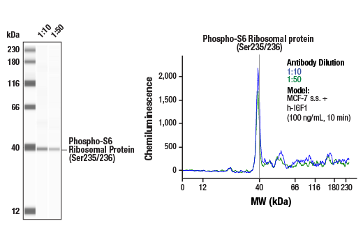
Simple Western™ analysis of lysates (1 mg/mL) from serum-starved MCF-7 cells treated with human IGF-1 (100 ng/mL, 10 min) using Phospho-S6 Ribosomal Protein (Ser235/236) (D57.2.2E) XP® Rabbit mAb #4858. The virtual lane view (left) shows the target band (as indicated) at 1:10 and 1:50 dilutions of primary antibody. The corresponding electropherogram view (right) plots chemiluminescence by molecular weight along the capillary at 1:10 (blue line) and 1:50 (green line) dilutions of primary antibody. This experiment was performed under reducing conditions on the Jess™ Simple Western instrument from ProteinSimple, a BioTechne brand, using the 12-230 kDa separation module.