Revision 1
#93130
Store at -20C
Stat Antibody Sampler Kit II
1 Kit
(6 x 20 microliters)
877-616-CELL (2355)
877-678-TECH (8324)
3 Trask Lane | Danvers | Massachusetts | 01923 | USA
For Research Use Only. Not for Use in Diagnostic Procedures.
| Product Includes | Product # | Quantity | Mol. Wt | Isotype/Source |
|---|---|---|---|---|
| Stat1 (D1K9Y) Rabbit mAb | 14994 | 20 µl | 84, 91 kDa | Rabbit IgG |
| Stat2 (D9J7L) Rabbit mAb | 72604 | 20 µl | 97, 113 kDa | Rabbit IgG |
| Stat3 (D1B2J) Rabbit mAb | 30835 | 20 µl | 79, 86 kDa | Rabbit IgG |
| Stat4 (C46B10) Rabbit mAb | 2653 | 20 µl | 81 kDa | Rabbit |
| Stat5 (D2O6Y) Rabbit mAb | 94205 | 20 µl | 90 kDa | Rabbit IgG |
| Stat6 (D3H4) Rabbit mAb | 5397 | 20 µl | 110 kDa | Rabbit IgG |
| Anti-rabbit IgG, HRP-linked Antibody | 7074 | 100 µl | Goat |
Please visit cellsignal.com for individual component applications, species cross-reactivity, dilutions, protocols, and additional product information.
Description
Stat Antibody Sampler Kit II provides an economical means to examine the complete Stat family: Stat1-6. The kit contains enough a primary antibody to perform two western blot experiments with each primary antibody.
Storage
Background
Janus kinases (Jaks) and signal transducers and activators of transcription (Stats) are utilized by receptors for a wide variety of ligands including cytokines, hormones, growth factors, and neurotransmitters. Jaks, activated via autophosphorylation following ligand-induced receptor aggregation, phosphorylate tyrosine residues on associated receptors, Stat molecules, and other downstream signaling proteins (1,2). The phosphorylation of Stat proteins at conserved tyrosine residues activates SH2-mediated dimerization followed rapidly by nuclear translocation. Stat dimers bind to interferon response element (IRE) and gamma interferon-activated sequence (GAS) DNA elements, resulting in the transcriptional regulation of downstream genes (1,2). The remarkable range and specificity of responses regulated by the Stats is determined in part by the tissue-specific expression of different cytokine receptors, Jaks and Stats (2,3), and by the combinatorial coupling of various Stat members to different receptors. Serine phosphorylation in the carboxy-terminal transcriptional activation domain has been shown to regulate the function of Stat1, Stat2, Stat3, Stat4, and Stat5 (1). Phosphorylation of Stat3 at Ser727 via MAPK or mTOR pathways is required for optimal transcriptional activation in response to growth factors and cytokines including IFN-gamma and ciliary neurotrophic factor (CNTF) (4,5). Jak/Stat pathways also play important roles in oncogenesis, tumor progression, angiogenesis, cell motility, immune responses, and stem cell differentiation (6-11).
Background References
- Darnell Jr., J. et al. (1994) Science 264, 1415-1421.
- Leonard, W.J. and O'Shea, J.J. (1998) Annu. Rev. Immunol. 16, 293-322.
- Caldenhoven, E. et al. (1996) J. Biol. Chem. 271, 13221-13227.
- Wen, Z. et al. (1995) Cell 82, 241-250.
- Yokogami, K. et al. (2000) Curr. Biol. 10, 47-50.
- Lim, C.P. and Cao, X. (1999) J. Biol. Chem. 274, 31055-31061.
- Bromberg, J. F. et al. (1999) Cell 98, 295-303.
- Su, L. et al. (1999) J. Biol. Chem. 274, 31770-31774.
- Dentelli, P. et al. (1999) J. Immunol. 163, 2151-2159.
- Cattaneo, E. et al. (1999) Trends Neurosci. 22, 365-369.
- Frank, D.A. (1999) Mol. Med. 5, 432-456.
Trademarks and Patents
Cell Signaling Technology is a trademark of Cell Signaling Technology, Inc.
U.S. Patent No. 7,429,487, foreign equivalents, and child patents deriving therefrom.
All other trademarks are the property of their respective owners. Visit cellsignal.com/trademarks for more information.
Limited Uses
Except as otherwise expressly agreed in a writing signed by a legally authorized representative of CST, the following terms apply to Products provided by CST, its affiliates or its distributors. Any Customer's terms and conditions that are in addition to, or different from, those contained herein, unless separately accepted in writing by a legally authorized representative of CST, are rejected and are of no force or effect.
Products are labeled with For Research Use Only or a similar labeling statement and have not been approved, cleared, or licensed by the FDA or other regulatory foreign or domestic entity, for any purpose. Customer shall not use any Product for any diagnostic or therapeutic purpose, or otherwise in any manner that conflicts with its labeling statement. Products sold or licensed by CST are provided for Customer as the end-user and solely for research and development uses. Any use of Product for diagnostic, prophylactic or therapeutic purposes, or any purchase of Product for resale (alone or as a component) or other commercial purpose, requires a separate license from CST. Customer shall (a) not sell, license, loan, donate or otherwise transfer or make available any Product to any third party, whether alone or in combination with other materials, or use the Products to manufacture any commercial products, (b) not copy, modify, reverse engineer, decompile, disassemble or otherwise attempt to discover the underlying structure or technology of the Products, or use the Products for the purpose of developing any products or services that would compete with CST products or services, (c) not alter or remove from the Products any trademarks, trade names, logos, patent or copyright notices or markings, (d) use the Products solely in accordance with CST Product Terms of Sale and any applicable documentation, and (e) comply with any license, terms of service or similar agreement with respect to any third party products or services used by Customer in connection with the Products.
Revision 1
#93130
Stat Antibody Sampler Kit II
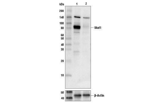
Western blot analysis of extracts from A549 cells (lane 1) or STAT1 knock-out cells (lane 2) using Stat1 (D1K9Y) Rabbit mAb #14994 (upper), and β-actin (D6A8) Rabbit mAb #8457 (lower). The absence of signal in the STAT1 knock-out A549 cells confirms specificity of the antibody for STAT1.
CUT&RUN was performed with HT-1080 cells treated with (hIFN-γ) #8901 (50 ng/ml, 30 min) and Stat1 (D1K9Y) Rabbit mAb or Rabbit (DA1E) mAb IgG XP® Isotype Control (CUT&RUN) #66362, using CUT&RUN Assay Kit #86652. The enriched DNA was quantified by real-time PCR using human AIM2 promoter primers, human FZD5 promoter primers, and SimpleChIP® Human α Satellite Repeat Primers #4486. The amount of immunoprecipitated DNA in each sample is represented as signal relative to the total amount of input chromatin, which is equivalent to one.

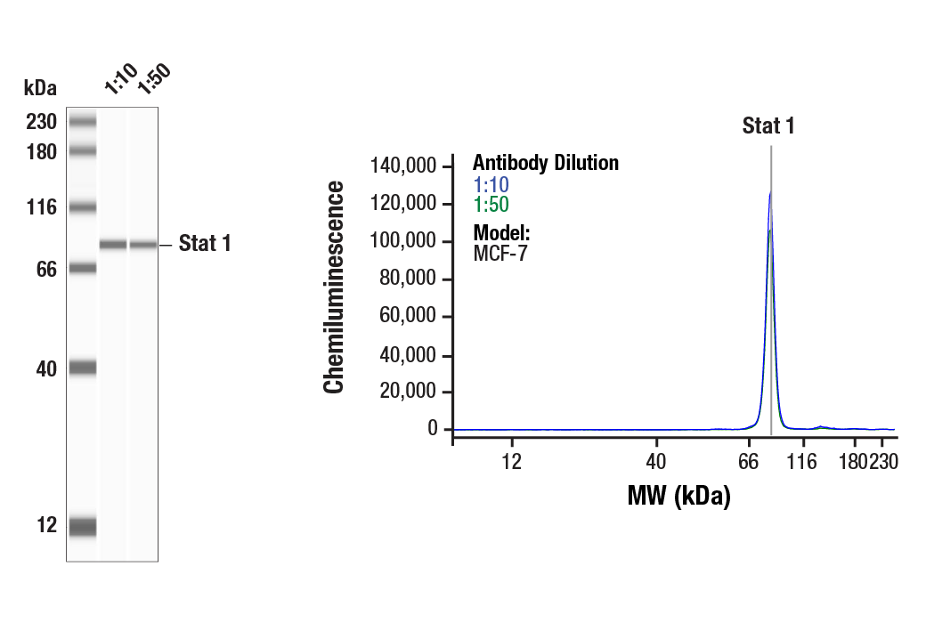
Simple Western™ analysis of lysates (0.1 mg/mL) from MCF-7 cells using Stat1 (D1K9Y) Rabbit mAb #14994. The virtual lane view (left) shows the target band (as indicated) at 1:10 and 1:50 dilutions of primary antibody. The corresponding electropherogram view (right) plots chemiluminescence by molecular weight along the capillary at 1:10 (blue line) and 1:50 (green line) dilutions of primary antibody. This experiment was performed under reducing conditions on the Jess™ Simple Western instrument from ProteinSimple, a BioTechne brand, using the 12-230 kDa separation module.
Revision 1
#93130
Stat Antibody Sampler Kit II
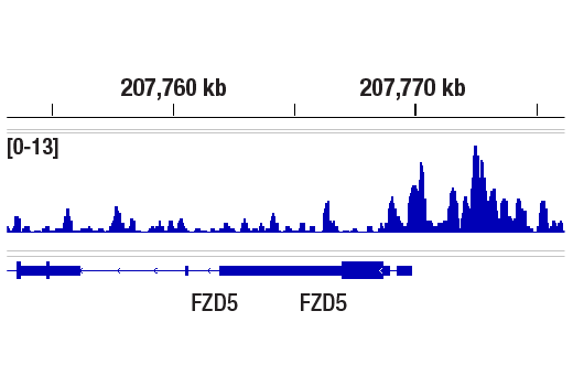
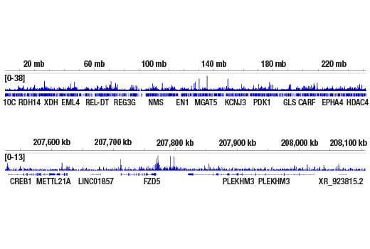
CUT&RUN was performed with HT-1080 cells and Stat1 (D1K9Y) Rabbit mAb, using CUT&RUN Assay Kit #86652. DNA library was prepared using DNA Library Prep Kit for Illumina (ChIP-seq, CUT&RUN) #56795. The figure shows binding across FZD5 gene, a known target gene of Stat1 (see additional figure containing CUT&RUN-qPCR data).
CUT&RUN was performed with HT-1080 cells and Stat1 (D1K9Y) Rabbit mAb, using CUT&RUN Assay Kit #86652. DNA Libraries were prepared using DNA Library Prep Kit for Illumina (ChIP-seq, CUT&RUN) #56795. The figures show binding across chromosome 2 (upper), including FZD5 gene (lower), a known target gene of Stat1 (see additional figure containing CUT&RUN-qPCR data).
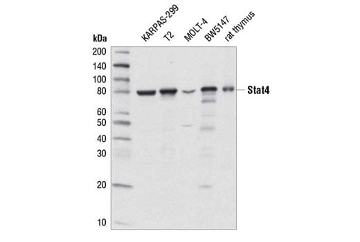
Western blot analysis of extracts from various cell lines and from rat thymus using Stat4 (C46B10) Rabbit mAb.
Revision 1
#93130
Stat Antibody Sampler Kit II
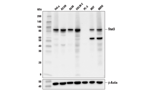
Western blot analysis of extracts from various cell lines using Stat3 (D1B2J) Rabbit mAb (upper) or β-Actin (D6A8) Rabbit mAb #8457 (lower).
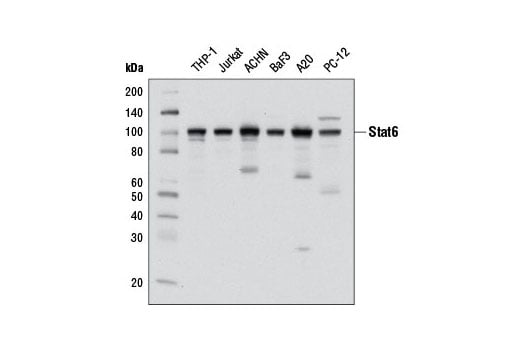
Western blot analysis of extracts from various cell lines using Stat6 (D3H4) Rabbit mAb.
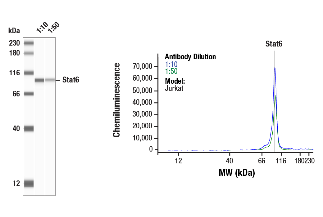
Simple Western™ analysis of lysates (0.1 mg/mL) from Jurkat cells using Stat6 (D3H4) Rabbit mAb #5397. The virtual lane view (left) shows the target band (as indicated) at 1:10 and 1:50 dilutions of primary antibody. The corresponding electropherogram view (right) plots chemiluminescence by molecular weight along the capillary at 1:10 (blue line) and 1:50 (green line) dilutions of primary antibody. This experiment was performed under reducing conditions on the Jess™ Simple Western instrument from ProteinSimple, a BioTechne brand, using the 12 – 230 kDa separation module.
Revision 1
#93130
Stat Antibody Sampler Kit II
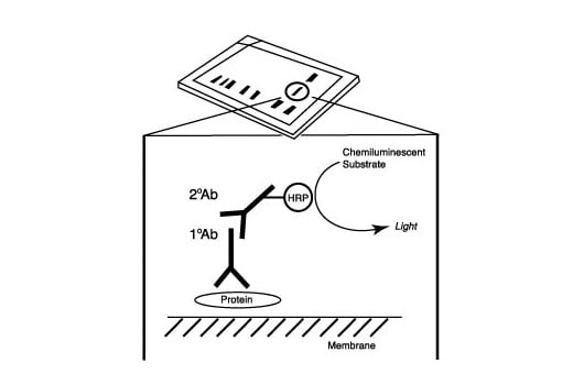
After the primary antibody is bound to the target protein, a complex with HRP-linked secondary antibody is formed. The LumiGLO® is added and emits light during enzyme catalyzed decomposition.
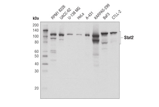
Western blot analysis of extracts from various cell lines using Stat2 (D9J7L) Rabbit mAb. KARPAS cell line source: Dr Abraham Karpas at the University of Cambridge.
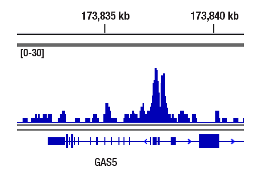
CUT&RUN was performed with U266 cells treated with Human Interferon-α1 (hIFN-α1) #8927 (10nM, 30min) and Stat2 (D9J7L) Rabbit mAb, using CUT&RUN Assay Kit #86652. DNA Library was prepared using DNA Library Prep Kit for Illumina® (ChIP-seq, CUT&RUN) #56795. The figure shows binding across GAS5 gene.
Revision 1
#93130
Stat Antibody Sampler Kit II
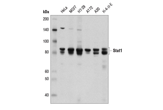
Western blot analysis of extracts from various cell lines using Stat1 (D1K9Y) Rabbit mAb.
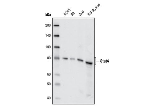
Western blot analysis of extracts from ACHN, SR and Caki cell lines and from rat thymus using Stat4 (C46B10) Rabbit mAb.
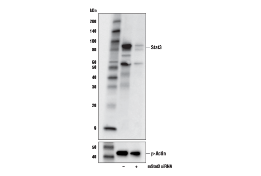
Western blot analysis of extracts from NIH/3T3 cells, mock transfected (-) or transfected with an siRNA against mouse Stat3 (mStat3 siRNA; +), using Stat3 (D1B2J) Rabbit mAb (upper) or β-Actin (D6A8) Rabbit mAb #8457 (lower).
Revision 1
#93130
Stat Antibody Sampler Kit II
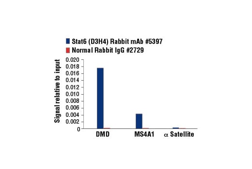
Chromatin immunoprecipitations were performed with cross-linked chromatin from Ramos cells starved overnight then treated with IL-4 (100 ng/ml, 30 min) and either Stat6 (D3H4) Rabbit mAb or Normal Rabbit IgG #2729 using SimpleChIP® Enzymatic Chromatin IP Kit (Magnetic Beads) #9003. The enriched DNA was quantified by real-time PCR using SimpleChIP® Human DMD Intron 2 Primers #7710, human MS4A1 promoter primers, and SimpleChIP® Human α Satellite Repeat Primers #4486. The amount of immunoprecipitated DNA in each sample is represented as signal relative to the total amount of input chromatin, which is equivalent to one.
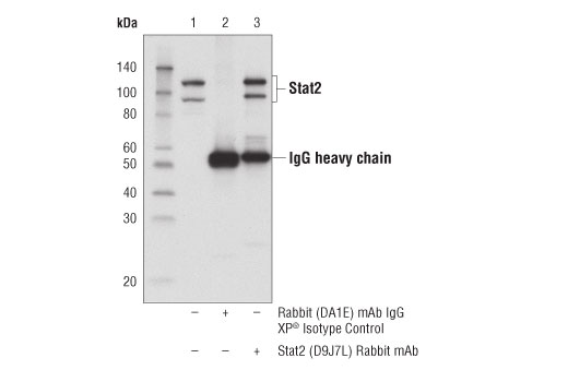
Immunoprecipitation of Stat2 from KARPAS-299 cell extracts. Lane 1 is 10% input, lane 2 is Rabbit (DA1E) mAb IgG XP® Isotype Control #3900, and lane 3 is Stat2 (D9J7L) Rabbit mAb. Western blot was performed using Stat2 (D9J7L) Rabbit mAb. KARPAS cell line source: Dr Abraham Karpas at the University of Cambridge.
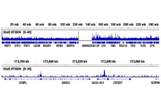
CUT&RUN was performed with U266 cells treated with Human Interferon-α1 (hIFN-α1) #8927 (10nM, 30min) and Stat2 (D9J7L) Rabbit mAb, using CUT&RUN Assay Kit #86652. DNA Library was prepared using DNA Library Prep Kit for Illumina® (ChIP-seq, CUT&RUN) #56795. The figures show binding across chromosome 1 (upper), including GAS5 gene (lower).
Revision 1
#93130
Stat Antibody Sampler Kit II
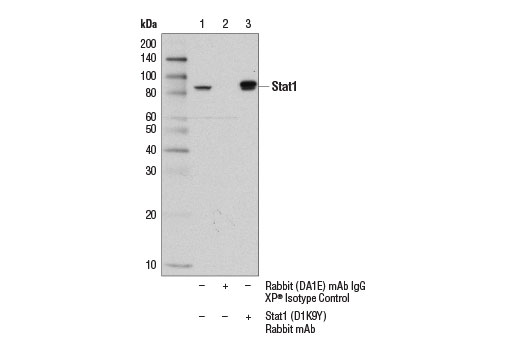
Immunoprecipitation of Stat1 from MCF7 cell extracts using Rabbit (DA1E) mAb IgG XP® Isotype Control #3900 (lane 2) or Stat1 (D1K9Y) Rabbit mAb (lane 3). Lane 1 is 10% input. Western blot was performed using Stat1 (9H2) Mouse mAb #9176.
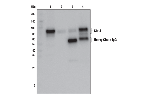
Immunoprecipitation of Stat4 protein from NK-92 cell extracts. Lane 1 is 10% input, lane 2 is beads only, lane 3 is Rabbit (DA1E) mAb IgG XP® Isotype Control #3900, and lane 4 is Stat4 (C46B10) Rabbit mAb. Western blot analysis was performed using Stat4 (C46B10) Rabbit mAb.
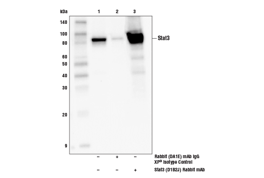
Immunoprecipitation of Stat3 from MCF7 cell extracts. Lane 1 is 10% input, lane 2 is Rabbit (DA1E) mAb IgG XP® Isotype Control #3900, and lane 3 is Stat3 (D1B2J) Rabbit mAb. Western blot was performed using Stat3 (D1B2J) Rabbit mAb. A conformation-specific secondary antibody was used to avoid reactivity with IgG.
Revision 1
#93130
Stat Antibody Sampler Kit II
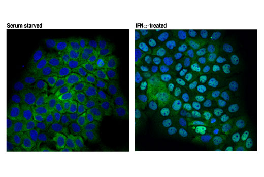
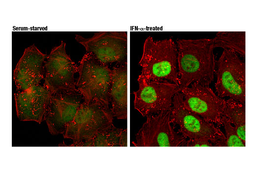
Confocal immunofluorescent analysis of A-431 cells, serum starved (left) or treated with IFNα (1000 U/ml for 30 mins; right) using Stat2 (D9J7L) Rabbit mAb (green). Blue pseudocolor = DRAQ5® #4084 (fluorescent DNA dye).
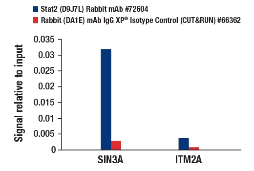
CUT&RUN was performed with U266 cells treated with Human Interferon-α1 (hIFN-α1) #8927 (10nM, 30min) and either Stat2 (D9J7L) Rabbit mAb or Rabbit (DA1E) mAb IgG XP® Isotype Control (CUT&RUN) #66362, using CUT&RUN Assay Kit #86652. The enriched DNA was quantified by real-time PCR using human SIN3A promoter primers and human ITM2A upstream primers. The amount of immunoprecipitated DNA in each sample is represented as signal relative to the total amount of input chromatin, which is equivalent to one.
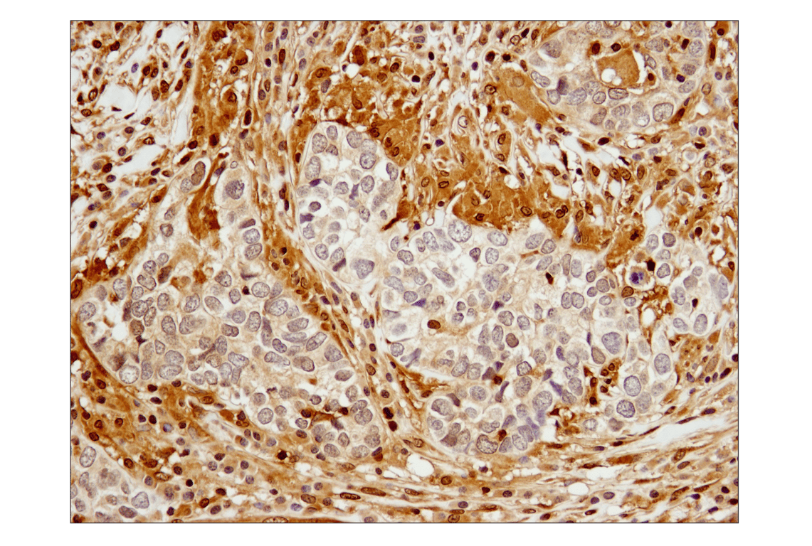
Immunohistochemical analysis of paraffin-embedded human breast carcinoma using Stat1 (D1K9Y) Rabbit mAb.
Revision 1
#93130
Stat Antibody Sampler Kit II
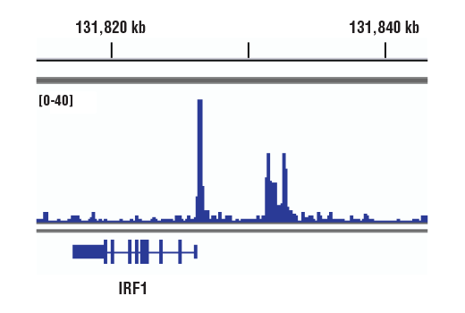
Chromatin immunoprecipitations were performed with cross-linked chromatin from NK-92 cells starved of IL-2 overnight then treated with IL-12 (10 ng/ml) for 4 hr and Stat4 (C46B10) Rabbit mAb, using SimpleChIP® Plus Enzymatic Chromatin IP Kit (Magnetic Beads) #9005. DNA Libraries were prepared using DNA Library Prep Kit for Illumina® (ChIP-seq, CUT&RUN) #56795. The figure shows binding across IRF1, a known target gene of Stat4 (see additional figure containing ChIP-qPCR data). For additional ChIP-seq tracks, please download the product datasheet.
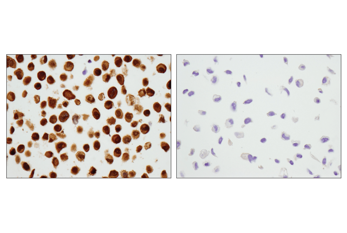
Immunohistochemical analysis of paraffin-embedded HeLa cell pellet (left, positive) or PC-3 cell pellet (right, negative) using Stat3 (D1B2J) Rabbit mAb.
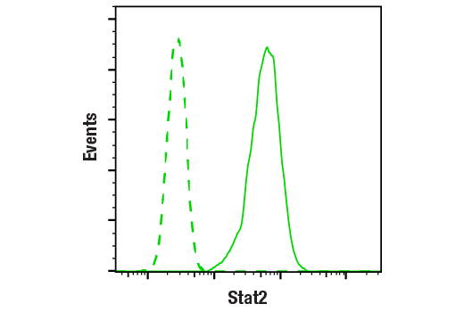
Flow cytometric analysis of K-562 cells using Stat2 (D9J7L) Rabbit mAb (solid line) compared to concentration-matched Rabbit (DA1E) mAb IgG XP® Isotype control #3900 (dashed line). Anti-rabbit IgG (H+L), F(ab')2 Fragment (Alexa Fluor® 488 Conjugate) #4412 was used as a secondary antibody.
Revision 1
#93130
Stat Antibody Sampler Kit II
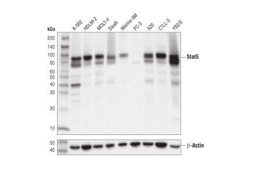
Western blot analysis of extracts from various cell lines using Stat5 (D2O6Y) Rabbit mAb (upper) and β-Actin (D6A8) Rabbit mAb #8457 (lower).
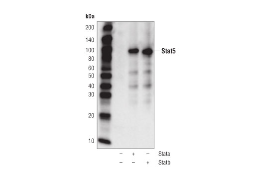
Western blot analysis of PC-3 cells, mock transfected (-) or transfected with constructs expressing full-length human Stat5a (+) or Stat5b (+) using Stat5 (D2O6Y) Rabbit mAb.
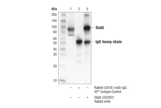
Immunoprecipitation of Stat5 from K-562 cell extracts. Lane 1 is 10% input, lane 2 is precipitated with Rabbit (DA1E) mAb IgG XP® Isotype Control #3900, and lane 3 is Stat5 (D2O6Y) Rabbit mAb. Western blot was performed using Stat5 (D2O6Y) Rabbit mAb.
Revision 1
#93130
Stat Antibody Sampler Kit II
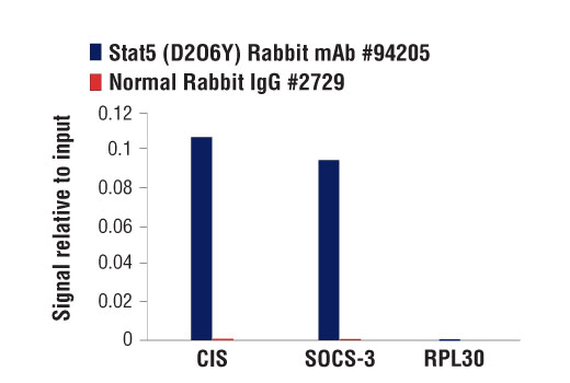
Chromatin immunoprecipitations were performed with cross-linked chromatin from BaF3 cells starved of IL-3 for 6 hours followed by induction with IL-3 for 45 minutes, and either Stat5 (D2O6Y) Rabbit mAb or Normal Rabbit IgG #2729 using SimpleChIP® Enzymatic Chromatin IP Kit (Magnetic Beads) #9003. The enriched DNA was quantified by real-time PCR using SimpleChIP® Mouse CIS Intron 1 Primers #5131, mouse SOCS-3 promoter primers, and SimpleChIP® Mouse RPL30 Intron 2 Primers #7015. The amount of immunoprecipitated DNA in each sample is represented as signal relative to the total amount of input chromatin, which is equivalent to one.
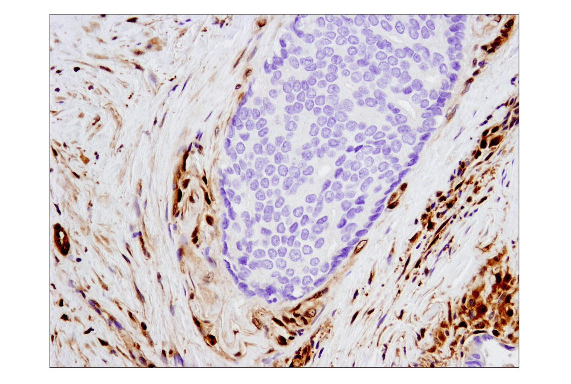
Immunohistochemical analysis of paraffin-embedded human lymphoma using Stat1 (D1K9Y) Rabbit mAb.
Chromatin immunoprecipitations were performed with cross-linked chromatin from NK-92 cells starved of IL-2 overnight then treated with IL-12 (10 ng/ml) for 4 hr and Stat4 (C46B10) Rabbit mAb, using SimpleChIP® Plus Enzymatic Chromatin IP Kit (Magnetic Beads) #9005. DNA Libraries were prepared using DNA Library Prep Kit for Illumina® (ChIP-seq, CUT&RUN) #56795. The figure shows binding across chromosome 5 (upper), including IRF1 (lower), a known target gene of Stat4 (see additional figure containing ChIP-qPCR data).
Revision 1
#93130
Stat Antibody Sampler Kit II
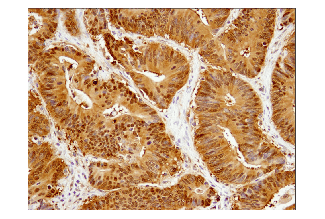
Immunohistochemical analysis of paraffin-embedded human colon carcinoma using Stat3 (D1B2J) Rabbit mAb.
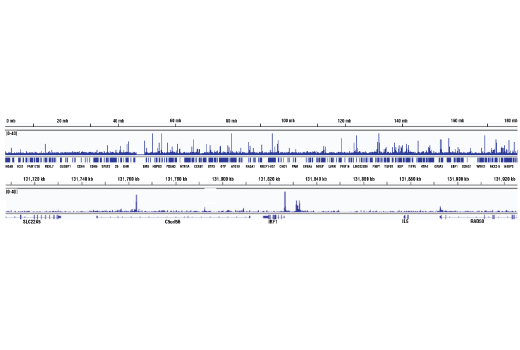
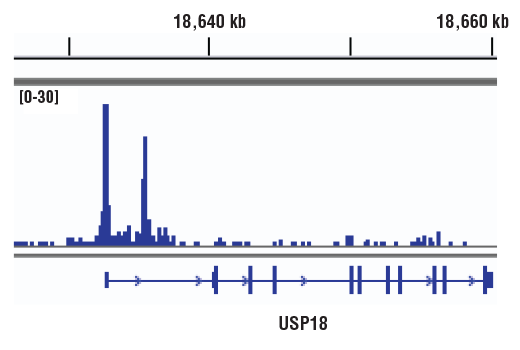
Chromatin immunoprecipitations were performed with cross-linked chromatin from U266 cells treated with Human Interferon-α (IFN-α) #9906 (10nM) for 30 min and Stat2 (D9J7L) Rabbit mAb, using SimpleChIP® Plus Enzymatic Chromatin IP Kit (Magnetic Beads) #9005. DNA Libraries were prepared using DNA Library Prep Kit for Illumina® (ChIP-seq, CUT&RUN) #56795. The figure shows binding across USP18, a known target gene of Stat2 (see additional figure containing ChIP-qPCR data). For additional ChIP-seq tracks, please download the product datasheet.
Confocal immunofluorescent analysis of HeLa cells, serum-starved overnight (left) or treated with Human Interferon-α1 (hIFN-α1) #8927 (1,000 units/ml, 30 min; right), using Stat1 (D1K9Y) Rabbit mAb (green) and β-Actin (8H10D10) Mouse mAb #3700 (red).
Revision 1
#93130
Stat Antibody Sampler Kit II
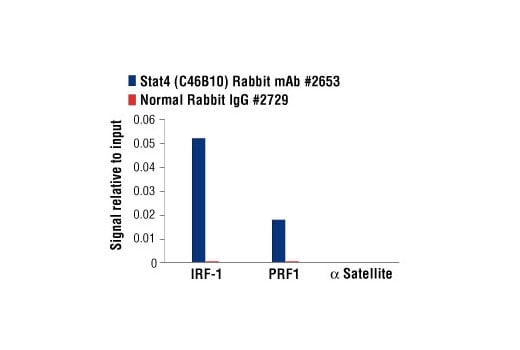
Chromatin immunoprecipitations were performed with cross-linked chromatin from NK-92 cells starved of IL-2 overnight then treated with IL-12 (10 ng/ml) for 4 hr and either Stat4 (C46B10) Rabbit mAb #2653 or Normal Rabbit IgG #2729 using SimpleChIP® Enzymatic Chromatin IP Kit (Magnetic Beads) #9003. The enriched DNA was quantified by real-time PCR using human IRF-1 promoter primers, human PRF1 promoter primers, and SimpleChIP® Human α Satellite Repeat Primers #4486. The amount of immunoprecipitated DNA in each sample is represented as signal relative to the total amount of input chromatin, which is equivalent to one.
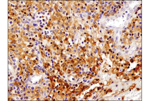
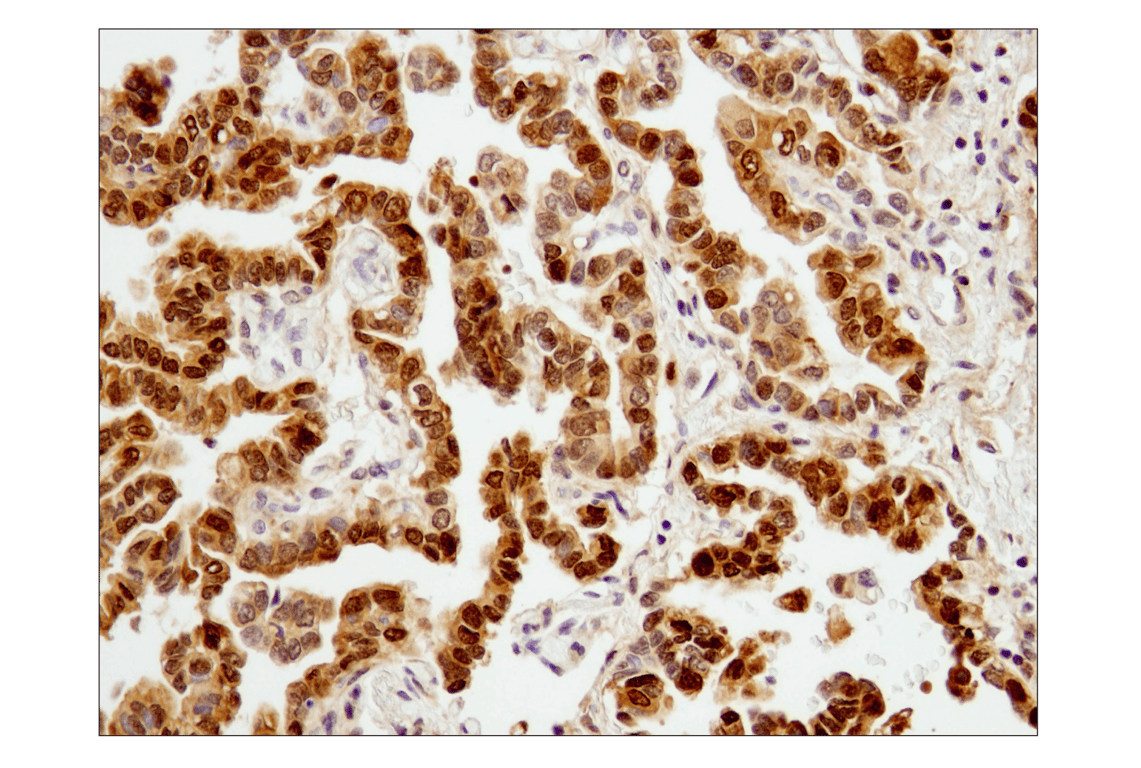
Immunohistochemical analysis of paraffin-embedded human non-small cell lung carcinoma using Stat3 (D1B2J) Rabbit mAb.
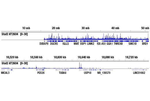
Chromatin immunoprecipitations were performed with cross-linked chromatin from U266 cells treated with Human Interferon-α (IFN-α) #9906 (10nM) for 30 min and Stat2 (D9J7L) Rabbit mAb, using SimpleChIP® Plus Enzymatic Chromatin IP Kit (Magnetic Beads) #9005. DNA Libraries were prepared using DNA Library Prep Kit for Illumina® (ChIP-seq, CUT&RUN) #56795. The figure shows binding across chromosome 22 (upper), including USP18 (lower), a known target gene of Stat2 (see additional figure containing ChIP-qPCR data).
Revision 1
#93130
Stat Antibody Sampler Kit II
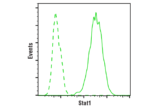
Flow cytometric analysis of ACHN cells using Stat1 (D1K9Y) Rabbit mAb (solid line) compared to concentration-matched Rabbit (DA1E) mAb IgG XP® Isotype Control #3900 (dashed line). Anti-rabbit IgG (H+L), F(ab')2 Fragment (Alexa Fluor® 488 Conjugate) #4412 was used as a secondary antibody.
Immunohistochemical analysis of paraffin-embedded human endometrioid adenocarcinoma using Stat3 (D1B2J) Rabbit mAb.
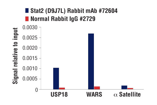
Chromatin immunoprecipitations were performed with cross-linked chromatin from U266 cells treated with Human Interferon-α (IFN-α) #9906 (100 ng/ml) for 30 min, and either Stat2 (D9J7L) Rabbit mAb or Normal Rabbit IgG #2729 using SimpleChIP® Enzymatic Chromatin IP Kit (Magnetic Beads) #9003. The enriched DNA was quantified by real-time PCR using human USP18 promoter primers, SimpleChIP® Human WARS Intron 1 Primers #30101, and SimpleChIP® Human α Satellite Repeat Primers #4486. The amount of immunoprecipitated DNA in each sample is represented as signal relative to the total amount of input chromatin, which is equivalent to one.
Revision 1
#93130
Stat Antibody Sampler Kit II
Immunohistochemical analysis of paraffin-embedded human prostate carcinoma using Stat3 (D1B2J) Rabbit mAb.
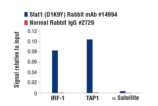
Chromatin immunoprecipitations were performed with cross-linked chromatin from HT-1080 cells treated with Human Interferon-γ (hIFN-γ) #8901 (50 ng/ml, 30 min) and either Stat1 (D1K9Y) Rabbit mAb or Normal Rabbit IgG #2729 using SimpleChIP® Enzymatic Chromatin IP Kit (Magnetic Beads) #9003. The enriched DNA was quantified by real-time PCR using human IRF-1 promoter primers, SimpleChIP® Human TAP1 Promoter Primers #5148, and SimpleChIP® Human α Satellite Repeat Primers #4486. The amount of immunoprecipitated DNA in each sample is represented as signal relative to the total amount of input chromatin, which is equivalent to one.
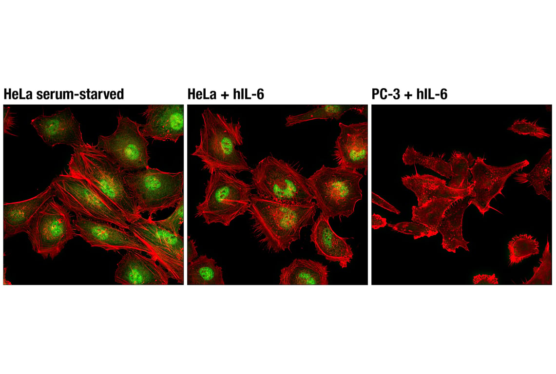
Confocal immunofluorescent analysis of HeLa cells (Stat3 positive), serum-starved (left) or treated with Human Interleukin-6 (hIL-6) #8904 (100 ng/mL, 20 min; center), or PC-3 cells (Stat3 negative) treated with Human Interleukin-6 (hIL-6) #8904 (100 ng/mL, 20 min; right), using Stat3 (D1B2J) Rabbit mAb (green) and β-Actin (8H10D10) Mouse mAb #3700 (red).
Revision 1
#93130
Stat Antibody Sampler Kit II
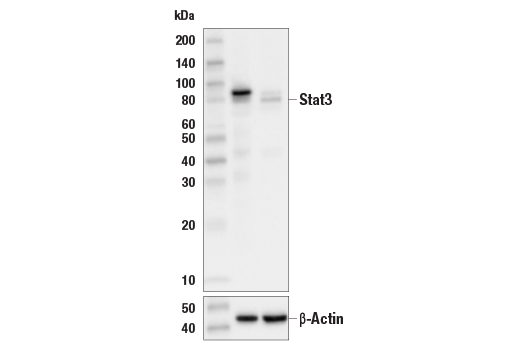
Western blot analysis of extracts from HeLa control cells (lane 1) or Stat3 knockout cells (lane 2) using Stat3 (D1B2J) Rabbit mAb #30835 (upper) or β-actin (13E5) Rabbit mAb #4970 (lower). The absence of signal in the Stat3 knockout cells confirms the specificity of the antibody for Stat3.
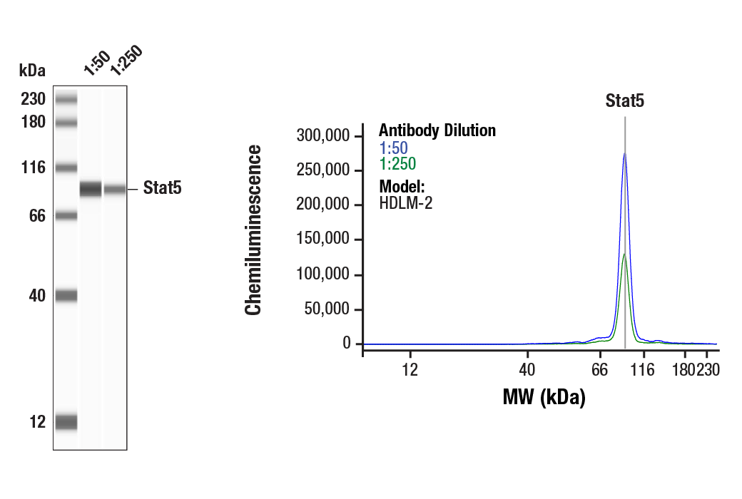
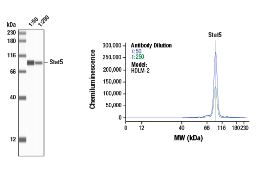
Simple Western™ analysis of lysates (0.1 mg/mL) from HDLM-2 cells using Stat5 (D2O6Y) Rabbit mAb #94205. The virtual lane view (left) shows the target band (as indicated) at 1:50 and 1:250 dilutions of primary antibody. The corresponding electropherogram view (right) plots chemiluminescence by molecular weight along the capillary at 1:50 (blue line) and 1:250 (green line) dilutions of primary antibody. This experiment was performed under reducing conditions on the Jess™ Simple Western instrument from ProteinSimple, a BioTechne brand, using the 12-230 kDa separation module.
Simple Western™ analysis of lysates (0.1 mg/mL) from HDLM-2 cells using Stat5 (D2O6Y) Rabbit mAb #94205. The virtual lane view (left) shows the target band (as indicated) at 1:50 and 1:250 dilutions of primary antibody. The corresponding electropherogram view (right) plots chemiluminescence by molecular weight along the capillary at 1:50 (blue line) and 1:250 (green line) dilutions of primary antibody. This experiment was performed under reducing conditions on the Jess™ Simple Western instrument from ProteinSimple, a BioTechne brand, using the 12-230 kDa separation module.