| Product Includes | Product # | Quantity | Mol. Wt | Isotype/Source |
|---|---|---|---|---|
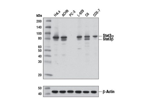
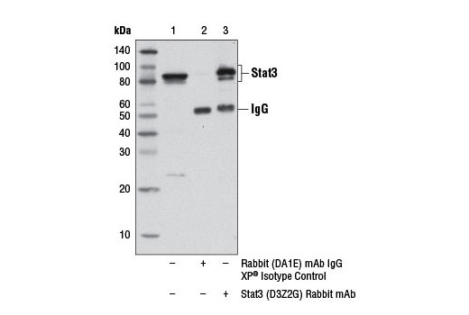
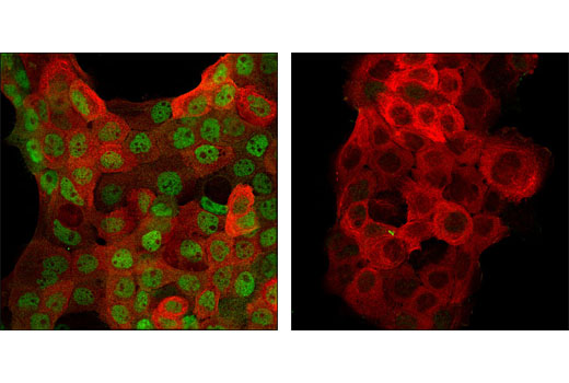
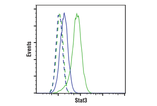
| Stat3 (D3Z2G) Rabbit mAb | 12640 | 20 µl | 79, 86 kDa | Rabbit IgG |
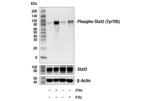
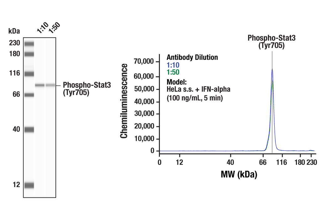
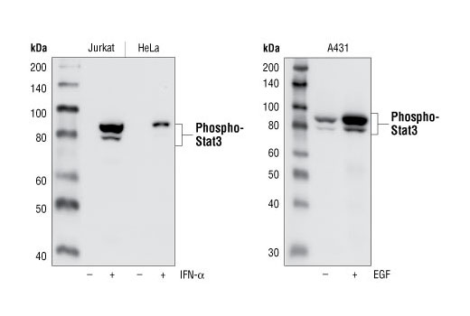
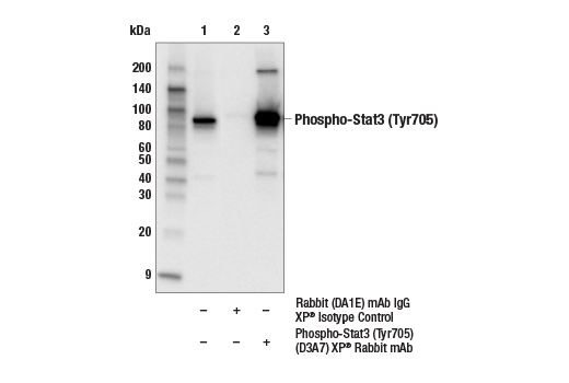
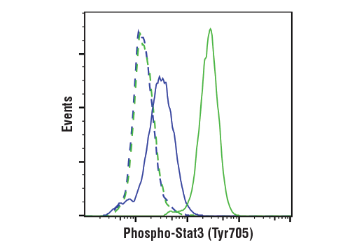
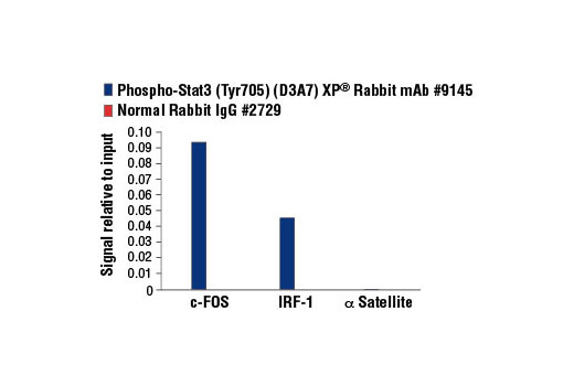
| Phospho-Stat3 (Tyr705) (D3A7) XP® Rabbit mAb | 9145 | 20 µl | 79, 86 kDa | Rabbit IgG |
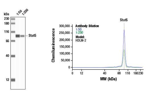
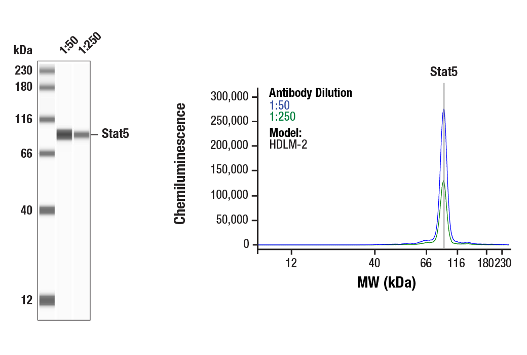
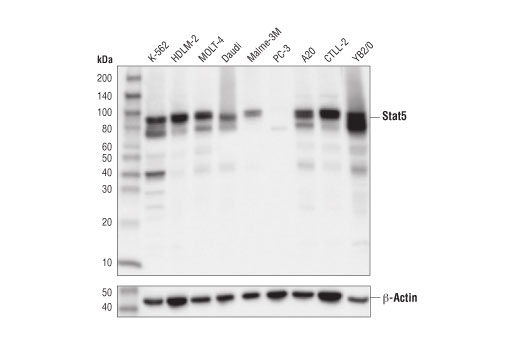
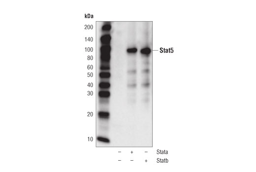
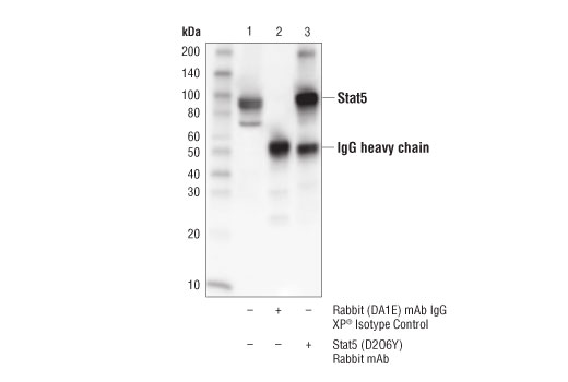
| Stat5 (D2O6Y) Rabbit mAb | 94205 | 20 µl | 90 kDa | Rabbit IgG |
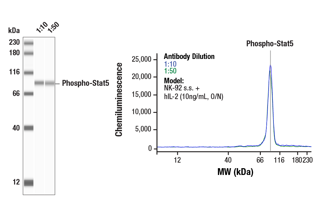
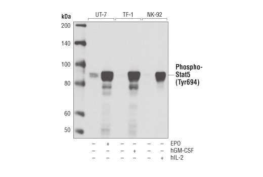
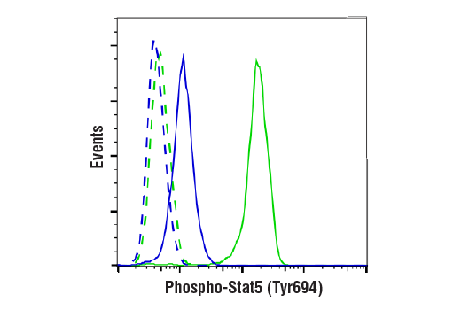
| Phospho-Stat5 (Tyr694) (D47E7) XP® Rabbit mAb | 4322 | 20 µl | 90 kDa | Rabbit IgG |
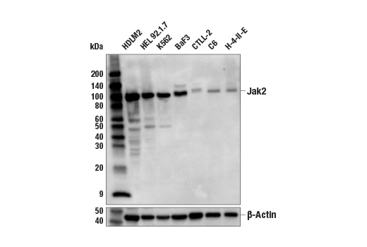
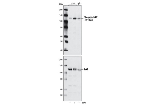
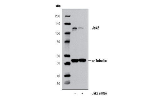
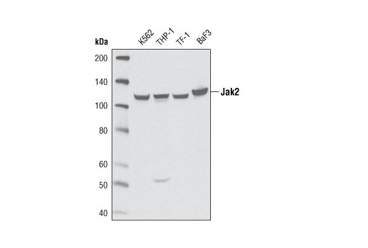
| Jak2 (D2E12) XP® Rabbit mAb | 3230 | 20 µl | 125 kDa | Rabbit IgG |
| Phospho-Jak2 (Tyr1007) (D15E2) Rabbit mAb | 4406 | 20 µl | 125 kDa | Rabbit IgG |
| Anti-rabbit IgG, HRP-linked Antibody | 7074 | 100 µl | Goat |
Please visit cellsignal.com for individual component applications, species cross-reactivity, dilutions, protocols, and additional product information.
Description
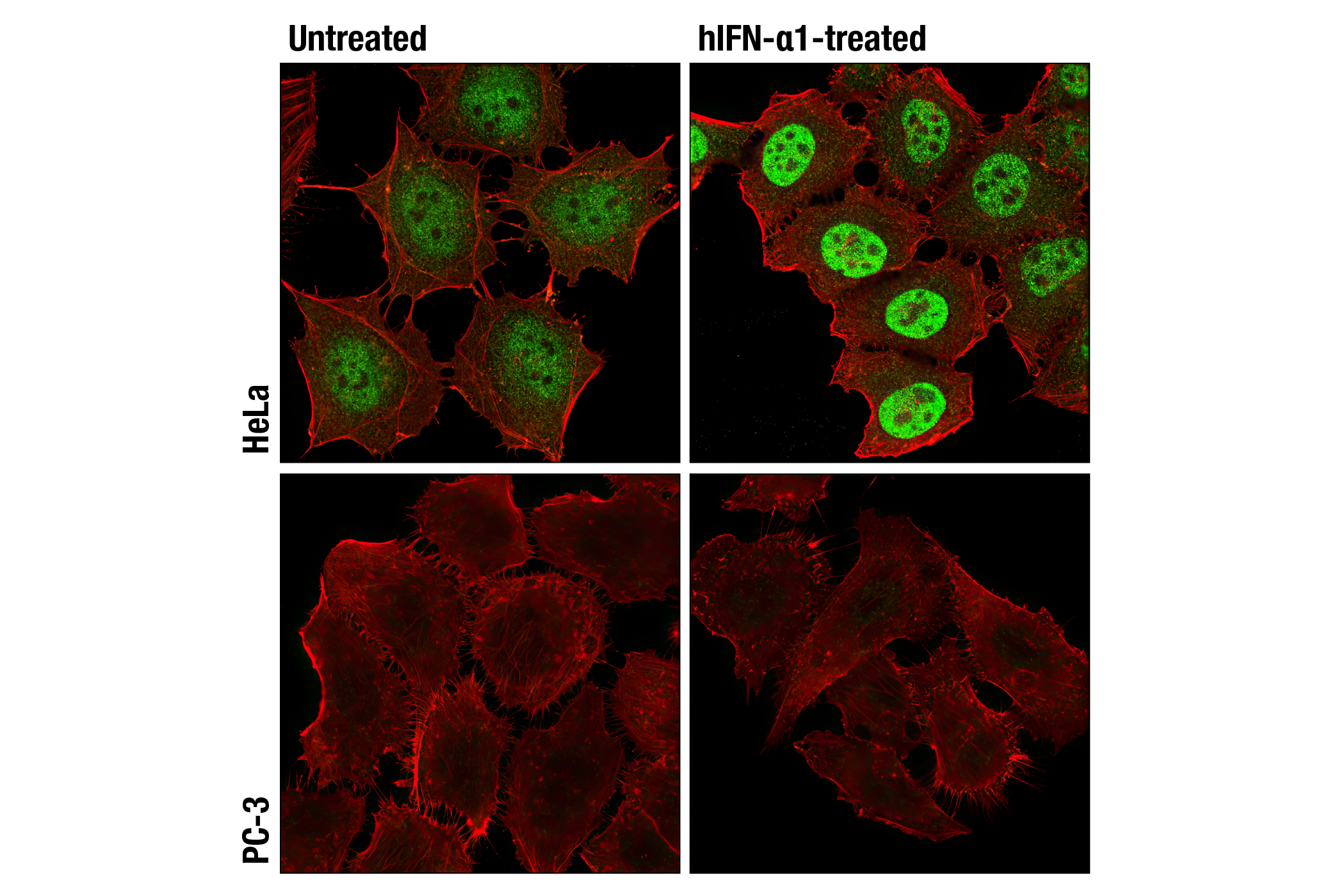
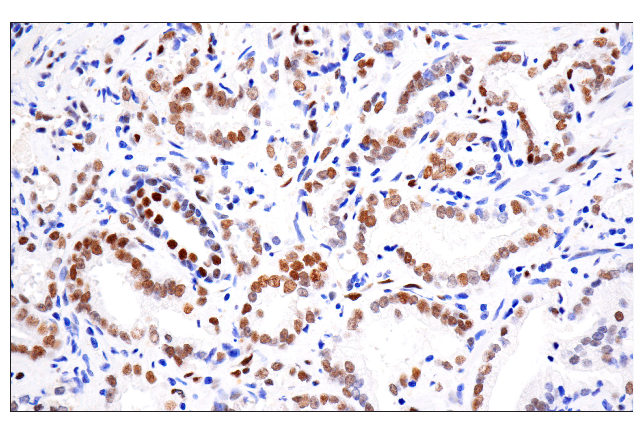
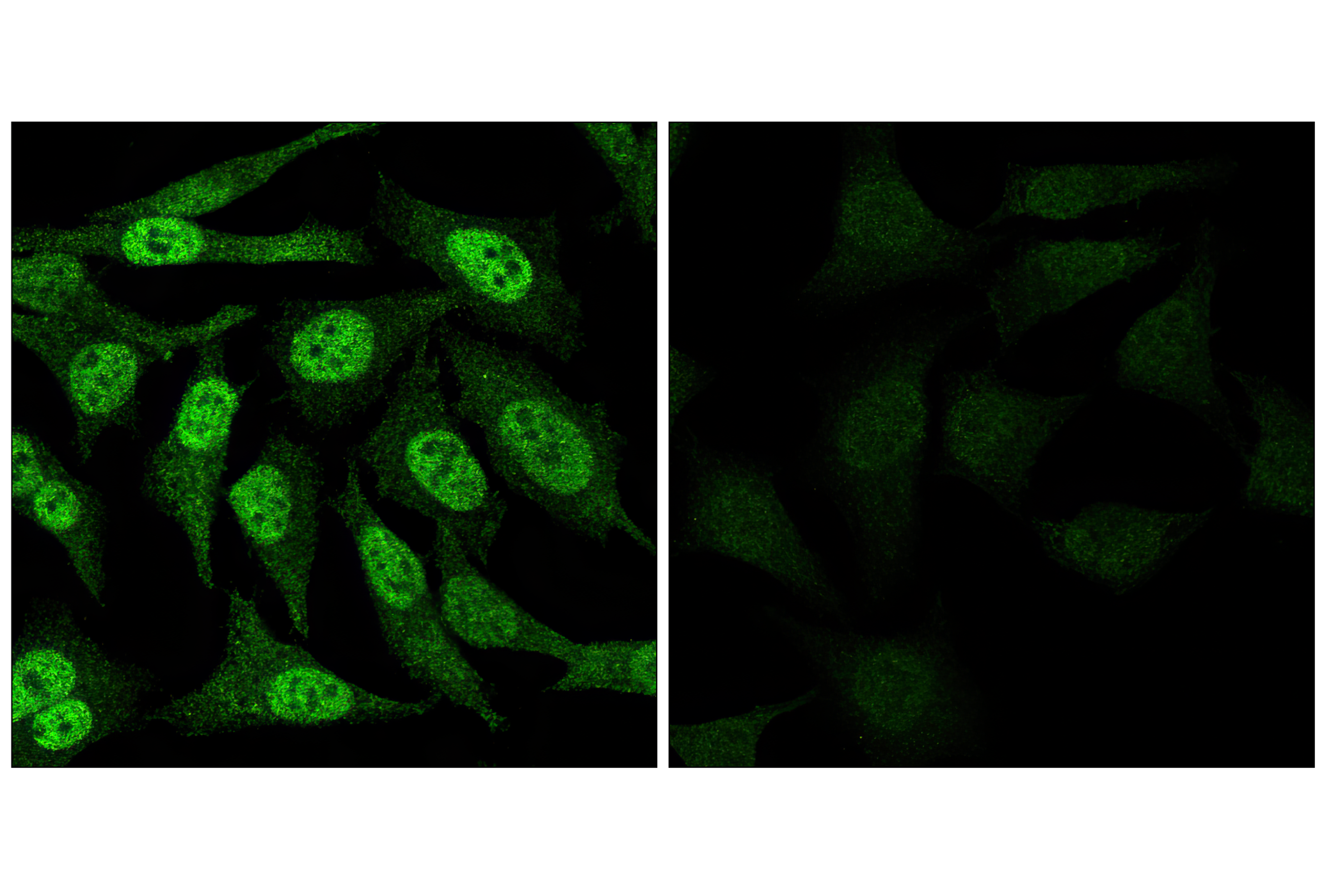
The Stat3/Stat5 Signaling Antibody Sampler Kit provides an economical means of detecting the activation of Stat3, Stat5, and Jak2 using phospho-specific and control antibodies. The kit includes enough antibodies to perform two western blot experiments with each primary antibody.
Storage
Background
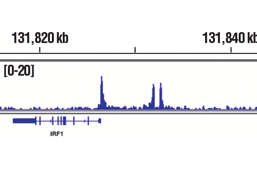
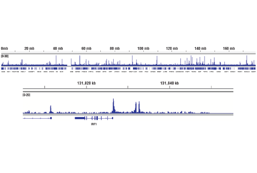
Janus kinases (Jaks) and signal transducers and activators of transcription (Stats) are utilized by receptors for a wide variety of ligands including cytokines, hormones, growth factors, and neurotransmitters. Jaks, activated via autophosphorylation following ligand-induced receptor aggregation, phosphorylate tyrosine residues on associated receptors, Stat molecules, and other downstream signaling proteins (1,2). The phosphorylation of Stat proteins at conserved tyrosine residues activates SH2-mediated dimerization followed rapidly by nuclear translocation. Stat dimers bind to interferon response element (IRE) and gamma interferon-activated sequence (GAS) DNA elements, resulting in the transcriptional regulation of downstream genes (1,2). The remarkable range and specificity of responses regulated by the Stats is determined in part by the tissue-specific expression of different cytokine receptors, Jaks and Stats (2,3), and by the combinatorial coupling of various Stat members to different receptors. Serine phosphorylation in the carboxy-terminal transcriptional activation domain has been shown to regulate the function of Stat1, Stat2, Stat3, Stat4, and Stat5 (1). Phosphorylation of Stat3 at Ser727 via MAPK or mTOR pathways is required for optimal transcriptional activation in response to growth factors and cytokines including IFN-gamma and ciliary neurotrophic factor (CNTF) (4,5). Jak/Stat pathways also play important roles in oncogenesis, tumor progression, angiogenesis, cell motility, immune responses, and stem cell differentiation (6-11).
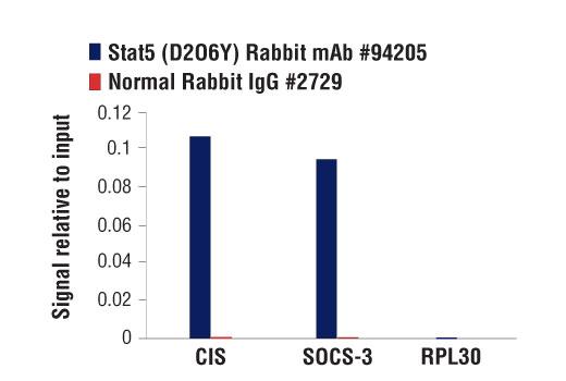
In the context of hematopoiesis, Stat3 and Stat5 may act antagonistically (12,13). Stat3 activity can promote differentiation of myeloid progenitor cells into neutrophils in a granulocyte colony-stimulating factor (G-CSF)-dependent manner, while Stat5 activation results in inhibition of this pathway in a granulocyte-macrophage colony-stimulating factor (GM-CSF)-dependent manner (13). Stat5 activity upregulates SOCS3, which subsequently inhibits Stat3 and results in differentiation to monocytes and macrophages (13). In addition to their roles in regulating gene expression as transcription factors, Stat3 and Stat5 are also capable of altering chromatin landscapes through recruitment of chromatin remodeling enzymes (14,15). While they serve many key functions in normal growth and development, if disrupted, the Jak2/Stat3 and Jak2/Stat5 signaling axes contribute to various diseases, including many types of cancer, non-alcoholic fatty liver disease, and eosinophilic cellulitis (16-20).
- Darnell Jr., J. et al. (1994) Science 264, 1415-1421.
- Leonard, W.J. and O'Shea, J.J. (1998) Annu. Rev. Immunol. 16, 293-322.
- Caldenhoven, E. et al. (1996) J. Biol. Chem. 271, 13221-13227.
- Wen, Z. et al. (1995) Cell 82, 241-250.
- Yokogami, K. et al. (2000) Curr. Biol. 10, 47-50.
- Lim, C.P. and Cao, X. (1999) J. Biol. Chem. 274, 31055-31061.
- Bromberg, J. F. et al. (1999) Cell 98, 295-303.
- Su, L. et al. (1999) J. Biol. Chem. 274, 31770-31774.
- Dentelli, P. et al. (1999) J. Immunol. 163, 2151-2159.
- Cattaneo, E. et al. (1999) Trends Neurosci. 22, 365-369.
- Frank, D.A. (1999) Mol. Med. 5, 432-456.
- Cohen, P.A. et al. (2008) Blood 112, 1832-43.
- Zhang, M. et al. (2023) Cell Death Discov. 9, 274.
- Wingelhofer, B. et al. (2018) Leukemia 32, 1713-1726.
- Orlova, A. et al. (2019) Cancers (Basel) 11, 1930.
- Warsch, W. et al. (2013) Blood 122, 2167-75.
- Halim, C.E. et al. (2020) Biomedicines 8, 316.
- Huang, B. et al. (2022) Front. Oncol. 12, 1023177.
- Kaltenecker, D. et al. (2019) Cytokine 124, 154569.
- Morot, J. et al. (2023) JAMA Dermatol. 159, 820-829.
Background References
Trademarks and Patents
Limited Uses
Except as otherwise expressly agreed in a writing signed by a legally authorized representative of CST, the following terms apply to Products provided by CST, its affiliates or its distributors. Any Customer's terms and conditions that are in addition to, or different from, those contained herein, unless separately accepted in writing by a legally authorized representative of CST, are rejected and are of no force or effect.
Products are labeled with For Research Use Only or a similar labeling statement and have not been approved, cleared, or licensed by the FDA or other regulatory foreign or domestic entity, for any purpose. Customer shall not use any Product for any diagnostic or therapeutic purpose, or otherwise in any manner that conflicts with its labeling statement. Products sold or licensed by CST are provided for Customer as the end-user and solely for research and development uses. Any use of Product for diagnostic, prophylactic or therapeutic purposes, or any purchase of Product for resale (alone or as a component) or other commercial purpose, requires a separate license from CST. Customer shall (a) not sell, license, loan, donate or otherwise transfer or make available any Product to any third party, whether alone or in combination with other materials, or use the Products to manufacture any commercial products, (b) not copy, modify, reverse engineer, decompile, disassemble or otherwise attempt to discover the underlying structure or technology of the Products, or use the Products for the purpose of developing any products or services that would compete with CST products or services, (c) not alter or remove from the Products any trademarks, trade names, logos, patent or copyright notices or markings, (d) use the Products solely in accordance with CST Product Terms of Sale and any applicable documentation, and (e) comply with any license, terms of service or similar agreement with respect to any third party products or services used by Customer in connection with the Products.